PICS
 Call Kai
 Learn
 Practice Test
 Spaced Repetition
 Match
 Flashcards
 Knowt Play
1/282

There's no tags or description
Looks like no tags are added yet.
283 Terms




[REVIEW] Valgus vs Varus


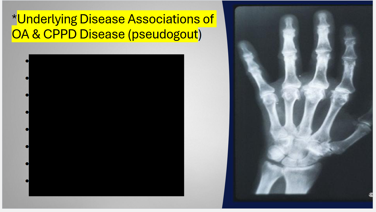
Radiograph of a hand affected by osteoarthritis Showing
(1) joint space narrowing,
(2) osteophytes,
(3) joint destruction.
(4) Changes @ carpometacarpal joint

Radiograph of the hips showing
(1) joint space narrowing and
(2) osteophyte formation.
What does a normal knee vs one w/ OA look like?


Radiograph of the knee in (A)anteroposterior and (B) lateral views showing
(1) joint space narrowing
(2) osteophyte formation.


How does the X ray look Different for OA vs RA






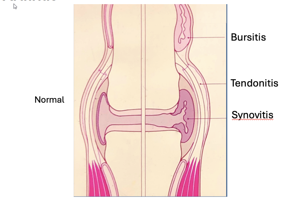
What are the Sites of Inflammation in Rheumatoid Arthritis


Moderate RA of PIPS, MCPS and wrists before treatment
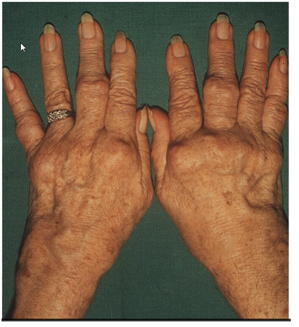
Severe RA with swelling of MCPs and wrists with PIP nodules
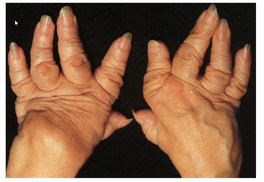
Severe RA: Arthritis Mutilans. A similar appearance is seen in Psoriatic Arthritis

L: RA: erosions in corners of a small joint
R: Synovial proliferation (pennus) causing erosions

RA: Erosions in the corners of a small joint
How does RA radiographs differ from OA?


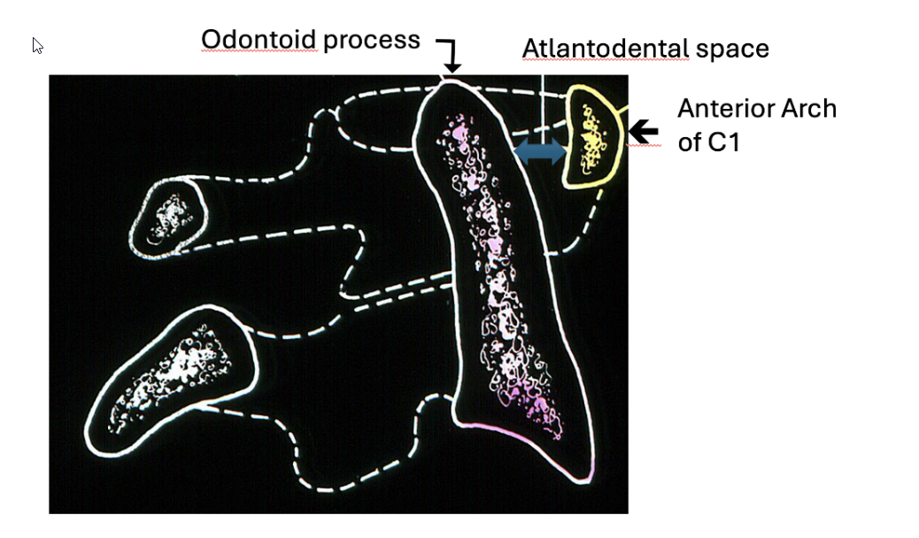

Atlantoaxial subluxation

Atlantoaxial Subluxation: Serial radiographs of the cervical spine, showing progressive widening of C1- C2 space. Flexion and extension xrays before surgery are important due to neurological risks of intubation in chronic RA patients

repair to stabilize C1-C2 subluxation

RA with nodules over PIP joints

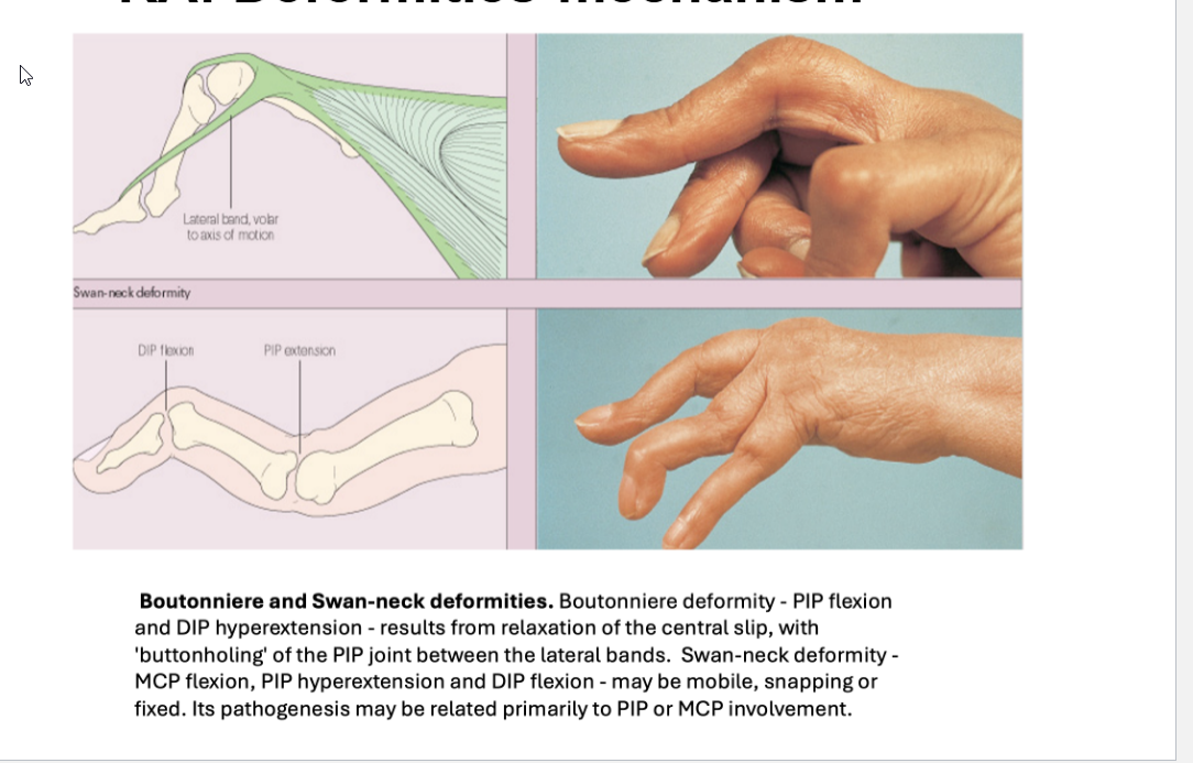
Swan Neck Deformity associated w/ RA
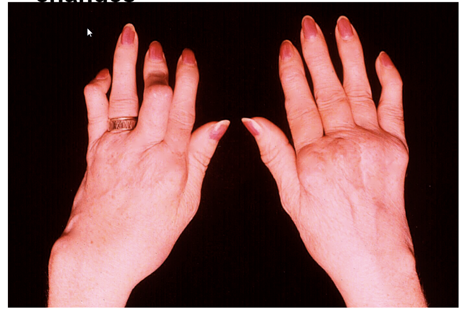
Swan neck, Boutonierre changes
Describe the Mechanism for Swan neck, Boutonierre changes
Describe the Foot Deformity associated w/ RA
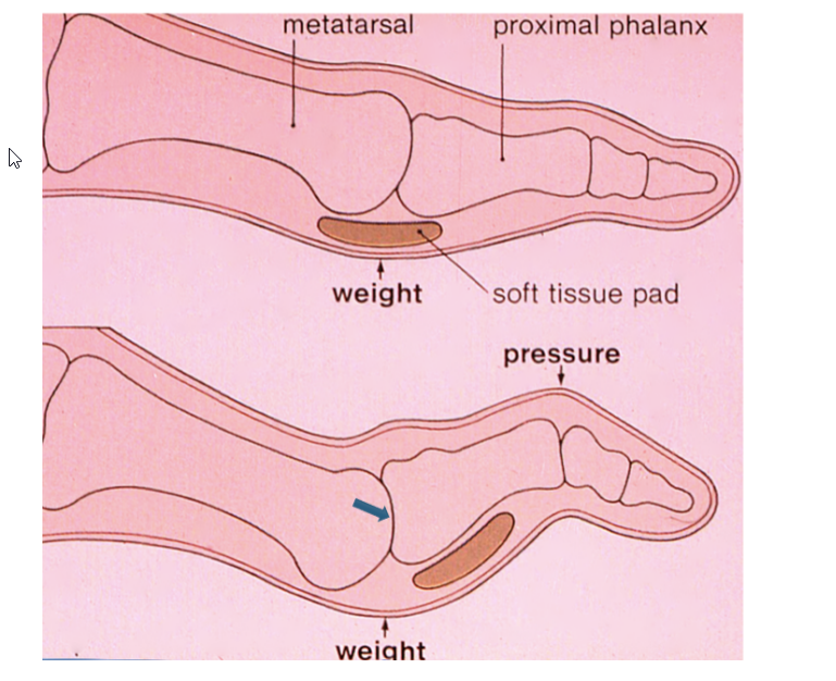

RA: Soft tissue ulcer due to deformity

RA: collapsed ankle joints

RA: extensor tendon rupture
Tendon retraction → bumps over wrist
Inability to straighten digits 4 and 5

Rheumatoid nodules on forearm

RA nodules: olecranon bursa

RA: Large Baker’s Cyst

RA nodule in the eye

RA: Inflammation of the sclera with thinning. (Scleromalacia perforans)

RA: Scleromalacia perforans
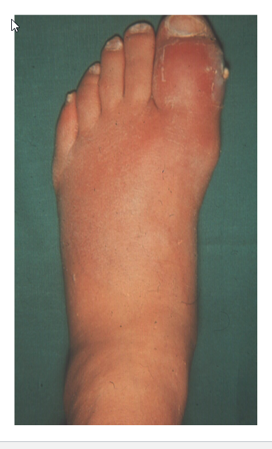
Gout: Acute Attack-Podagra
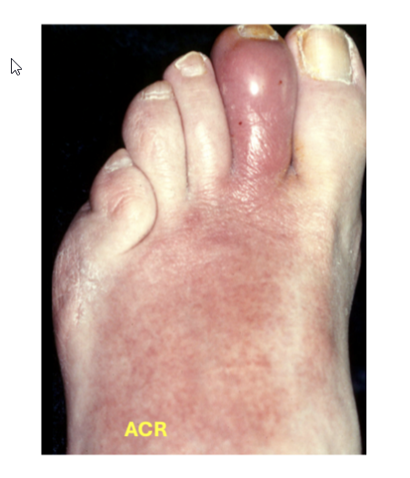
Gout: acute attack toe
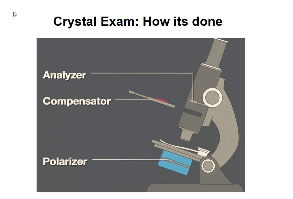
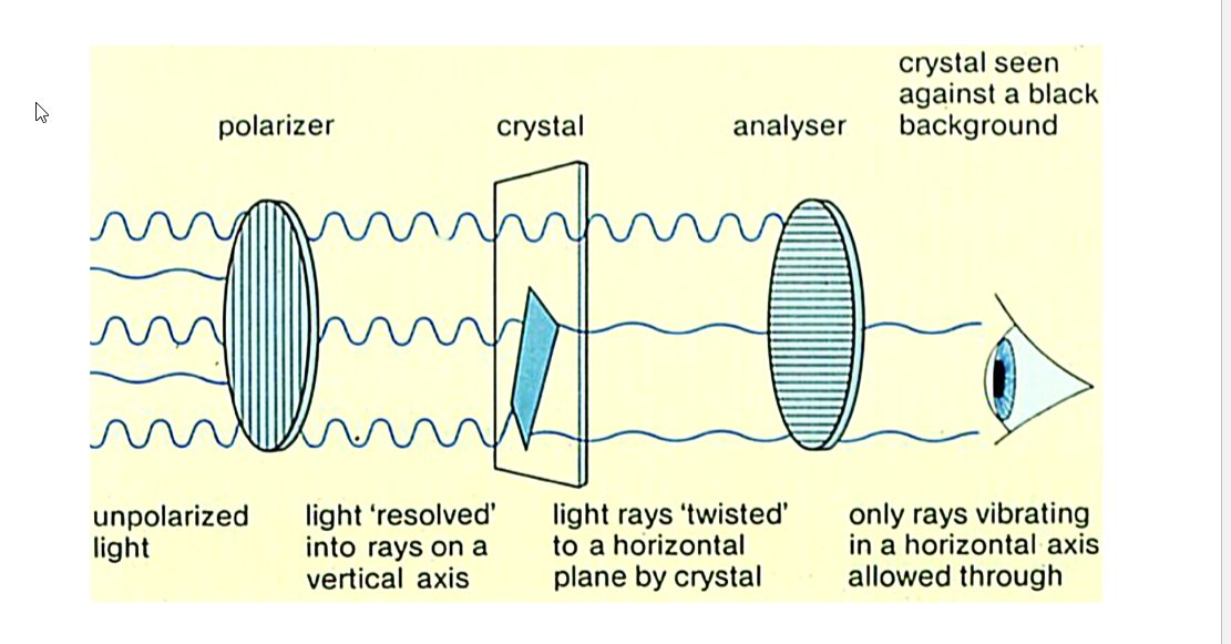
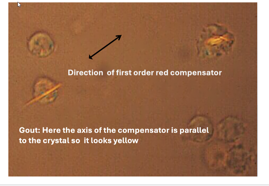
In a crystal exam for gout or pseudogout, what do you see?


Needle Shape Monosodium urate
Calcium pyrophosphate rhomboid shape

Gout: Intracellular monosodium urate crystals


Large Tophus in ear

Several tophi-ear

Gout – Tophi in Olecranon bursitis

Gout- tophi, uric acid leaking from skin
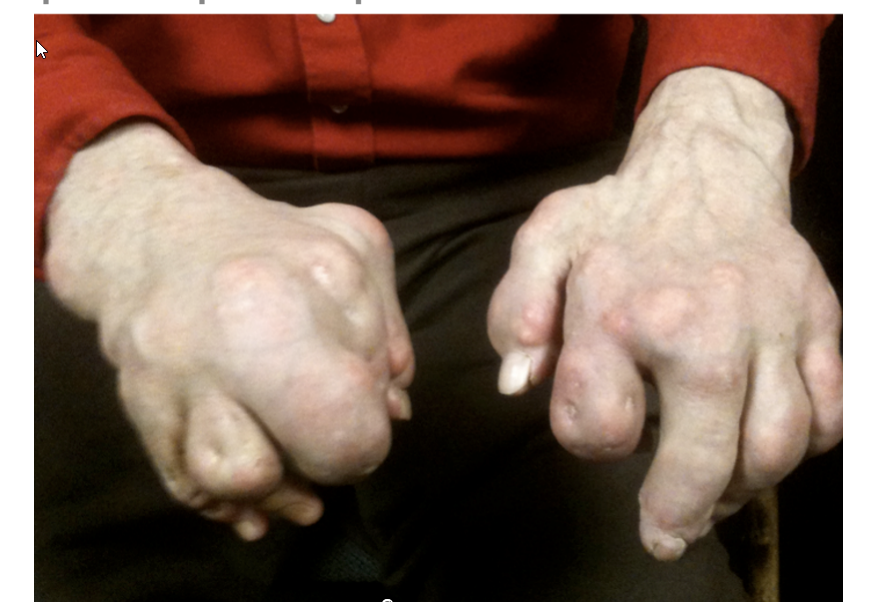
Gout: with tophi and scars where tophi have drained

Milwaukee shoulder
Top,
Diffuse swelling of left shoulder is present.
Bottom,
Radiograph of the same patient
diffuse joint- space narrowing of the glenohumeral joint associated with cortical erosions of the humeral head and distal clavicle.
upward displacement of the humerus, indicating a rotator cuff tear.
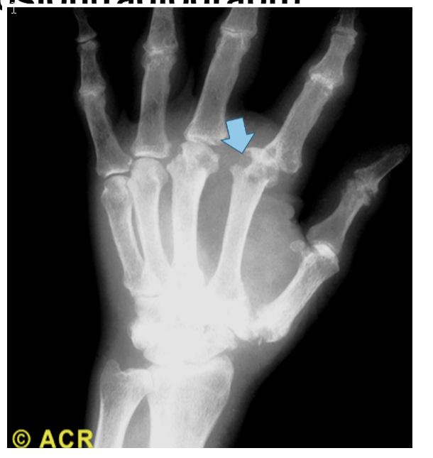
Gout: hand erosion(radiograph)

Gout: erosion of toe (radiograph)

Gout: Large erosion of great toe with overhanging edge (arrow)

Gout: Long standing with multiple erosions

Chondrocalcinosis: hand (radiograph)

Chondrocalcinosis: knee

Ankylosing Spondylitis: Limited lumbar flexion, limited chest excursion

Ankylosing Spondylitis: Limited lumbar flexion. Hip flexion contracture
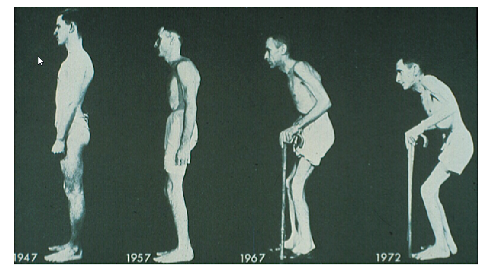
Ankylosing Spondylitis: Disease progression
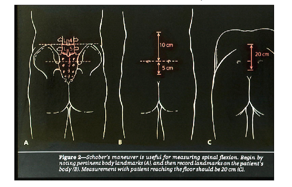
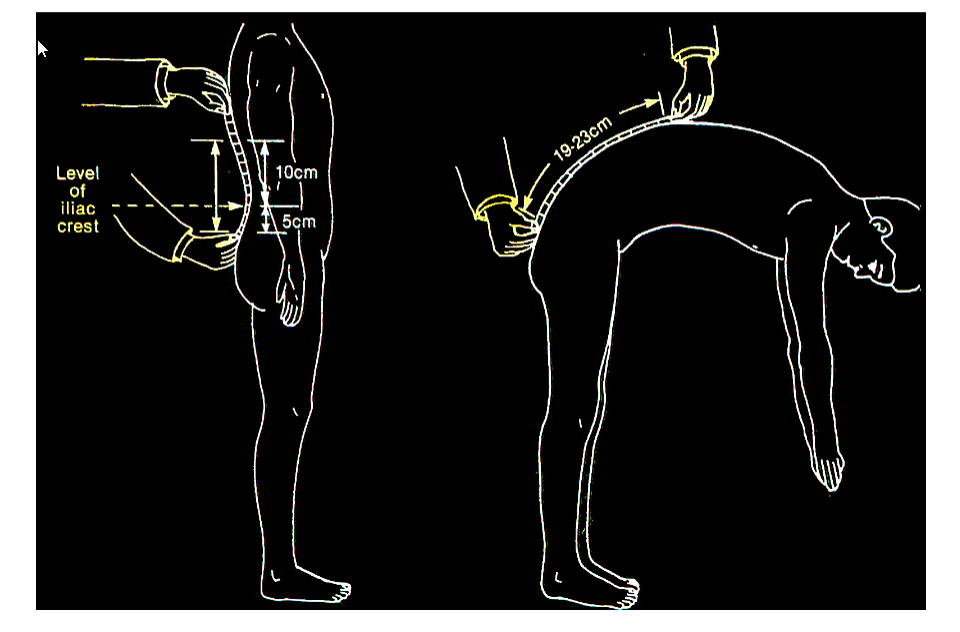


Ankylosing Spondylitis- Fusion of Sacroiliac joints

Pelvis: Bilateral Sacroiliitis

Pelvis: Tilt view: Bilateral erosive sacroiliitis

AS: Anterior Longitudinal Ligament Calcification, Marginal syndesmophytes, osteoporosis

AS- Bamboo spine, Sacroiliac joints fused and not well seen

AS-Cervical spine- lateral view complete fusion in a patient with ankylosing spondylitis for 40 years.
He later suffered a neck fracture (C6-7 level) after a trivial fall.

AS-Fracture through disc space
Lateral view of the lower thoracic and upper lumbar area.
There is ankylosis of the entire spine except at T12- L1.There is distraction (widening) of the disc space and fracture (arrow) through the posterior elements.

Psoriatic Arthritis- Severe Hand involvement

Psoriatic Arthritis

Psoriatic Arthritis- significant nail involvement
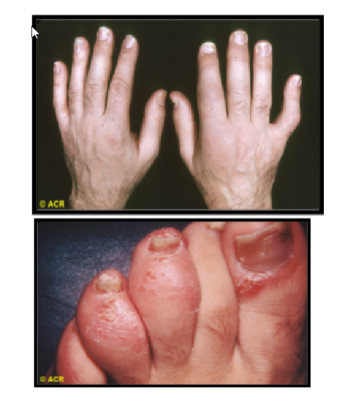
Psoriatic arthritis: musculoskeletal features
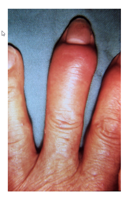


Psoriatic Arthritis - Arthritis Mutilans
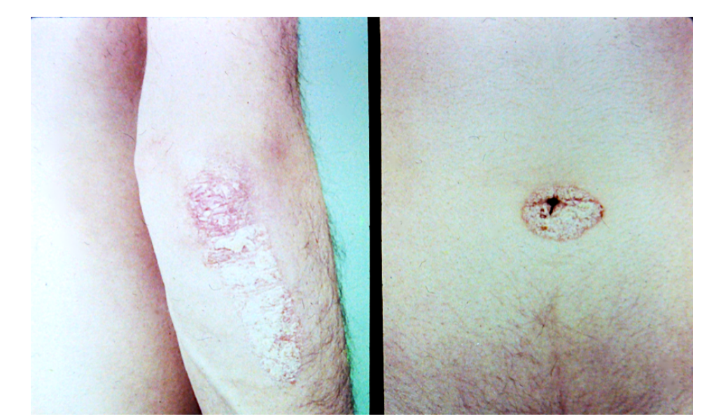
Psoriasis- may need to look carefully for rash

Psoriatic arthritis- Pencil in cup deformity

Psoriatic arthritis - bony ankylosis (fusion) of the DIP joint

Marginal VS Non-marginal syndesmophyte
Marginal = Ankylosing Spondylitis
Non-Marginal = PsA


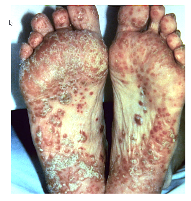
Reactive Arthritis:
Keratoderma Blenorrhagicum

Reactive arthritis: Keratoderma Blennorrhagicum
Skin biopsy often comes back from the dermatopathologist “psoriasiform dermatitis.
Discrete, circinate, scaly, and plaque-like lesions on the foot in reactive arthritis resemble secondary syphilis and psoriasis.
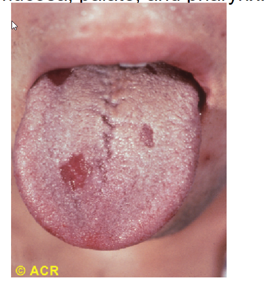
Reactive arthritis: erosions, tongue. Superficial erosions of the tongue in reactive arthritis are usually asymptomatic. Similar erosions occur on the buccal mucosa, palate, and pharynx.

Ankylosing spondylitis: iridocyclitis with synechiae
Ciliary injection and irregularity of the pupil are present. Adhesions between the iris and lens (synechiae) appear at the 1-o'clock and 4-o'clock positions. Synechiae are caused by repeated episodes of iritis and may eventually lead to glaucoma and blindness. The pupil is dilated from medication. The medial aspect of the sclera has been blanched by topical medication.

Arachnodactyly, Steinberg thumb sign, and Walker–Murdoch wrist sign in Marfan syndrome.

Thumb/Wrist Sign
Pec. Carinatum
Pec. Excavatum

Hindfoot Deform
Pes Planus
Protrusio Acetabuli
What are the SPINAL/ NEUROLOGICAL FINDINGS of Marfans? (2)

SPINAL/ NEUROLOGICAL FINDINGS
Dural ectasia,
widening of lumbosacral dural sac
Scoliosis or thoracolumbar kyphosis
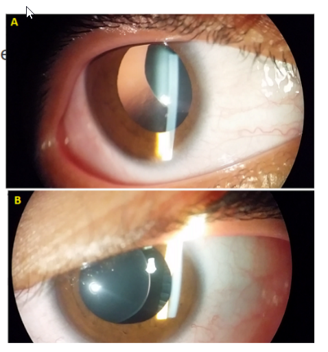
Ectopia lentis


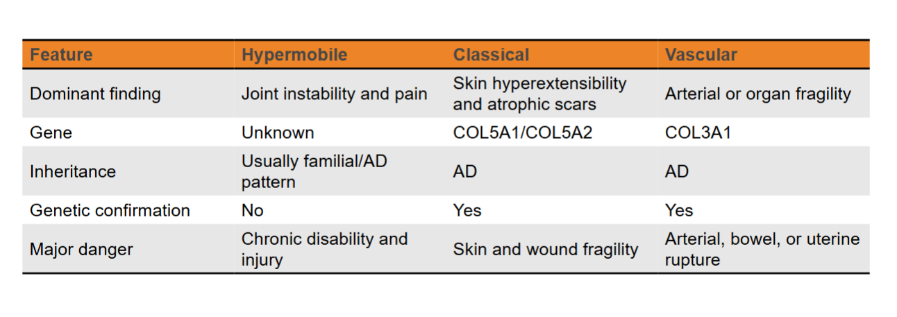
Left figure: signs of diffuse osteoporosis, anterolateral bowing deformity of the femur (shepherd's crook – red arrow), complete fracture of proximal femoral diaphysis (green arrow) White arrows pointing to zebra stripe sign from bisphosphonate therapy
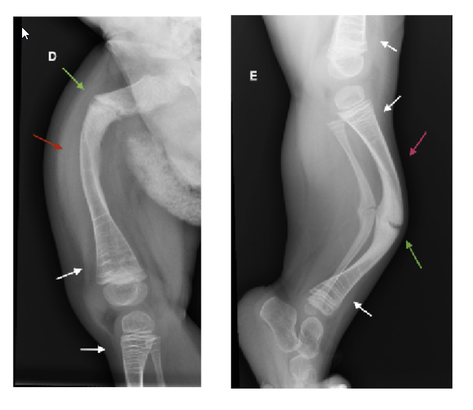
Right figure: anterior bowing deformity of the tibia known as saber shin (pink arrow) and incomplete tibia fractures (green)

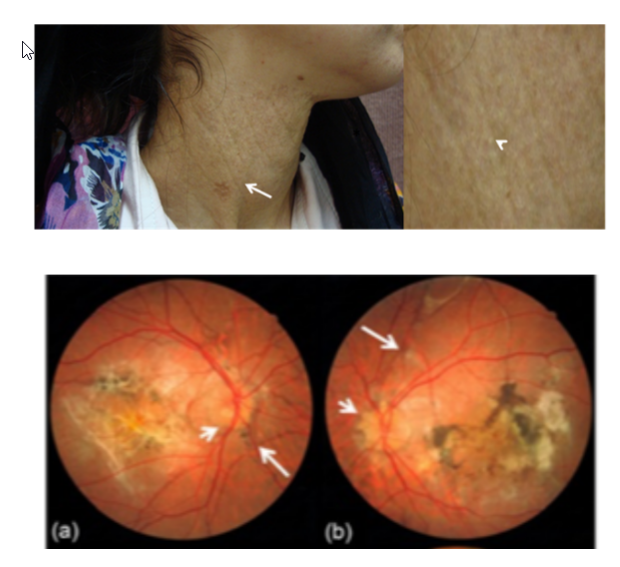
Small yellow papules coalesce into reticular or cobblestone plaques on the lateral neck. Affected skin may become lax and wrinkled
Angioid streaks radiate from the optic disc and represent breaks in the calcified Bruch membrane. Choroidal neovascularization may cause hemorrhage and macular scarring