Objectives: 15-16 and 7
 Call Kai
 Learn
 Practice Test
 Spaced Repetition
 Match
 Flashcards
 Knowt Play
1/53

There's no tags or description
Looks like no tags are added yet.
54 Terms
What are Acute Leukemias?
malignant disease of hematopoietic tissue, characterized by replacement of normal bone marrow elements with neoplastic blood cells
What are features of Acute Leukemia?
age: all ages
onset: sudden
course (untreated): < 6 months
Leukemic cells: immature
anemia: mild-severe
thrombocytopenia: mild-severe
WBC count: variable
organomegaly: mild
What is the main cytochemistry finding that indicates ALL
negative peroxidase and sudan Black B
What is the main cytochemistry finding that indicates AML
positive peroxidase and sudan black-B
positive for myeloid cells
What is Myeloperoxidase (MPO)
detects enzyme myeloperoxidase, which is found in granules of myeloid cells
used to identify myeloid differentiation
positive MPO strongly supports AML
rules out ALL
What are the results of Myeloperoxidase (MPO) stain?
Peroxidase is present in granules of myeloid cells
Promyelocytes/ seg neutrophils: pos
Eosinophils: pos
Monocytes: weak pos
RBC precursors / lymphs: neg
positive in myeloid cells
POSITIVE MPO rules out ALL
What does Sudan Black B (SBB) stain do?
detects fats and lipids within cells
stains graules of myeloid cells
Positive SBB supports AML
rules out ALL
What are the results of Sudan Black (SBB) stain?
fats/lipids: pos
mature segmented neutrophils: pos
eosinophils: pos
monocytes: variable
lymphocytes: negative
myeloid cells: pos
POS SBB rules out ALL
What is naphthol AS-D Chloroacetate (NASDA) esterase stain?
identifies granulocytic differentiation
neutrophils and their precursors stain positive
monocytes and lymphocytes stain negative
What happens in naphthol AS-D Chloroacetate (NASDA) esterase s
neutrophils/precursors: pos
monocytes/lymphocytes: neg
good for myeloid differentiation (granulocytes)
Describe Alpha-nephthl butryate esterase.
identifes monocytic differentiation
enzyme is found in:
monocytes
macrophages
T lymphocytes
Describe Acid Phosphatase stain.
produces red color in granules
found in all hematopoietic cells, highest level in: macrophages/osteoclasts
What happens in Acid Phosphatase stain?
monocytes: pos
neutrophils / precursors: weak pos
lymphocytes: pos
reaction can be inhibited by tartrate
TRAP is an isoenzyme found in lymphoid cells of hair cell leukemia
Describe PAS (periodic acid-schiff)
detects glycogen and carbohydrates in the cytoplasm
positive reaction produces a red color
What happens in PAS stain?
erythroblasts - pos
monocytes - faint staining
lymphocytes - few pos granules in ALL
lymphoblasts may show coars block pattern of activity
megakaryocytes - pos
Describe TRAP stain.
stain cells with acid phosphatase
in most cells the red color disappears
in Hairy cell leukemia → red color persists due to tartrate-resistant isoenzyme
primarily associated with hairy cell leukemia rather than AML
What is associated with Prefibrotic Primary Myelofibrosis (PMF)?
myeloproliferative of WBCs and megakaryocytes
atypical megakaryocytes
no BM fibrosis
increased megakaryocytes
PB:
immature granulocytes / normoblasts
tear drop cells
increased collagen due to increased PDGF (plt derived growth factor)
What is associated with Fibrotic Primary Myelofibrosis (PMF)?
BM fibrosis
Immature WBCs
nRBCs
What are the major criteria of PrePMF?
megakaryocyte proliferation without reticulin fibrosis > grade 1 with increased age-adjusted BM cellularity, granulocytic proliferation, decreased erythropoiesis
not meeting WHO criteria for BRC-ABL1, CML, PV, ET, MDS
presence of JAK2,CALR,MPL mutation or absence of these, presence of another clonal marker or absence of minor reactive BM reticulin fibrosis
What are the minor criteria of PrePMF?
anemia not attributed to a comorbid condition
Leukocytosis > 11 × 10^9/L
palpable splenomegaly
LDH> above upper limit
What is needed to diagnoseprePMF?
all 3 major criteria + 1 minor criteria
What are the major criteria of Overt PMF?
presence of megakaryocyte proliferation accompanied by either reticulin and/or collagen fibrosis grade 2 or 3
not meeting WHO criteria for ET,PV, BRC-ABL1, CML, MDS
presence of JAK2,CALR,MPL mutation or in absence of these, presence of another clonal marker, or absence of reactive myelofibrosis
What are the minor criteria of Overt PMF?
anemia not attributed to a comorbid condition
leukocytosis > 11 × 10^9/L
palpable splenomegaly
LDH > upper limit
Leukoblastosis
What is needed to diagnose Overt PMF?
meets all 3 major criteria + 1 minor
What AML subtype patches this description?
routine cytochemical stains negative
SBB, MPO, and esterases negative
M0 - minimally differentiated
What AML subtype patches this description?
MPO positive
M1 - without maturation
What AML subtype patches this description?
MPO positive
Auer rods may be present
M2 - with maturation
What AML subtype patches this description?
MPO+
SBB+
NASDA+
abundant granules + Auer rods
M3 - Acute promyelocytic leukemia (APL)
What AML subtype patches this description?
both granulocytic + monocytic differentiation
M4 - Acute myelomonocytic leukemia
What AML subtype patches this description?
monocytic differentiation
alpha-naphthyl butyrate esterase helps identify monocytic cells
M5 - Acute monocytic leukemia
What AML subtype patches this description?
PAS positive
Abnormal erythroid precusors
M6 - Acute erythroid leukemia
What AML subtype patches this description?
Megakaryocytic differentiation
identified with markers:
CD41, CD42b, CD61
M7 - Acute Megakaryoblastic Leukemia

Describe AML-M0
BM > 30% blasts
lack evidence of differentiation
no Auer rods
SBB-, MPO-, NASDA-
CD13+, CD33+, CD34+

Describe AML-M1
myeloblasts usually predominate in BM with minimal maturation
MPO +

Describe AML-M2.
myeloid precursor cells of all stages found in BM
Auer rods present
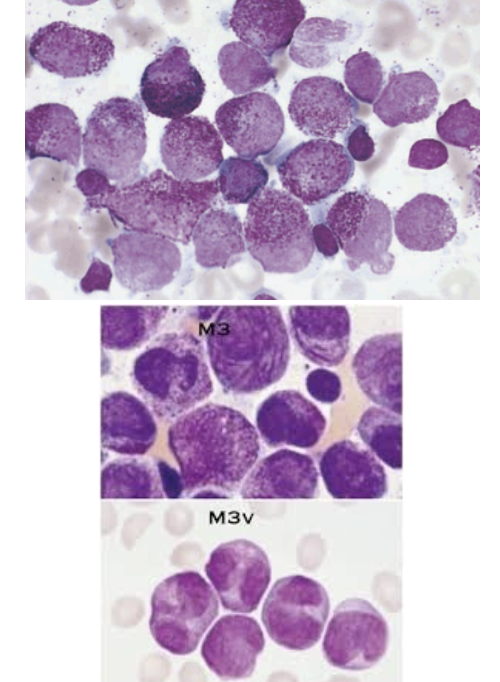
Describe AML-M3-APL
promyelocytes predominate
abundance of azurophilic granules
Auer rods present
correlation to DIC
t(15;17)
SBB+, MPO+, NASDA+
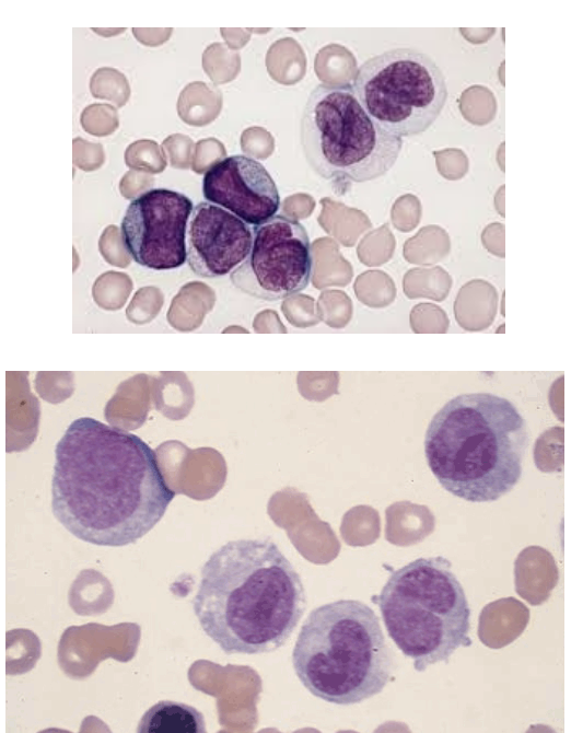
Describe AML-M4
monocytic and granulocytic precursor cells

Describe AML-M5a
monocytic leukemia
poorly differentiated
1-2 nucleoli

Describe AML-M5b
differentiated monocytic cells
monoblasts, promonocytes, monocytes
Low N:C

Describe AML-M6
abnormal erythroid precursors predominate
Bizarre nuclear shapes
Multinucleated giant forms
vacuoles in cytoplasm
may have Auer rods
Myeloblasts increased
PAS+

Describe AML-M7
> 20% of megakaryocytes and megakaryoblasts in Bm with increase in reticulum fibers, fibrotic growth
children with Down Syndrome
Giant PLTs seen
What major classification systems are used to classify acute leukemias?
FAB (French-American-British)
distinguishes lymphoblasts from myeloblasts
morphological / cytochemical characterization
divides AML into M0-M7 and ALL into L1 and L3
WHO (World Health Organization)

Describe FAB ALL-L1
morphology
small, monotonous lymphocytes, scant cytoplasm
high nuclear to cytoplasmic ratio
features:
uniform size, no prominent nucleoli
clinical features
common in childhood

Describe FAB ALL-L2
morphology
large, heterogenous lymphoblasts, variable size/shape
features
mixed L1-L3 type features, more cytoplasm than L1
irregular nuclear contours
clinical note
common in adult ALL

Describe FAB ALL-L3
morphology
large, uniform blast cells, abundant cytoplasm + vacuoles
features
“stary sky” appearance, vacuoles represent lipid droplets
associated with Burkitt lymphoma
clinical note
rare in ALL
What is in the WHO classification for ALL?
B lymphoblastic leukemia/lymphoma t(9;22)(q34.1;q11.2);BCR-ABL1
B lymphoblastic leukemia/lymphoma t(v;11q23.3);KMT2A rearranged
B lymphoblastic leukemia/lymphoma t(12;21)(p13.2;q22.1);ETV6-RUNX1
B lymphoblastic leukemia/lymphoma with hyperdiploidy
B lymphoblastic leukemia/lymphoma with hypodiploidy
B lymphoblastic leukemia/lymphoma t(5;14)(q31.1;q32.3) IL3-IGH
B lymphoblastic leukemia/lymphoma t(1;19)(q23;p13.3);TCF3-PBX1
Describe B lymphoblastic leukemia/lymphoma:
t(9;22)(q34.1;q11.2);BCR-ABL1
t(9;22)
children / adults
poorer prognosis
immunophenotyping
CD10+, CD19+, CD34+
treatment: chemo
Describe B lymphoblastic leukemia/lymphoma:
t(v;11q23.3);KMT2A rearranged
t(11q23.3)
CNS involvement, organomegaly
occurs in utero / after birth
immunophenotyping
CD10-, CD15, CD19+, CD24-
treatment: chemo
Describe B lymphoblastic leukemia/lymphoma:
t(12;21)(p13.2;q22.1);ETV6-RUNX1
20-30% of childhood preB ALL
good prognosis, 90% remissions
t(12,21)
immunophenotyping
CD10+, CD19+, HLA-DR+,CD13+, CD33+
CD9-, CD20-
Describe B lymphoblastic leukemia/lymphoma:
with hyperdiploidy
affects 25-33% of children (age 3-5), 5% of adults with B-ALL, not seen in infants
favorable prognosis
extra chromsomes: 4,6,17
blasts contain> 50 chromosomes, usually without transolocations
immunophenotype:
CD19+, CD10+, CD34+, CD45-
Describe B lymphoblastic leukemia/lymphoma:
with hypodiploidy
neoplasm of blasts committed to B cell lineage
comprises 5% of all ALL
blasts contain < 46 chromosomes
poor prognosis
except if patient has 44-45 chromosomes
immunophenotyping
CD10+, CD19+
treatment: chemo
Describe B lymphoblastic leukemia/lymphoma:
t(5;14)(q31.1;q32.3) IL3-IGH
Neoplasm of Blasts committed to B cell lineage
translocation between IL3 and IgH gene
variable eosinophilia
affects children / adults
< 1% ALL
immunophenotyping
CD10+, CD19+
treatment: chemo
Describe B lymphoblastic leukemia/lymphoma:
t(1;19)(q23;p13.3);TCF3-PBX1
neoplasm with blasts of B cell lineage
translocation between TCF3 on chromosome 19 and PBX1 on chromosome 1
more common in children than adults
6% of all B-ALL
immunophenotyping
CD9+, CD10+, CD19+, CD34-
Describe T-ALL
15-25% of patients with ALL
immunophenotype: CD7+, CD5+, CD2+, TdT+
CD4 and CD8 can be coexpressed
high WBC count > 100 × 10^9/L in 50% of cases
M>F
mass in chest area possible