anatomy and physiology practical midterm 1
 Call Kai
 Learn
Learn Practice Test
 Spaced Repetition
 Match
Match Flashcards
 Knowt Play
1/80

There's no tags or description
Looks like no tags are added yet.
Name | Mastery | Learn | Test | Matching | Spaced | Call with Kai | Chat |
|---|
No analytics yet
Send a link to your students to track their progress
81 Terms
what is mitosis
cell division where cell nucleus divides and creates two identical daughter cells
what are the stages of mitosis
prophase, metaphase, anaphase, telophase, and cytokinesis
what happens before mitosis
interphase
what happens in interphase and name the different stages
g1 phase - cell recovers from previous division and increase in size, synthesizes the enzymes organelles needed fro DNA replication
s phase - DNA replication happens
g2 phase - cell continues to grow and replenish energy, cell checks for any error or damage in replicated DNA before entering mitosis
what happens in prophase
chromatin coils into visible chromosomes
nuclear envelope disappears
spindle fibers attach to kinetochore of centromere
what happens in metaphase
chromosomes line up in middle of cell
what happens in anaphase
sister chromatids are pulled apart from each other to form individual chromosomes
what happens in telophase
Chromosomes reach the opposite poles and begin to decondense into chromatins
New nuclear envelope form around each new set of chromosomes
mitotic spindle breaks down
what happens in cytokinesis
cytoplasm divides
a cleavage furrow pinches the cell into two
happens during anaphase and telophase

what stage is this and what makes it obvious
cytokinesis since it looks like two cells are about to form/are divided and the chromosomes are decondenses to chromatin (become a lighter color/not noticeable)

what stage is this and what makes it obvious
telophase since DNA is still in chromosome form (the darker purple color/noticeable) and the nuclear envelope is beginning to form/cell is in middle of dividing

what stage is this and what makes it obvious
anaphase since it is still one cell but the sister chromatids are separated from each other to become individual chromosomes/are being pulled to opposite poles

what stage is this and what makes it obvious
metaphase since chromosomes are lined up in the middle of cell

what stage is this and what makes it obvious
prophase since it is a singular cell and the chromosomes are being condense to chromosomes/have a dark purple color
describe structure of Simple cuboidal epithelial tissue
one layer and cubed-shaped cells
one layer of circular nulcei located in the middle
location of simple cuboidal epithelial tissue
Kidneys tubules and surfaces of ovaries
function of simple cuboidal epithelial tissue
Secretion, Absorption, and move mucus

what is this
simple cuboidal epitheium

name the different parts of this simple cuboidal epithelium
A = Basement Membrane
B = Nucleus
C = Lumen
describe the structure of Simple columnar epithelial tissue
one layer and tall/column/rectangle shaped cells
oval shaped nuclei located near the basement membrane
have goblet cells
location of Simple columnar epithelial tissue
Stomach, Intestines and Uterus
function of Simple columnar epithelial tissue
Absorption and Secretion

what is this
simple columnar epithelial

name the different parts of a simple columnar epithelial tissue
A = simple columnar epithelial tissue
B = Nucleus
C = Goblet Cell
Function of goblet cell
create muscus
structure of Pseudostratified columnar epithelial tissue
look like stratified but is actually simple (one layer)
tall, thin cells
different heights of adjacent cells and positions of nuclei
almost always have cilia and goblet cells
function of Pseudostratified columnar epithelial tissue
secretion, movement of mucus, and protection
location of Pseudostratified columnar epithelial tissue
Respiratory tract

what is this
pseudostratified columnar tissue

label the different parts of this tissue
A = pseudostratified columnar
B = goblet cell
C = Cilia
structure of Stratified squamous epithelial tissue
multiple layers
flat cells near the apical surface
near basement layers, the cells look cuboidal
can be keratinized (no organelles or nucleus present) or non-keratinized (nucleus and organelles present)
function of Stratified squamous epithelial tissue
keratinized = waterproof, helps prevent water loss
non-keratinized = Remains moist and protects internal surfaces from abrasion
location of stratified squamous epithelial tissue
keratinized = epidermis
non-keratinized = Mouth, Esophagus and Vagina

what is this
stratified squamous epithelium
describe structure of stratified cubodial
multiple layers
cube-shaped cells
circular nuclei
function of stratified cubodial
Secretion and Protection
location of stratified cubodial
Sweat glands ducts and Salivary glands ducts

what is this
stratified cuboidal epithelium
describe structure of Transitional epithelial tissue
layers with all kinds of shaped cells, looks messy
on apicial surface, cells look like dome-shaped
looks cuboidal when unstretched and squamous when stretched
function of Transitional epithelial tissue
Stretching and Protection
location of Transitional epithelial tissue
Urinary bladder; stretches as bladders fills with urine

what is this
Transitional epithelial tissue
describe structure of Reticular connective tissue
Loose = Fewer fibers and more ground substance
decliate and mesh like framework
function of reticular connective tissue
Creates a supportive network
location of reticular connective tissue
Leukocytes, Lymph nodes, and Bone marrow

what is this
loose connective tissue, reticular
describe structure of adipose connective tissue
contains adipocytes, which are lipid-filled cells
function of adipose connective tissue
Protecting, insulation, and storage of energy

what is this
loose connective tissue, adipose
structure of dense regular connective tissue
protein fibers run parallel/looks like a straight line
more fibers and less ground substance
fiber protein = collagen and elastic
location of dense regular connective tissue
Tendons and Ligaments

what is this
dense regular connective tissue
describe structure of Hyaline cartilage
smooth
chondrocytes look less compact
fibers = collagen
function of hyaline cartilage
Provides smooth surfaces for joints
Allows for growth of long bones
Supports structures while maintaining flexibility
Helps keep airways open
location of hyaline cartilage
Nose, Trachea, end of bones at joints

what is this
Hyaline cartilage
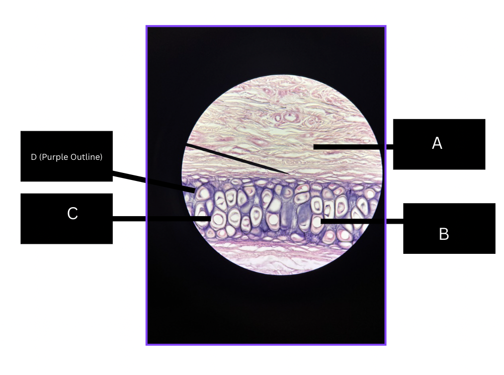
describe structure of Elastic cartilage
A = Perichondrium (dense connective tissue that surrounds cartilages)
B = Chondrocytes (cartilage cells that maintain cartilage tissue)
C = Lacunae (small cavities where chondrocytes live in)
D = Elastic Fibers
function of elastic cartilage
flexible but maintains shape
location of elastic cartilage
External ear, and Epiglotti

what is this
Elastic cartilage

what is this
compact bone

describe structure of the bone
A = oteocytes within lacunae
B = Lamae (bone matrix)
C = central canal of oseton
fibers = collagen

what is this
skeletal muscle
what are the layers of the Integumentary System (superficial to deep)
epidermis, dermis, and Subcutaneous tissue
what are the layers to the epidermis layer (superficial to deep)
stratum corneum, stratum lucidum, stratum granulosum, stratum spinosum, and stratum basale

what is this
stratum basale of the epidermal layer

what is this
stratum spinosum of the epidermal layer

what is this
stratum granulosum of the epidermal layer
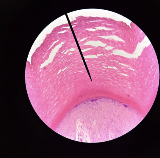
what is this
stratum corneum of the epidermal layer
what are the layers to the dermis layer (superficial to deep)
papillary dermis and reticular dermis
what happens in stratum basale
where skin cells (keratinocytes) form and divide cells
Produces new skin cells through mitosis - every 19 days
2 daughter cells: 1 stays at basement layer (attaches to hemidesmosomes), 1 goes superficially up (40-56 days)
Replaces old, damaged, or dead skin cells
what happens in stratum spinosum
8-10 layers
Still alive, stay metabolically active
Provide strength and flexibility to the skin
Helps cells resist stretching and physical stress
what happens in stratum granulosum
3-5 layers
Keratinization becomes more pronounced
Cells release lipids that help form a water resistant barrier
begin to die since no enough nutrients are provided here
what happens in stratum lucidum
thin layer
found only in thick skin
Provides protection in areas exposed to friction/palms and soles
what happens in stratum corneum
layers of dead, flattened, keratin filled cells
25+ layers squamous cells, joined by desmosomes
Maintain protective barrier and prevents excess water loss
Protects against bacterias, chemicals, and physical damage
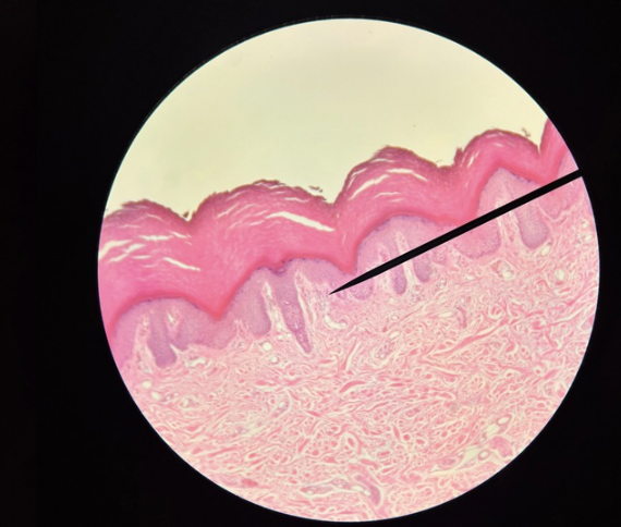
what is this
Papillary dermis of the dermis layer
what is the papillary dermis
thin and superficial layer of the dermis
located directly beneath the epidermis
Helps connect the epidermis to dermis to provide nutrients
Supplies oxygen, Temperature control, Sensation (detects touch, pain, temperature)

what is this
Reticular dermis of the dermis layer
what is the reticular dermis
deep, thick bottom layer of the dermis
sitting right above the subcutaneous tissue
Composed of dense irregular connective tissue

describe the layers of the integumary system
A = Stratum Corneum
B = Stratified Squamous Keratinized
C = Loose connective tissue
D = Dense connective tissue
function of the integumary system