UNIT 8. DNA SEQUENCING
 Call Kai
 Learn
 Practice Test
 Spaced Repetition
 Match
 Flashcards
 Knowt Play
1/80

There's no tags or description
Looks like no tags are added yet.
81 Terms
cDNA
DNA synthesized from a single stranded RNA
catalyzed by reverse transcriptase enzyme from retroviruses (e.g. HIV)
more common and efficient way because it does not contain introns unlike gDNA
1.Ethanol precipitation
2.Phenol-chloroform extraction
3.Minicolumn purification
3 DNA Isolation Techniques
Ethanol precipitation
3 DNA Isolation Techniques
usually by ice-cold ethanol or isopropanol.
Since DNA is insoluble in these alcohols, it will aggregate together, giving a pellet upon centrifugation. Precipitation of DNA is improved by increasing of ionic strength, usually by adding sodium acetate.
Phenol-chloroform extraction
3 DNA Isolation Techniques
phenol denatures proteins in the sample.
After centrifugation of the sample, denaturated proteins stay in the organic phase while aqueous phase containing nucleic acid is mixed with the chloroform that removes phenol residues from solution.
toxic reagents
Minicolumn purification
3 DNA Isolation Techniques
that relies on the fact that the nucleic acids may bind (adsorption) to the solid phase (silica or other) depending on the pH and the salt concentration of the buffer.

dNTPs can undergo chemical modification such as methylation, hydroxylation, and oxidation, forming a carboxylic acid
What is the challenge with dNTPs in DNA sequencing
Maxam-Gilbert Sequencing
Sanger Sequencing
Early DNA Sequencing Methods
Maxam-Gilbert Sequencing
Early DNA Sequencing Methods
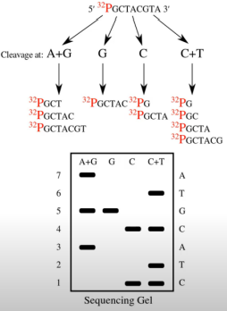
Radioactive label at the 5'-end
And then chemically treat the DNA to break off 1 or 2 bases at a time.
Visualization was done by gel electrophoresis (PAGE)
Difficult, time-consuming, toxic chemicals, and was NOT high accuracy.
Sanger Sequencing Method/ Chain Termination Sequencing
Early DNA Sequencing Methods
DNA sequencing method based on selective chain termination
etermines the nucleotide order by incorporating fluorescently labeled chain-terminating ddNTPs during DNA synthesis, separating the resulting fragments by capillary electrophoresis, and detecting their terminal bases through laser fluorescence.
ddNTPs (dideoxynucleotide triphosphates)
Modified bases with no 3' OH group, labeled with different fluorochromes for each base (A, T, G, C)
ause chain termination because no further nucleotide can be added once they’re incorporated.
Reaction mixture
Primer elongation & chain termination
Capillary gel electrophoresis separation of DNA fragments
Laser detection of fluorophores & computational sequence analysis
Chromatograph
Steps in Sanger Sequencing
Microfluidics
the science of manipulating very small volumes of fluids (microliters to picoliters) in tiny channels, often thinner than a hair.
It allows precise control of fluid movement, mixing, and reactions on a miniature scale.
requires less sample, less reagent, and gives faster results.
Lab-on-a-chip (LOC) Technologies
devices use microfluidics to integrate multiple laboratory processes—such as sample preparation, reaction, separation, and detection—onto a single small chip.
Faster
Portable
Cost-effective
Suitable for point-of-care testing
Illumina Sequencing
Roche 454 Sequencing
Ion Torrent Technology
Next Generation Sequencing
Illumina sequencing
Next Generation Sequencing
Genome fragmentation (100–150 bp reads)
The DNA is broken into short fragments (typically 100–150 base pairs). Adapters are attached to both ends to allow binding to the flow cell.Binding to the flow cell (not tiny wells)
The fragments attach to a glass slide called a flow cell, which contains oligonucleotides complementary to the adapters.
These fragments undergo bridge amplification, forming clusters of identical copies. Each cluster originates from a single DNA fragment.Addition of fluorescent nucleotides and DNA polymerase
The flow cell is flooded with DNA polymerase and four fluorescently labeled reversible terminator nucleotides (A, T, G, C).Terminator incorporation (one base at a time)
Only one nucleotide can be incorporated per cycle because each nucleotide has a reversible terminator that blocks further extension.Imaging
A camera captures the fluorescent signal emitted from each cluster. The color corresponds to the base added.Terminator removal and repeat cycle
The terminator and fluorescent label are chemically removed, allowing the next cycle to proceed.Same-length reads
Since one base is added per cycle across all clusters simultaneously, all fragments are extended one base at a time, making reads uniform in length.
Illumina sequencing
Next Generation Sequencing
uses bridge amplification and reversible fluorescent terminators to determine DNA sequences one base at a time through repeated imaging cycles.

Roche 454 Sequencing
Next Generation Sequencing
DNA fragments are attached to beads and amplified using emulsion PCR using adaptor-specific primers
Each bead is placed in a tiny well of a slide.
Nucleotides are added one at a time.
When a nucleotide is incorporated, pyrophosphate (PPi) is released.
This triggers a chemical reaction producing light, which is detected.
Roche 454 Sequencing
Next Generation Sequencing
Longer read lengths than early Illumina (~1 Kb)
sequencing multiple reads at once via reading optical signals (light) as bases are added
DNA varies in length
Disadvantages:
Difficulty reading long homopolymer regions (e.g., AAAA)
Expensive
Discontinued commercially

Ion Torrent sequencing
Next Generation Sequencing
DNA is fragmented and amplified on beads (emulsion PCR).
Beads are placed on a semiconductor chip.
When a nucleotide is incorporated, a hydrogen ion (H⁺) is released.
The release changes the pH, which is detected electrically (no fluorescence).
Faster sequencing time
No need for fluorescent labels or cameras
Lower instrument cost
30, 000 bases
Ion torrent technology uses a small USB that can read _________, however, is less accurate than the others.
Artificial Gene synthesis (DNA Printing)
Artificial gene synthesis is the process of chemically creating a DNA sequence from scratch, without using a natural DNA template. Instead of copying existing DNA (like PCR), scientists design the sequence on a computer and then chemically build it base by base.
Detritylation
Coupling
Capping
Oxidation
Steps in Artificial gene synthesis
Bioinformatics
•Collect statistics from biological data
•Build a computational model
•Solve a computational modeling problem
•Test and evaluate a computational algorithm
Bioinformatics
•Types of biological information and databases
•Sequence analysis and molecular modeling
•Genomic analysis
•Systems biology
Genomics
is the study of an organism’s entire genome — all of its DNA, including genes and non-coding regions.
It focuses on:
DNA sequencing
Gene structure and function
Gene regulation
Genetic variation
elps us understand diseases, evolution, personalized medicine, and biotechnology.
Comparative Genomics
compares the genomes of different organisms to identify:
Similar genes
Evolutionary relationships
Conserved sequences
Genetic differences
scientists can determine how species evolved and identify genes responsible for specific traits or diseases.
Proteomics
is the large-scale study of all proteins (the proteome) produced by a cell, tissue, or organism.
It focuses on:
structure
function
interactions
Post-translational modifications
Nutrigenomics
is the study of how nutrition affects gene expression and how genes influence the way the body responds to nutrients.
Cloning Vectors
are DNA molecules used to carry foreign DNA fragments into a host cell (usually bacteria) so the DNA can be replicated and sometimes expressed.
commonly as plasmids, bacteriophages, and artificial chromosomes
Origin of Replication (Ori)
Multiple cloning site (MCS) / Polylinker
Selectable Markers
Components of cloning vector
Origin of Replication (ori)
Components of cloning vector
is a specific DNA sequence where replication begins.
Allows the vector to replicate independently inside the host cell.
Ensures that the inserted gene is copied every time the cell divides.
Multiple Cloning Site (MCS) / Polylinker
Components of cloning vector
is a short DNA region containing several unique restriction enzyme recognition sites.
Allows insertion of foreign DNA using restriction enzymes.
Provides flexibility because different enzymes can be used.
Selectable Markers
Components of cloning vector
are genes that allow identification of cells that successfully took up the vector.
Antibiotic resistance genes
Ampicillin resistance (ampR)
Kanamycin resistance (kanR)
After transformation, cells are grown on antibiotic-containing media.
Only cells containing the plasmid survive.
Antibiotic resistance genes
Most Common Type of Selectable markers
Palindrome
a sequence that reads the same in the 5′ → 3′ direction on both strands (when complementary base pairing is considered).
5′-GAATTC-3′
3′-CTTAAG-5′
Restriction enzymes
______________ bind to palindromic DNA as two identical units and cut both strands at specific positions, breaking the DNA backbone.
Bacillus amyloliquefaciens strain H
BamHI was isolated from the bacterium:
Buts after the first Guanine base
5′-G↓GATCC-3′
3′-CCTAG↑G-5′
BamHI cuts where?
sticky ends with 5’ overhang (GATC)
BamHI produces what type of cuts
Reporter Genes
are genes attached to a regulatory sequence or gene of interest to indicate whether that gene is being expressed.
They produce an easily measurable signal, such as color, fluorescence, or luminescence.
Green fluorescent protein (GFP)
Common Reporter Genes
Produces green fluorescence under UV/blue light.
lacZ
Common Reporter Genes
Encodes β-galactosidase, which turns X-gal blue (used in blue-white screening).
Luciferase
Common Reporter Genes
Produces light in the presence of its substrate (luciferin).
Plasmids
Types of Cloning Vectors
small extrachromosomal DNA
can replicate independently
Small, circular double-stranded DNA molecules naturally found in bacteria, or archea
carry genes for survival and advantage of the organism (e.g. antibiotic resistance)
Bacteriophage
Types of Cloning Vectors
viruses that infect bacteria & archaea
more than 131 bacteriophage in the planet
Example is lambda phage
DNA packaged into viral particles
Efficient infection of host cells
Cosmids
Types of Cloning Vectors
Hybrid vectors combining plasmid features with bacteriophage λ cos sites.
Can be packaged into phage particles
Replicate like plasmids in bacteria
Used for large DNA fragment cloning
BACs (Bacterial Artificial Chromosomes)
Types of Cloning Vectors
Engineered plasmids based on the F plasmid of bacteria.
Very stable
Low copy number
Suitable for large DNA fragments
YACs (Yeast Artificial Chromosomes)
Types of Cloning Vectors
Vectors that replicate in yeast cells such as Saccharomyces cerevisiae.
Contain yeast centromere, telomeres, and ori
Behave like real chromosomes
Cloning very large DNA fragments
HACs (Human Artificial Chromosomes)
Types of Cloning Vectors
Synthetic chromosomes designed to function in human cells.
Contain centromere, telomeres, replication origins
Can carry very large genes
Dolly the sheep in 1996.
The first successfully cloned mammal was ___________________
Reproductive cloning
is the process of creating a new organism that is genetically identical to another (multicellular) organism.
CRISPR (Clustered Regularly Interspaced Short Palindromic Repeats)
is the process of creating a new organism that is genetically identical to another organism.
bacterial antiviral defense mechanisms
Origin of CRISPR
Guide RNA (gRNA)
Cas9 enzyme
PAM sequence (Photospacer Adjacent Motif)
Components of CRISPR-Cas9
gRNA
Components of CRISPR-Cas9
Designed to match a specific DNA target.
Guides Cas9 to the exact DNA sequence.
Cas9 enzyme
Components of CRISPR-Cas9
Acts as molecular scissors.
Cuts double-stranded DNA at the target site.
PAM sequence (Protospacer Adjacent Motif)
Components of CRISPR-Cas9
Short DNA sequence required for Cas9 binding.
Ensures proper targeting.
gRNA binds to the target dsDNA
Cas9 creates a double strand break in the dsDNA
Cell repairs break either through
NHEJ (Non-Homologous End Joining) - dresults to deletions/insertions
HDR (Homologous-Directed repair) - allow insertion or correction of genetic material
Steps of CRISPR
Nobel Prize Award in Chemistry (2020) to Jennifer Doudna and Emmanuelle Charpentier.
Nobel Prize won by CRISPR
Gene therapy (e.g., sickle cell disease & β-thalassemia)
Cancer immunotherapy
Functional genomics
Agriculture improvement
Disease modeling
Applications of CRISPR
Microarray
laboratory tool used to measure the expression of thousands of genes at the same time or to detect specific DNA sequences in a sample.
based on the principle of complementary base pairing (A–T and C–G).
a small solid surface (usually a glass slide or silicon chip) that contains thousands of microscopic DNA spots arranged in a grid pattern.
probes
Each spot in the microarray chip contains many copies (10-12 moles) of a known DNA sequence called _________
Isolation of mRNA
mRNA —→ cDNA using RT enzyme
cDNA is labeled with fluorophores
Red = diseased
Green = normal
Hybridization in the microarray chip
Washing of unbound sequences
Laser scanning & Imaging
Bright signal = high gene expression
Weak signal = low gene expression
Yellow (red + green) = equal expression
Steps in Microarray
• Cancer research (tumor profiling)
• Drug development
• Disease diagnosis
• Pharmacogenomics
• Toxicology studies
• Genotyping
Applications of Microarray
In situ hybridization (ISH)
is a laboratory technique used to detect specific DNA or RNA sequences directly within cells or tissue sections, while preserving their original location
allows us to see where a gene is located or expressed inside tissues.
Shows location of the gene
Maintains tissue structure
Provides spatial information
Tissue preparation (fixation w formaldehyde)
Permeabilization
Probe Hybridization
Washing
Detection
Steps of In Situ Hybridization
DNA In Situ Hybridization
Types of In Situ Hybridization
Gene mapping
Detecting chromosomal abnormalities
Detects specific DNA sequences in chromosomes.
RNA In Situ Hybridization
Types of In Situ Hybridization
Which cells express a gene
Level and pattern of gene expression
Detects mRNA expression.
temperature
salt
detergent concentration
Solution parameters in ISH that can be manipulated
radioactive —→ autoradiography
fluorophores —→ fluorescence microscopy
antigen —→ immunohistochemistry
ISH Probes are labeled with? and detected using?