oncology revision and cytology cases
 Call Kai
 Learn
 Practice Test
 Spaced Repetition
 Match
 Flashcards
 Knowt Play
1/23

There's no tags or description
Looks like no tags are added yet.
24 Terms
what does hitology examine
organisation of tissue structure
degree of cellularity
nuclear to cytoplasmi ratio
nuclear morphology
necrosis
mitotic index
individualisation of cells
invasivness of cells
for benign tumours describe
differentitation
growth rate
local invasion
metastasis
host consequences
well differentiated, similar to tissue of origin
slow, progressive
no invasion, caspsule
none
none, space ocupying lesion effects
for malignant tumours describe
differentitation
growth rate
local invasion
metastasis
host consequences
lack of differentiation, atypical structures
slow to rapid growth
local invasio, infiltrative, no capsule
frequent mets
life threatening
for bening tumours describe these cytological features
cellular morphology
nuclear to cytoplasmic ratio
nuclear morphology
mitotic count
necrosis
invasion of surrounding tissue
minimal variation in size and shape of cells (anisocytosis/ pleomorphism)
normal to reduced
minimal variation in size and shape of nuclei (anisokaryosis)
low mitotic rate, rare mitotic figures
none
none
for malignant tumours describe these cytological features
cellular morphology
nuclear to cytoplasmic ratio
nuclear morphology
mitotic count
necrosis
invasion of surrounding tissue
marked anisocytosis/ cellulr pleomorphism
increased (big nuclei)
marked anisokaryosis, multiple nuclei
high mitotic rate, lots of mitotic figures and abnormal
present
invasion, incl bv and lymphatics
what are the cytological features for criteria of malignancy
pleomorphism/ anisocytosis
anisokaryosis
muliple, bizarre shape nuclei
macrocytosis, macrokaryosis, macronuclei
anisonuceliosis
increased N:C ratio: less cytoplasm, more nuclei
nuclear molding (huggin nuclei)
coarse chromatin
abnormal mitotic figures
what is neoplasia
diesease characterised by abnormal cell growth
result of imbalance in mechs that regulate cell proliferation
what contributes to neoplastic growth
genetic and epigenetic alterations of somatic cell dna
multiple alterations are required
gain of functions in genes that promote cell proliferations (oncogenes)
loss of function in genes that oppose cell proliferation (tumour supressor genes)
§How you might expect neoplasia to present in our patients
see the lump
palpate the lump → reactive?
abnormal body functions → depends where the cancer is
How will you diagnose it?
fna
imaging
incisional and excisional biopsy→ histopathology
what is grading
Derived from histopathologic analysis of biopsy
May be predictive of behaviour, depending on tumour type
Helps answer the question of “what”
what is staging
Describes the extent of a tumour, often accounting for tumour size and presence of local and distant metastasis
Helps answer the question of “where”
for sarcomas
tissue of origin
most common route of metastasis
most common metastasis site
intial staging diagnostic
Mesenchymal
Haematogenous
lungs
thoracic radiography
for carcinomas
tissue of origin
most common route of metastasis
most common metastasis site
intial staging diagnostic
epithelial
lymphatic, haematogenous
regional LN, lungs, liver, spleen
regional/draining LN aspirate, thoracic xray, abd US
for round cell
tissue of origin
most common route of metastasis
most common metastasis site
intial staging diagnostic
immune system cells
lymphatic
regional LN, liver, spleen
regional/draining LN aspirate, abd US with liver/ spleen aspirate
list some round cell tumours
mast cell tumour, lymphoma, plasma cell tumour, histocytic tumour
what genes prevent cancer?
tumour suppressor genes
what genes promote cancer
oncogenes

What type of cell is this?
describe its appearance
Round cell
mortar lines

What type of cell is this?
describe its appearance
epithelial cell
- exfoliative
- clump together
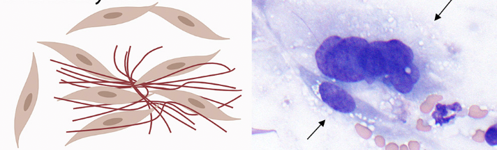
What type of cell is this?
describe its appearance
mesenchymal
- spindle shaped

what cell type is this ?
mesenchymal

what cell type is this ?
epithelial

what cell type is this ?
round