A6-A7 Pelvic Structure & Reoroductive Organs
 Call Kai
 Learn
 Practice Test
 Spaced Repetition
 Match
 Flashcards
 Knowt Play
1/11

There's no tags or description
Looks like no tags are added yet.
12 Terms

Pelvis

Pelvic Ligaments


Pelvic Muscles
Pelvic floor has:
Levator ani: pubo, ilio, puborectalis
Separate muscle: coccygeus
Fascia: connective tissue


Male Reproductive System
1. Epididymis: arches over the posterior and lateral sides of the testis. Sperm matures here
2. Ductus/vas deferens: Duct to transport sperm, the end expands to form the ampulla.
3. Ejaculatory duct: formed by Ampulla of the ductus deferens & seminal gland. Carries semen toward the urethra.
4. Urethra (terminal portion): Transports urine and semen (at different times).
Three regions of the urethra:
1. Prostatic urethra
Portion that passes through the prostate gland.
2. Intermediate (membranous) urethra
Portion located within the urogenital diaphragm.
3. Spongy urethra
Runs through the penis. Opens to the outside through the external urethral orifice.


Accessory Glands
Seminal Glands: posterior to the bladder. Secrets citric acid, fructose, coagulating enzyme and prostaglandins
Prostate: inferior to bladder, encircles urethra. Secretions contain nutrient source: citrate and enzymes
Bulbo: produces mucus for lubricant, neutralizes acidic of urine


Structure of the Testis

Erectile Tissue (transverse section)
Internal structures
Contains the spongy urethra and three cylindrical bodies of erectile tissue, covered by a sheath of dense fibrous connective tissue.
There are two types of erectile tissue:
A. Corpus spongiosum (singular)
The midventral erectile body. Surrounds the spongy urethra.
Distally: Forms the glans penis.
Proximally: Forms the bulb, which is part of the root.
B. Corpora cavernosa (two)
Two paired dorsal erectile bodies. Extend along the body of the penis.
Makes up most of the penis.
Their proximal ends extend backward and form the crura, which attach to the pelvic bones and make up part of the root.

Female Reproductive System (Vagina, Ligaments, Pouches, Cervix)
a thin-walled tube that lies between the bladder and the rectum and extends from the cervix to the body exterior
provides a passageway for birth and menstrual flow
Ligaments of Ovaries
The ovaries flank (lie on either side of) the uterus held by ligaments
Ovarian ligament: Anchors the ovary medially to the uterus.
Suspensory ligament: Anchors the ovary laterally to the pelvic wall.
Broad ligament
Mesosalpinx: Supports the uterine (fallopian) tubes.
Mesovarium: Suspends the ovary between the uterus and pelvic wall.
Mesometrium: supports the uterus (largest)
Round ligament
A cord-like structure on each side of the uterus. Helps keep the uterus positioned in the pelvis.
Uterosacral Ligament
Extends from the cervix and upper vagina to the sacrum (back of the pelvis).

External Female Reproductive System
also called the vulva or pudendum; external to the vagina
The mons pubis - a fatty, rounded area overlying the pubic symphysis
Labia majora - Runs posteriorly from the mons pubis are two elongated, hair-covered fatty skin folds, it covers the labia minora.
Labia Minora - The labia minora covers vestibule, that has external partsurethra and the vagina
Clitoris - a small, protruding structure (anterior to the vestibule) composed largely of erectile tissue; richly innervated with nerve endings sensitive to touch. It becomes swollen with blood and erect during tactile stimulation.
greater vestibular glands - flanking the vaginal opening; release mucus to keep the vestibule moist and lubricated
external urethral orifice - the opening of the urethra

Uterine/Fallopian Tube
start of the duct system
Each uterine tube begins laterally near an ovary and ends medially where it empties into the superior part of the uterus
infundibulum - the opening of the tube into the peritoneal cavity; its margin surrounded by ciliated, fingerlike projections called fimbriae that drape over the ovary
2. Ampulla: Middle of uterine tube, where fertilization usually occurs.
the isthmus - the narrow medial third of the tube, empties into the superior part of the uterus
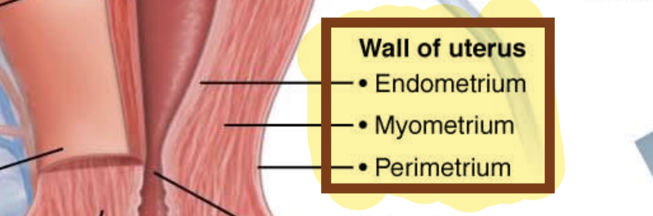
Parts of uterus and uterine wall
Layers:
The perimetrium - the outermost serous layer
The myometrium - the bulky middle layer; has bundles of smooth muscle
The endometrium - has mucosa, consists epithelium and a thick lamina propria
Parts of the Uterus:
Uterus
A hollow, muscular organ located: in front of the rectum & behind and above the bladder
Function: Receives, retains, and nourishes a fertilized ovum.
Regions of the uterus
1. Fundus
Rounded, uppermost region of the uterus. Located superior to where the uterine tubes enter.
2. Body
The major (largest) portion of the uterus.
Cervix
Narrow neck or outlet of the uterus. Projects into the vagina.
Isthmus
Slightly narrowed region between the body and cervix.
Cervix and cervical canal
Cervical canal: The hollow passage inside the cervix.
Connects the uterine cavity to the vagina through two openings:
Internal os: Opening between the cervical canal and the uterine cavity.
External os: Opening between the cervical canal and the vagina.
Cervical glands: secret mucus that fills cervical canal & external os. Helps block bacteria
