GENET Quiz #2
 Call Kai
 Learn
 Practice Test
 Spaced Repetition
 Match
 Flashcards
 Knowt Play
1/61

There's no tags or description
Looks like no tags are added yet.
62 Terms
Regulon
A regulon is a group of genes or operons that are regulated
coordinately in response to the same signal by the same regulatory
protein. The genes/operons comprising the regulon are usually
dispersed throughout the genome and have separate promoters
and other regulatory sequences. This does not mean that the
genes/operons are not also regulated by other regulatory proteins.
Regulons occur in both bacteria and eukaryotes.
The Major Players of the Gal Operon
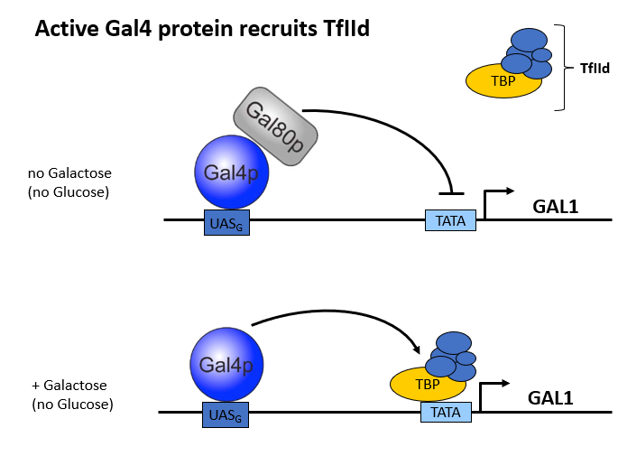
Gal 4p: Activator of the Gal Operon
Gal80p: Inhibitor of Gal Operon. ON average, it’s bound to Gal4 unless removed.
Gal3p: Activator: stops Gal80, makes it leave Gal4 alone, which then allows transcription to go forward
Mig1: In presence of Glucose, mig1 enters the cytoplasm and inhibts Gal4 as well as shutting down gal 1 and other transriptional spots
TfIID is the transciption factor recruited by Gal4
When is Gal regulon inhibited vs. activated?
Both of there processes only occur in absence of glucose because if glucose is present mig1 shuts the whole thing doen and none of this happens
Upstream Activator Sequences
Only work upstream of the transcription start site, never downstream or withing the gene. So they don't act like enhancers.
Binding site for activators, which then recruit transciription factors, TBP, and RNA Pol II
The dissociation model
• In the dissociation model, Gal80p and Gal3p bind to each other, and as a result, Gal80p
is removed from the Gal4p activation domain.
• Note: The dissociation could also occur in the nucleus, if Gal3p were able to enter thenucleus (unclear). However, if Gal3p were indeed exclusively located in the cytoplasm,then this would suggest that the dissociation model is correct and that Gal3p bindingto Gal80p occurs in the cytoplasm.

The non-dissociation model
• In the non-dissociation model, Gal80p and Gal3p bind to each other, but do so by staying attached to the Gal4 protein.
• Note: The non-dissociation postulates an interaction between Gal3p and Gal80p in the nucleus. If Gal3p is unable to enter the nucleus (unclear), the model would be incorrect, because Gal4p is always nuclear and always bound to DNA.

chIP
Chromatin Imunopreciation
“Enriched” = Isolated

Heparin
Molecule with a similar sugar phosphate backbone to DNA which makes it similar enough for binding. It goes like this:
1. Isolate nuclei
2. Nuclear extract (remove proteins from DNA into your tube)
3. Run isolated proteins through heparin sepharase so all nuclear proteins that aren't DNA binding will run thrrough it, and the ones that are DNA binding will stick
Now you only have DNA binding proteins
Major vs. Minor Groove of DNA
Major groove is taller than minor groove. Alpha helices fit perfectly into major groove but not minor groove, so most DNA binding proteins use alpha helices as recognition helices and are major groove binding proteins. Also, there's more chemical information in the major groove.
What's a domain?
- Usually folds independently (doesn't require rest of protein)
- Autonomous function. Often works independently when fused to other proteins, or even in isolation
- Usually evolutionarily conserved, and gene family members have domains in common (except activation domains). In addition some structurally similar domains exist that aren't evolutionaily conserved (ex. Helix-turn-helix is a structural motif)
Helix-Turn-Helix (HTH)
- Structural Motif (Domainish)
- VERY common in bacteria
- Evlutionarily reinvented several times: similar structures found in multiple species despite having no evolutionary relation
EX.
- Lac repressor
- Region 4 of sigma factor (binds to -35 element)
- Homeodomains in eukaryotes

Basic Region Leucine Zipper (bZip)
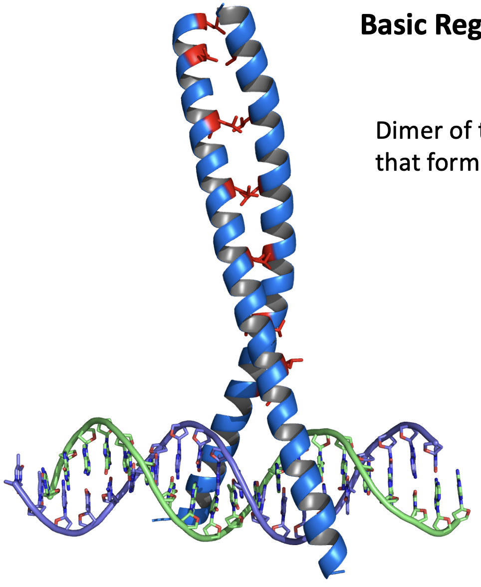
Dimer of two alpha helices that form a coiled coil
- At the top, the two helices are joined by a leucine every ~7 amino acids
- The top zipper part has nothing to do with DNA binding, and is just a dimerization motif
- The bottom part of the zipper is rich in Basic amino acids and binds to DNA
- The Basic Region is positively charged, which helps it bind to the negatively charged DNA
Basic Helix-loop-Helix domain (bHLH)
- Has an two sets of alpha helixes joined by a loop
- The two strands are joined into a leucine zipper
- At the bottom of the dimer is a basic region where DNA can be bound
C2H2 Zinc Finger Domain (ZNF-C2H2)
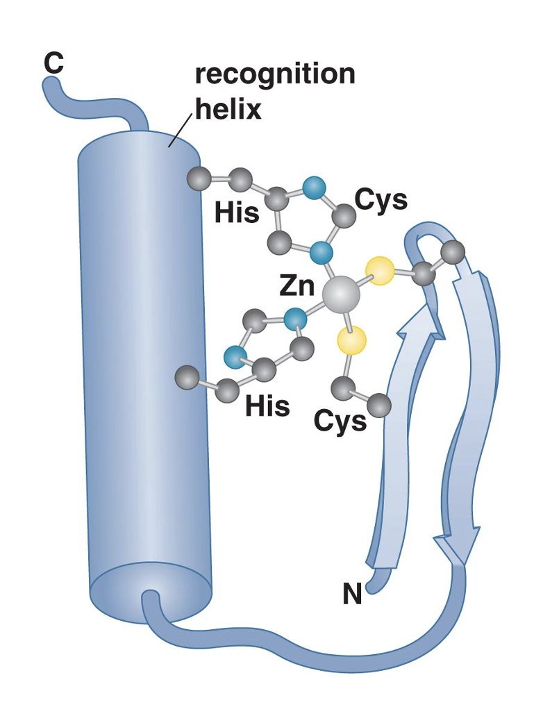
- Two anti-parallel beta-sheets and an alpha helix are stabilized bya zinc atom
- Often in multiple repeats, from 1 to >30
- Spacing between repeats highly variable
- 1 repeat contacts ~3 bp
The four most common DNA Binding Domains
ZNF-C2H2: Zinc Finger
Homeodomain (have helix-turn-helix motif)
BHHLH: Basic Region Helix-Loop-Helix
bZip: Leucine zipper
NHR: Nuclear Hormone Receptor
Nuclear Receptors
- Not membrane bound, localize to the nucleus
- Bind to DNA via a pair of C4 (because 4 cystines) zinc fingers which form a dimer
- Bind Hormone Resonse Elements (short DNA sequence in the promoter that tells hormone receptor when to turn off transcription)
found in all multicellular animals, absent in plants & fungi
48 proteins in humans, 21 in Drosophila
bind steroid hormones, certain lipids and some other small lipophilic molecules
some nuclear receptors bind drugs and toxins
Critical regulators of:
inflammation
maintaining salt concentrations
development
detoxification pathways
cholesterol homeostasis
Pathways very simple and fast (comparitively)
The 3 Types of Hormone Respone Elements
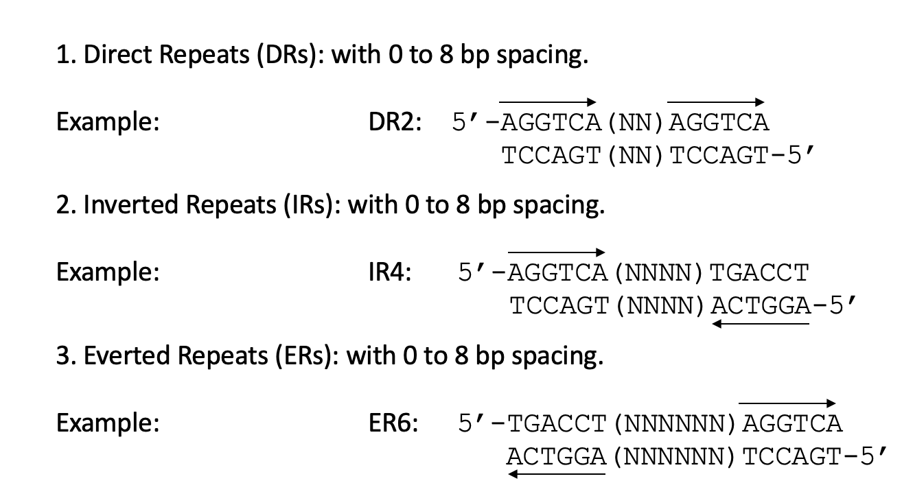
What are some methods we can use to test binding or Regulatory DNA Elements?
- DNAse I Footprinting
- EMSA: Electrophoretic Mobility Shift Assay
- ChIP: Chromatin Immunopreciation
Electrophoretic Mobility Shift Assay
- Strictly works in vitro
BASIC IDEA: Free DNA runs faster than protein bound DNA, so it will appear further down on the gel
- Incubate a protein (purified or mixture of thousands of proteins) with a radioactive piece of DNA
- Obviously DNA binding proteins want to bind to DNA
- Gel is non-denaturing, so protein-DNA complexes remain intact throughout the procedure
- Expose the gel to detection device that detects radioactivity and can tell you where all the DNA ends up
THE TYPES OF DNA: In this method, along with the radioactive DNA, two other types of DNA must be added for the method to work.
- Cold (meaning unlabeled) Specific Competitor DNA: fragment of DNA containing the suspected dna recognition seuqence, usually slightly mutated. Whether or not the proteins bind to this lets you know if it can compete with the unaltered DNA. If it can, then the mutation didn't affect the binding site and the band will disappear. If it didn't compete, the mutation is messing with the binding site.
- Nonspecific Competitor DNA: Added in high amoints to saturate non-specific binding. Required because the proteins have non specific binding activity as well as specific, so if you put only specific DNA, that doesn't say much because without anything more specific to bind to, the protein would bind to non-specific DNA too, giving you no information about where it binds ideally. Adding competitors tells you which one the protein would actually choose.
THE ANTIBODIES:
A way to test if a sample of mixed proteins contains a specific one. If you have an antibody of the suspected protein, you can throw it in and find the protein, weighing down the protein as well as the DNA it's bound to, and leaving a band high up on the gel (called a Supershift).
DNAase I Footprinting
BASIC IDEA: DNA-bound protein protect DNA from digestion
- RADIOLABEL dna fragment at one end (must be on one end or you won't get unique fragments)
- Incubate protein w/ DNA
- Digest DNA with low concentration of DNAse I (such a low concentration that there is one cut per fragment)
- The presence of a protein means the DNA can't cut in that spot, so it must move up or down and cut there, which means there's unique lengths of DNA that act like a fingerprint
- Basically tells you where the protein is on the DNA
- Run fragments on high resolution gell (gel is denaturing, so the protein is separated from the DNA and the double helix is melted into two strands)
- Map location of "footprint" based on size of fragments
The Three Types of Activation
- True Activators: have activation domains composed of either i) acidic residues, ii) glutamine, or iii) proline. These activation domains interact with GTFs
- Chromatin Factors: remodellers and histone modifiers
- Architectural: bending and bridging
Domain Swapping
- The only real way to tell if a protein has a distinct activation domain

Co-Activators And Their Two Main Modes of Action
Job is to bind to a DNA bound transcription factor and increase rate of transcription and boost gene expression. Bacteria don't have them.
Mode 1: "True" activation via direct interaction with GTFs to recruit RNA Pol II.
Mode 2: Acting or recruiting proteins that open chromatin, makign DNA accessible for other DNA-binding proteins
Activation Domains
- In contrast to DNA-binding domains, activation domains (ADs)noften don't have a conserved three dimensional structure and arne't well defined
- Rather, activation domains are defined by amino acid content and often small repeats. For example, Gal4 AD is rich in acidic and hydrophobic amino acids
- The potency of an activation domain depends on its overall charge, resulting in sticky surfaces. This variation from domain to domain and allows fine tuning of interactions between ADs and Mediator and/or GTFs.
What do activation domains interatc with?
They contact Mediator and specific TAF subunits of TFIID. There's no evidence that ADs contact RNA Pol directly
VP16
- Present in the virus that causes herpes
- Hijacks Host Cell Factor I and a transcription factor called OctI. Oct I has its own activation domain.
- VP16 is an activation domain, so it can't bind to DNA alone, but once its a part of this (hijacked) complex it can bind
- The complex then activates first genes needed by virus to make more viruses

The Three Types of True Activation Domains
Acidic (D, E): Example: GAL4 or VP16
Glutamine-Rich (Q): Example SP1 or Jun (binding sites in many proximal promoters) or Oct-1.Widespread motif in transcription factors
Proline-Rich (P): CTF/NF1
Squelching
Overexpression of a known activator reduces actvating strength of other activators
Mediator
- Interacts with the CTD tail of RNA Pol ii and stimulates TFIIH to initiate CTD Phosphorylation
- Individual subunits only affects expression of a small subset of genes, function of many subunits unclear
- Likely exists in various forms,s dpeending on cell type, or even within cells (promoter context)
- Four functional modules: head, middle, and tail form the core, and then the kinase.
- Head and tmiddle interact with Pol II and the tail with activators
- Med 17: head, Med 15: tail

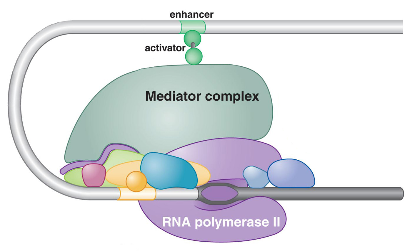
The Activator Bypass Experiment
- Usually we need Gal4 to activate Gal1.
- In this experiment, they placed lexA (bacterial DNA binding domain) site in the Gal operon
- They attached lexA to the Med15 of the mediator to see the operon would function
- Usually Gal4 would bind to UAS and the activation domain interacts with Med15 and then recruits the mediator and turns on Gal1, but here, we don't have any activation domain, only Med15 fused with lexA.
- Is this enough to turn on Gal1? YES!
- This proves you don't need toe activation domain in the context and also that the main job of the activation domain is to interact with the mediator and recruit with RNA pol to the promoter
Reporter Genes
A reporter system uses a proxy signal when the endogenous biological process is difficult or impossible to measure directly.
2 types: translational and transcriptional
What are they being used for?
Reporter genes allow you to easily study the activity of genes or proteins.
In vivo, they recapitulate some or all aspects of the native gene
Some are suitable for live-imaging (typically fluorescent proteins) or labelling selected cells (e.g., to visualize specific neuronal connections)
Others require the addition of substrates to detect the reporter gene product.
In most cases, they reside in the genome as an independent transgene.
Sometimes, they are designed to replace the endogenous gene (“knock-in”).
Transcriptional Reporter Genes
Idea: You want to know when and where a specific gene is expressed.
Approach:
Make a construct to fuse the reporter gene to the gene of interest.
Fused to transcriptional region
In most cases, one would replace the gene body with the reporter.
Typically, one would leave the 5’ UTR of the gene in place, but one could
also fuse the first codons of the gene in frame with the reporter gene.
One would insert the finished reporter gene construct into the host genome/cell (transgenic organism or transfection of cells)
Using Reporter Genes to identify tissue-specific roles of enhancers
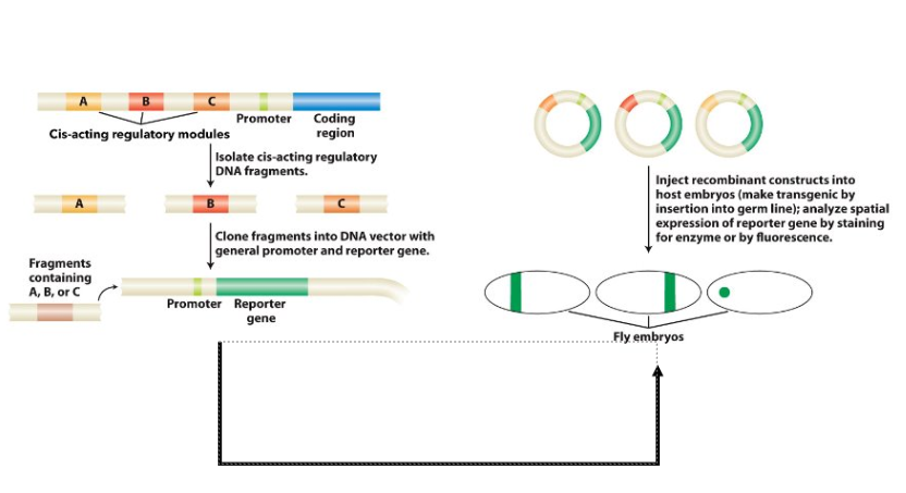
The regulatory region of Drosophila even-skipped (eve) gene

Lethal Reporter Genes (how Mig1 was discovered)

It was clear there had to be a glucose responsive site somewhere here to shut down Gal1
Translational Reporter Genes
Idea: You want to know when and where the protein is made.
Approach:
Make a construct to fuse the reporter gene to the coding region.
In most cases, the reporter is added to the N- or C-terminal end of the protein.
Typically, the resulting chimeric protein is the sum of both proteins, but sometimes
only parts of the endogenous protein (e.g. conserved domains) are being used.
Translational Reporters can in theory double-function as transcriptional reporters, but they are harder to make and may be less useful in some cases (e.g. protein is quickly secreted or unstable).
To be useful, “Translational Reporter Genes” must retain some or all of the original properties present in the endogenous protein.
Promoter Bashing

Basically you shrink the promoter of the gene (after hooking it up to reporter) to identify which regions are important for full gene expression
GFP
Green Fluorescence Protein (GFP).
Mode of detection: Fluorescence
Works in many organisms (codon bias is the only known issue in some species)
b-galactosidase (lacZ) as reporter
Mode of detection: histochemical (colored product, e.g., from X-gal)
Gene isolated from E. coli
Not used in plants because of endogenous b-galactosidase activity
b-glucuronidase (GUS)
Mode of detection: histochemical (colored product, e.g., from X-gluc)
Gene isolated from E. coli
Not normally used in vertebrates because of background b-glucuronidase activity
Luciferase
Mode of detection: luminescence (here: bioluminescence)
Most commonly used luciferase gene isolated from the firefly Photinus pyralis
Key Concept of Promoter, Enhancer, and Gene Traps
Use transposon that carries
a reporter gene
Let transposon insert
randomly into host genome
Generate 10,000s of lines,
resulting in one transposon
insertion per line (these lines
are called transgenic lines)
Screen lines for reporter
activity that have expression
pattern that is of interest
to you (e.g. specific neurons)
Type 1 Enhancer Trap

Type 1: Single reporter transgene inserts near enhancer (pic)
We hunt enhancers. Problem with this technique is that enhancers can act on very large distances so it can be hard to find the gene. An enhancer could be acting over 10 genes and you would have no idea which gene it was working on. Potentially quite slow. You have no idea where the enhancer is.
Lots of positive, you'll find lots of enhancers, but then you have to find out which gene its associated with.
Promoter Trap

TATA box missing, so the reporter gene doesn't have its own promoter. This trap requires that the reporter gene jumps into an exon. So the reporter is inserted in the middle of an exon, but it must be in the correct orientation (5'-3') and in the correct reading frame. So there's a 1 in 6 chance that it can be produced properly, which is why the protein synthesis cuts off in the diagram. If this works, you only have to sequence the flanks of your promoter gene and can be immediately sure what gene it is. So lots more screening to do because it's so rare that the reporter fits properly into a gene to be expressed (remember no promoter) but if it works, you know the gene.
A mutant is generated.
Gene Trap
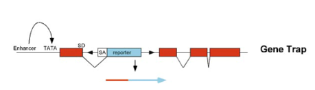
Similar problems to promoter trap: may be inserted upside down (3' to 5') and maybe out of reading frame. The big difference is that this time its inserted into an intron, which are much bigger than exons. So higher probability you get into a gene because of the bigger size. So you have to do a lot more work up front, but you are more likely to hit and again, you would automatically know what the gene is. No promoter, but the SA and SD tells you how it continues transcription.
A mutant is generated
Type 2 Enhancer Trap
Type II: Binary reporter system composed of Gal4 (transgene #1 – the driver) and
UAS-reporter (transgene #2 - the responder)
Gal4 driver line is controlled by a nearby enhancer
To make Type II work:
The Gal4 driver is crossed to “responder” line
The responder line harbors another transgene under UAS control
The transgene can be a reporter or a cDNA or construct that has
inverted repeats leading to RNA interference (RNAi, see later lecture)
Gene X in the responder line can be:
Reporter Gene (reveal activity, label cells)
cDNA (e.g. overexpress a gene)
RNAi construct (interfere with function)

The eyeless Gene in Drosophila
Reasearchers took gal4 driver line. What they wanted to do was express a gene called "eyeless" since they had a theory that "eyeless" was the master control gene, the top gene of eye developement.
What they found was that just the expression of this gene was sufficient to turn on eye develpoment on a flies leg. This means the gene can develop eyes wherever its expressed .
Eyeless gene has close homolog in vertebrates called pax6. The asked, can the trasnplantation of pax6 gene in flies create eyes too? Yes! Pax6 is known to be the master control gene of eye development in the mouse
These experiments proved that insect and vertebrate eyes evolved from a commonancestral eye, which was completely opposite of what the belief was at that time.
The two groups of nuclear receptors
Group 1: reside in cytoplasm when unliganded
ex. steroid hormone receptors (ex. Estrogen receptor, androgen receptor, etc.)
Homodimers
Inverted repeats
Group 2: bound to DNA even when unliganded
ex. VDR, SXR, CAR also orphan receptors
Heterodimers/homodimers/monomers
Direct repeats
Everted repeats
Classes of Ligands for Nuclear Receptors
AGONIST: Most nuclear receptors have this sort of agnist structureto be activated. Binding of ligand removes co repressor and recruits co acivator, reulting in transcription/gene activation
ANTAGONIST: Displaces agonist and also co activator. Neither co-activator nor co-repressor found, so it's basically a deactivation. Blocking agonist, but not blocking any constitutive activity .
INVERSE AGONIST: Few nuclear receptors don't require an agonist to recruit co-activator and are activated by default. Din't look at this diagram; its messed up. Basically, after the first step two things can happen: Either the agonist comes in and removes the activator, leading to basal transcription, or the agonist comes in and recruits a co-repressor, leading to no activation

Sex hormones

Xenobiotics vs. Endobiotics
Xenobiotics: Foreign substance that would never exist naturally in our body and have dangerous properties
Endobiotics: toxins produces in our bodies
St. John’s Wort Drama
Some emdications can have interactions with other medications
In the case of a woman who was on Cyclosporin for a recent organ transplant, St. John’s Wort makes the cyclosporin more inert and caused transplant rejection
Drug (St. John’s Wort in this example) binds to PXR (nuclear receptor that’s a dimer with RXR) and recruits co-activator
This leads to transcription of CYP3A gene, making theCYP3A enzyme
CYP3A enzyme lives in liver and intestines, and is extremely verstile and can detoxify ~50%60%of chemicals in existence. IT detoxifies other drugs, like Cyclosporin in this example.
Because St. John’s Wort binds to PXR and increases transcription, it increases production of the CYP3A enzyme which then increases dtoxification of Cyclosporin
The 2 possible consequences of drug interactions
Underdose Risk: One drug makes the other less effective/causes the other to be processed too quickly, leaving too little in your system for its purposes
Overdose risk: Grapefruits contain ferranocoumarins which bind directly to CYP3A in an irreversible inhibition, making the enzyme non functional. This means the drugs cannot be broken downs, which means i stay in your system for longer than it should, causing overdose isk
Vitamin D
STEROID PROHORMONE
once metabolized, a ligand for VDR
Consequences of severe Vitamin D3 deficiency (“classic symptoms”)
- Rickets (children)
Osteomalacia (adults):
“softening of the bone”
…due to reduced absorption of
calcium
Risk factors for Vitamin D3 deficiency:
Malnutrition
pigmented skin
Living in Canada and other Northern countries
Obesity
Advanced age
Vitamin D Metabolism

Lithocholic Acid vs. Vit. D
Low affinity ligand for VDR

Slow vs. Fast Twitch Muscle
Type I fibers (slow twitch): are mitochondria-rich and mainly use oxidative metabolism for energy production and are fatigue-resistant.
Type II fibers (fast twitch): comprise three subtypes, IIa, IIx, and IIb. Type IIb fibers have the lowest levels of mitochondrial content and oxidative enzymes, rely on glycolytic metabolism as a major energy source, and are susceptible to fatigue Type IIa and IIx lie between type I and IIb.
The Three important molecules of Excersize
AMPK = AMP-activated protein kinase (AMPK), AMP = Adenosine Monophosphate
PGC1a = Peroxisome proliferator-activated receptor gamma (PPAR) coactivator-1 alpha
PPARd = Peroxisome proliferator-activated receptor delta
SuperMice
PPARdelta = PPARd is normally highly expressed in skeletal muscle.
Researchers fused VP16 to PPARd and expressed the transgene specifically in skeletal muscle tissue of mice.
This resulted in increased Oxidative Type I Fibers in the Transgenic Mice.
The transgenics were dubbed the “marathon mouse”, because they had double the endurance without prior training.
The natural ligand for PPARd is not known.
GW1516, a synthetic ligand acts as an agonist for PPARd (leads to ~70% increased performance). Another compound, AICAR, activates PPARd indirectly via AMPK and PGC-1a (see next slide).
AMPK and its role in metabolism
AMPK has three binding sites for ATP. The mosr AMP you have, the more likely all three sites will be full
When you excerise and burn a lot of energy, the last thing you wanna do is save that energy as glycogen. (we need the energy). So we inhibit anabolic processes which build up glycogen and thus fat. What we do want is a breakdown process to utilise the nutrients and energy rich molecules we do have stored (anabolic). So we stimulate AMPK via excersize to process these up.
Obviously, catabolic processes stop when AMP is gone, which is when we stop excersizing. But what if we could create a molecule which stricks the body into thinking AMP is still present even when its not? Thus we have AICAR (agonist) which is highly carcinogenic

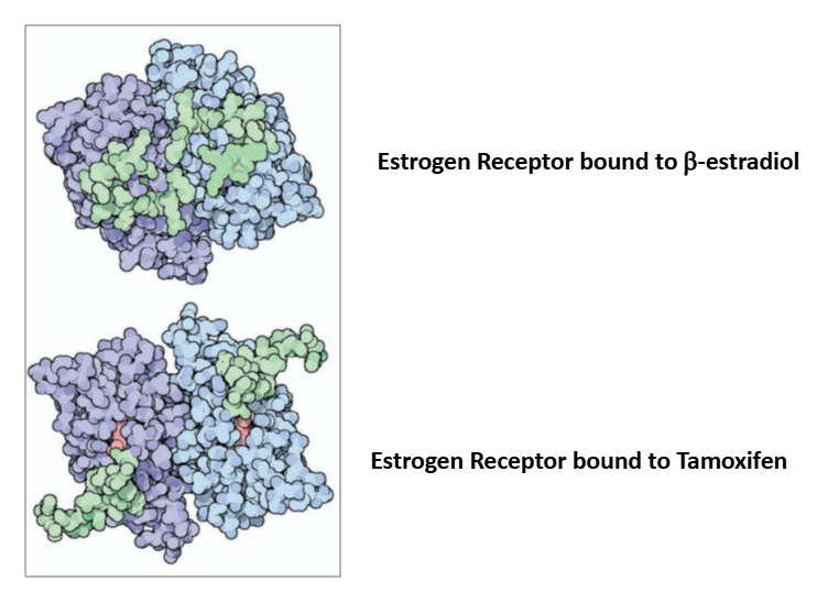
Tamoxifen
It was known that removing the ovaries of women with breast cancer
would stop tumor growth in one out of three patients, however the molecular
mechanism remained unknown'
The Jensen lab then developed a drug called tamoxifen, which mimics the effects
of removing ovaries. Later it was shown that tamoxifen acts as an anti-estrogen
and blocks the activity of the estrogen receptor in cancer cells.