Chapter 10- The Plasma Membrane
 Call Kai
 Learn
 Practice Test
 Spaced Repetition
 Match
 Flashcards
 Knowt Play
1/84

There's no tags or description
Looks like no tags are added yet.
85 Terms
cell membranes
the semipermeable membrane surrounding the cytoplasm of a cell; all biological membranes
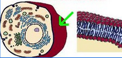
location of cell membranes
surround cell
create compartments within the cell

cell membranes consist of a
thin film of lipids and proteins

cell membranes are held together by
noncovalent interactions
transient, not locked down, like a mosh pit!

lateral diffusion
lipids exchange position and move around each other

rotation
lipids spin within the membrane

flexion
fatty acid tails move

flip flop
rare; requires an enzyme/energy; phospholipids flip between layers

lipid structure
glycerol and 3 fatty acids; amphiphilic molecules;

lipid head
hydrophilic (polar)
consists of a glycerol, phosphate, and a variable/identifiable component

lipid tails
hydrophobic (nonpolar)
hydrocarbon tails

saturated carbon chain
Fat containing fatty acids with only SINGLE carbon bonds

unsaturated carbon chain
Fat containing fatty acids with a double bond carbon bond, forming a kink in the chain

saturated lipid membranes
thicker and more rigid

thinner, more packed, and looser
unsaturated lipid membranes

common membrane phospholipids
phosphatidylcholine
phosphatidylethanolamine (-)
phosphatidylserine
sphingomyelin (neural cells - myelination)
sphingosine (+)

where are phospholipids generated
where are phospholipids generated

carrier proteins
bring fatty acids to RER and are embedded

acyl-CoA ligase
will bind CoA group to fatty acid

acyl transferase
removes CoA and attaches modified, unique lipid head groups

vesicle
transports phospholipids are specially brought/transported within the cell (typically to golgi apparatus or membrane)

flipase
enzyme catalyzes transfer of specific phosphorylases to the appropriate side of the bilayer

glycosylated lipids
exclusively in layer facing away from the cytosol; a sugar is attached

sphingosines
glycosylated lipids are common with

ganglosides
complex fat-and-sugar molecules located on the outer surface of cell membranes; signaling
net (-) charge; attached sialic acid, typically glucocerebroside (in neural cells)

plasma membrane lipid distribution
asymmetrical

plasma membrane lipid distribution is important for
cellular signaling
protection
distinguishing live/dead cells

cholesterol
in 1:1 ratio with phospholipids in cell membrane
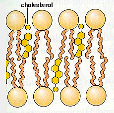
polar head group
rigid planer steroid ring structure
nonpolar hydrocarbon tail
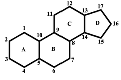
cholesterol structure

rigid planer steroid ring structure
can stiffen phospholipid region; full space between acyl chains - this decreases permeability
phospholipid organization in aqueous (aq) environment (one tail)
tails want to hide/tuck into themselves, even in a monolayer

micelle
conal hydrophobic region
one tail

phospholipid organization in aqueous (aq) environment (two tails)
cylindrical hydrophobic region
hydrophobic tails hide from water by forming into a scaled compartment ball

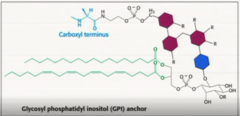
GPI anchors
lipids can serve as anchors in the membrane for proteins
associated with the membrane but not permanent

lipid rafts
concentrated areas of specific lipids and proteins or saturated fatty acid tails
- signaling site

formation of lipid rafts
formed by phospholipid bilayer budding off of the ER, expanding from the outer leaflet as it fills with triglycerides and cholesterol esters

strategic lipid rafts
outer leaflet fills with specific membrane proteins and can have associated membrane proteins on its outer surface

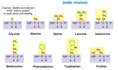
amino acids
building blocks of proteins

peptide bonds
carboxyl group and amino group of amino acids bond by dehydration synthesis
- rigid, NOT flexible
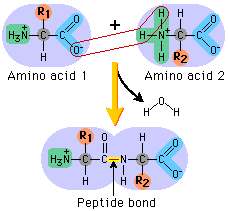
Bonds within amino acids
flexible

N-terminus to C-terminus
the direction in which the peptides are read

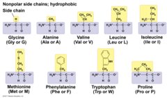
R group
a functional group that defines a particular amino acid and gives it special properties.

R group categories
nonpolar (no charge)
+ charge
- charge
polar
primary structure
The first level of protein structure; the specific sequence of covalently bonded amino acids making up a polypeptide chain.

secondary structure
The second level of protein structure; the regular local patterns of coils (alpha helixes) or folds (beta sheets) of a polypeptide chain.
made from h-bonds in the backbone

tertiary structure
The third level of protein structure; the overall, three-dimensional shape of a polypeptide due to interactions of the R groups of the amino acids making up the chain.

quaternary structure
The fourth level of protein structure; the shape resulting from the association of two or more polypeptide subunits.
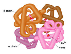
tertiary and quaternary structure
are dependent on side chain and their properties and noncovalent bonding (EN, differential charges, phenomena)

polar side chains
turn outward to h-bond with H2O

nonpolar side chains
hide in the protein's core
phenomena (electrostatic interactions, hydrogen bonds, can der waal interactions)
nonpolar groups bond when pushes very close together (such as in tight corners in protein structure)

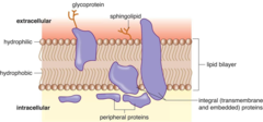
integral proteins
transmembrane protein
alpha helixes
beta barrel
gray area protein

transmembrane protein
An integral membrane protein that spans the phospholipid bilayer.
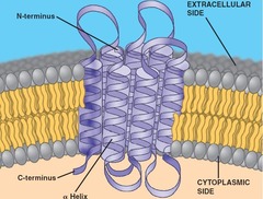
alpha helixes
can open access across the membrane; single or multiple

beta barrel
created when beta sheets are extensive enough to fold back on themselves; hollow cylinders in the membrane

traits of beta barrels
selective
if side chains in membrane (hydrophobic)
if side chains external (hydrophilic)

protein plug
beta barrel containing protein filter

gray area protein
refers to the monotopic membrane proteins or proteins that partially anchor into the lipid bilayer

transient proteins
containing polar domain/simple anchored
GPI anchored
inner leaflet
outer leaflet

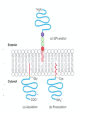
lipid anchored proteins
containing polar domain/simple anchored
GPI anchored

simple anchored protein
a protein attached by a covalent bond to a lipid molecule that holds it tightly to the cell membrane

GPI anchored protein
exclusively on the outer face and are clustered in certain regions
peripheral protein
inner leaflet
outer leaflet

inner leaflet protein
a membrane-associated protein that resides exclusively on the cytoplasmic, inside-facing layer of a cell's lipid bilayer

outer leaflet protein
molecules attached to or embedded in the outer layer of a cell membrane that face the outside environment

Hydropathy plots
reveal hydrophobic and hydrophilic amino acid regions
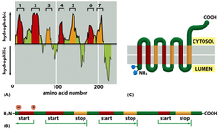
hydrophobic regions
above the x axis

hydrophilic regions
below the x axis

complex Hydropathy plots readings
minor hydrophobic structures may not fully dip below the graph

Glycosylation
non-cytosolic face glycocalyx (add sugar)
identification and protection

Disulfide bonds
non-cytosolic face stabilize protein structures (within some proteins or different proteins)

reducing bonds in cytosol prevent formation
why Disulfide bonds don't form in cytosol

Detergents
amphiphilic molecules that disrupt hydrophobic interactions

job of Detergents
disrupt lipid bilayer
form micelles
destroy
Individually form

detergent micelles and membrane proteins
displace membrane proteins by forming a water soluble protein lipid detergent complex

detergent micelles and phospholipids
clump around phospholipids in water soluble lipid detergent micelles

very strong detergent
denature proteins

to isolate functional proteins
use a milder detergent to remove proteins without denaturing
add lipids to wash away undesired elements and form vesicles with the desired protein
proteins remain functional in liposome

spontaneous liposome formation
decrease detergent concentration and increase phospholipid concentration

embedded proteins
mobile
proteins clustered into complexes
less mobile

proteins anchored by extra/intracellular proteins
less mobile
proteins anchored to proteins between cells
functionally immobile

tracked protein movement with fluorescence
Scientists labeled mouse and human cell-surface proteins with distinct green and red fluorescent antibodies.
These were fused into distinct cells into a single hybrid cell.
After 40 minutes of incubation at 37°C, the proteins completely intermixed, providing the first definitive visual proof that membrane proteins diffuse laterally within a fluid lipid bilayer

photobleaching
Scientists label the target protein with a fluorescent tag like GFP to establish a baseline of uniform cellular brightness.
A high-intensity laser pulse is then shot at a specific region, permanently destroying the fluorescence to create a distinct, dark "bleached" zone.
By taking a time-lapse video, researchers measure how quickly surrounding unbleached proteins diffuse back into the spot, calculating the protein's mobility and diffusion rate
