Central Nervous System (Slideshow 2) (HSF)
 Call Kai
 Learn
 Practice Test
 Spaced Repetition
 Match
 Flashcards
 Knowt Play
1/107

There's no tags or description
Looks like no tags are added yet.
108 Terms
Central NS consist of …
brain and spinal cord
Cephalization
evolutionary development of anterior portion of CNS
due to increased number of neurons
highest level reached in human brain
embryologically, the brain and spinal cord develop from…
the neural tube
Posterior end of neural tube forms…
spinal cord
Anterior end of neural tube forms …
brain
brain in embryological development is caused by…
constrictions of 3 primary vesicles
3 Primary Constrictions of Vesicles to form Brain (in embryo)
Prosencephalon forms forebrain
Mesencephalon forms midbrain
Rhombencephalon forms hindbrain
Brain grows faster than…, so …
surrounding membranous skull, it must fold to occupy available space.
Folding in brain development
forebrain moves towards brain stem
cerebral hemispheres double back, envelop diencephalon and midbrain while creasing and folding to increase surface area.
4 Regions of Adult Brains
Cerebral Hemispheres, Diencephalon, Brain Stem, Cerebellum
Gray Matter
short, nonmyelinated neurons and cell bodies
White Matter
myelinated and nonmyelinated axons
Basic Pattern found in CNS
central cavity surrounded by gray matter with white matter external to the gray matter
Cerebral Hemispheres (basic definition)
forms the superior part of brain, accounts of 83% of brain mass
Surface markings of Cerebral Hemispheres
Gyri, Sulci, Fissures (longitudinal, transverse cerebral)
Gyri (surface marking of cerebral hemispheres)
ridges
Sulci (surface marking of cerebral hemispheres)
shallow grooves
Fissures (surface marking of cerebral hemispheres)
deep grooves
Longitudinal Fissure of Cerebral Hemispheres
separates 2 hemispheres
Transverse Cerebral Fissure
separates cerebrum and cerebellum
Sulci divide each hemisphere into how many lobes?
five
5 lobes that sulci divide
frontal, parietal, temporal, occipital, insula (1st 4 are named after overlying skull bones)
Cerebral Cortex
“Executive suite” of brain, site of conscious mind, thin (2-4mm) superficial layer of gray matter, 40% of mass of brain
Functions of the Cerebral Cortex
Conscious Mind - awareness, sensory perception, voluntary motor initiation, communication, memory storage, understanding
4 General Consideration of Cerebral Cortex
3 types of functional areas
contralateral
lateralization/specialization
conscious behavior
3 Types of Functional Areas of Cerebral Cortex
Motor (voluntary movement), Sensory (awareness of sensation), Association (diverse information)
Motor Areas of Cerebral Cortex
located in frontal lobe, controls voluntary movement
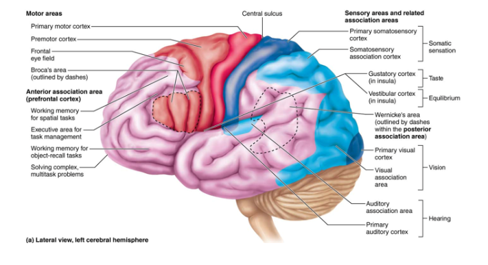
Primary (somatic) motor cortex of Cerebral Cortex
located in precentral gyrus of frontal lobe
Pyramidal Cells
large neurons that allow conscious control of precise, skilled, skeletal muscle movements
Pyramidal Tracts
formed from long axons that project down spinal cord
Somatotopy
all muscles of body that can be mapped to area on primary motor cortex
Premotor Cortex
helps plan movements
controls learned, repetitious, patterned motor skills
coordinates simultaneous or sequential actions
controls voluntary actions that depend on sensory feedback
Broca’s Area of Cerebral Cortex
present in one hemisphere (usually the left)
motor speech area that directs muscles of speech production
planning speech and voluntary moto activities
Frontal Eye Field of Cerebral Cortex
controls voluntary eye movement
Lateralization
division of labor between hemispheres (they are not identical)
Cerebral Dominance
refers to hemisphere that is dominant for language
90% of humans have left-sided dominance (resulting in right handiness)
Other 10% the roles are reversed
Left Hemisphere
controls language, math, logic
Right Hemisphere
visual-spatial skills, intuitions, emotion, artistic, and musical skills
how do hemispheres communicate?
instantaneously by fiber tracts and functional integration
3 Gray Matter Structures of the Diencephalon
Thalamus, Hypothalamus, Epithalamus
ALL enclosed in third ventricle
Thalamus (makeup)
Bilateral egg-shaped nuclei that form superolateral walls of third ventricle (connected by intermediate mass called interthalamic adhesion)
80% of diencephalon
Thalamus (function)
relay station for information coming into cortex
acts to mediate sensation, motor activates, cortical arousal, learning, and memory
Infundibulum
stalk that connects to pituitary gland
Hypothalamus (location & makeup)
below thalamus
forms cap over brain stem and inferolateral walls of third ventricle
contains important nuclei such as mammillary bodies
mammillary bodies
paired anterior nuclei that acts as olfactory relay stations
Hypothalamus (main function)
main visceral control and regulating center that is vital to homeostasis
controls ANS (such as blood pressure, rate and force of heartbeat, pupil size)
initiates physical responses to emotion (part of limbic system, perceives pleasure, fear, rage, drives and biological rhythms)
Hypothalamus (other functions)
regulates body temp (sweating/shivering)
regulates hunger and satiety in response to nutrient blood levels
regulates water balance and thirst
regulates sleep-wake cycles
endocrine system functions (such as secretions of anterior pituitary gland and production of posterior pituitary hormones
Epithalamus
most dorsal portion of diencephalon
forms roof of third ventricle
contains pineal gland (extends from posterior border, secretes melatonin, helps regulate sleep-wake cycle)
3 Regions of Brain Stem
midbrain, pons, medulla oblongata (in order from top to bottom)
Brain Stem (description)
similar in structure to spinal cord, but nuclei embedded in white matter, nuclei are associated with 10 of 12 pairs of cranial nerves
control automatic behaviors needed for survival
contain fiber tracts connecting high and low neural centers
Midbrain
between diencephalon and pons
relay center for visual, auditory and motor information
cerebral peduncles, form pillars that hold up cerebrum, and cerebral aqueduct
Cerebral Peduncles
2 ventral bulges that contain pyramidal motor tracts, forms pillars that hold up cerebrum
Cerebral Aqueduct
channel running through midbrain that connects third and fourth ventricles
Pons
located between mid-brain and medulla
4th ventricle separates pons from cerebellum
composed of conduction tracts
origin of cranial nerves V (trigeminal), VI (abducens), and VII (facial)
some nuclei play role in reticular formation and maintaining normal rhythm of breathing
Conduction Tracts
acts as a bridge relaying sensory and motor signals
longitudinal fibers - connect higher brain centers and spinal cord
transversal/dorsal fibers - relay impulses between motor cortex and cerebellum
Medulla (description)
essential to survival
blend into spinal cord at foreman magnum
contain 4th ventricle, continuation of central canal of spinal cord, medulla and pons form ventral wall, choroid plexus, cranial nerves VIII, IX, X, XII
Choroid Plexus
capillary-rich membrane that forms cerebral spinal fluid
Medulla (Function)
Autonomic Reflex Center (overlap with functions of hypothalamus, hypo relays instructions via medulla)
Cardiovascular Center (adjust force and rate of heart contractions)
Vasomotor Center (adjusts blood vessels diameter for blood pressure regulation)
Respiratory Center (generate rhythm, control rate and depth of breathing
Also regulate… vomiting, hiccupping, swallowing, coughing, sneezing
Cerebellum
11% of brain mass
located dorsal to pons and medulla
major role in balance (effected by alcohol easily)
processes input from cortex, brain stem, sensory receptors to provide precise and coordinated movements of skeletal muscles
Cerebellar Hemispheres connected by…
wormlike vermis
Arbor Vitae
Thin cortex of gray matter with distinctive treelike pattern of white matter
Meninges (function)
cover and protect CNS
protect blood vessels and enclose venous sinuses
cerebrospinal fluid (CSF)
forms partition in skull
3 Layers of Meninges
external to internal
dura mater, arachnoid mater, pia mater
Dura Mater (3 Layers of Meninges)
strongest
made up of 2 layers of fibrous CT
periosteal - inner surface of skull (only in brain)
meningeal - true external covering brain (into vertebral canal as spinal dura mat)
Arachnoid Mater (3 Layers of Meninges)
middle layer with spiderweb-like extensions
separated from dura mater by subdural space
subarachnoid space contains CSF and largest blood vessels of brain
Pia Mater (3 Layers of Meninges)
delicate CT that clings tightly to brain, following every convolution
many tiny blood vessels that feed brain
Meningitis
inflammation of the meninges (spread to CNS, leading to inflammation of brain)
diagnosed by observing microbes in sample of CSF obtained via lumbar puncture
Cerebrospinal Fluid (CSF) (definition)
forms a liquid cushion of constant volume around brain
composed of watery solution formed from blood plasma, with less protein and diff ion concentrations from plasma.
Function of CSF
gives buoyancy to CNS structures (reduces weight of brain by 97% by floating it so it’s not crushed under its own weight)
protects CNS from blows
nourishes brain and carries chemical signals
Choroid Plexus (makeup)
cluster of capillaries that hangs from roof of each ventricle, enclosed by pia mater and surrounding layer of ependymal cells
volume of 150ml (replaced every 8 hours)
Choroid Plexus (function)
CSF is filtered from plexus at constant rate
ependymal cells use ion pumps to control composition of CSF and help cleanse CSF by removing waste
cilia in ependymal cells help keep CSF in motion
hydrocephalus
obstruction blocks CSF circulation or drainage, increased pressure, compressing blood vessels and crush soft nervous tissue. treatment to drain CSF with shunt in abdominal cavity.
Brain Waves
reflect electrical activity of higher mental functions
Normal Brain Function are…
continuous and hard to measure
electroencephalogram (EEG)
records electrical activity that accompanies brain function, by electrodes placed on scalp measuring electrical potential differences between various cortical areas.
used for diagnosing epilepsy, sleep disorders, in research and brain death, localizing tumors, infection, and abscesses.
each person’s brain waves are…
unique, patterns change with age, sensory stimuli, brain disease, and chemical state of body
Brain waves are measured in…
frequency in Hertz (Hz) = number of peaks per second
Frequency waves are based on…
Hertz
4 Classes of Brain Waves
Alpha, beta, theta, and delta waves
alpha waves
8-13 Hz
regular and rhythmic, low-amplitude, synchronous waves
awake but relaxed
beta waves
14-30 Hz
rhythmic, less regular
awake and alert
theta waves
4-7 Hz
more irregular
common in kids, uncommon in awake adults
delta waves
4Hz or less
high amplitude
deep sleep, reticular activating system is suppressed
brain damage is awake adult
Spinal Cord (description)
enclosed in the vertebral column, begins at the foramen magnum, ends at the L1 or L2 vertebra
Spinal Cord (functions)
2-way communication to and from brain and body
major reflex center, reflex are initiated and completed at spinal cord
Protection of Spinal Cord
by bone, meninges, CSF, epidural space
Epidural Space
cushion of fat and network of veins in space between vertebrae and spinal dura mater
where does CSF go?
fills the subarachnoid space between arachnoid and pia maters
spinal cord terminates into…
conus medullaris (cone-shaped structure)
Filum terminale
fibrous extension of conus covered with pia mater, anchor the spinal cord, extending to the coccyx
Denticulate Ligaments
extension of pia mater that secure cord to dura mater
Cervical and Lumbar Enlargements
areas where nerves serving upper and lower limbs arise from spinal cord
Spinal Nerves
part of PNS, attached to spinal cord by 31 paired roots, each spinal cord segment is designated by paired spinal nerves that arise form it
Cauda Equina
collection of nerve roots at interior end of vertebral canal
two lengthwise grooves that run length of cord partially divided it into right and left halves are called
ventral median fissure (anterior)
dorsal median sulcus (posterior)
Gray matter is located in …, white matter is …
core, outside
Central Canal
runs length of cords and filled with CSF
Gray Matter and Spinal Roots
cross section of cord resembles butterfly
three areas on each side of center are mirror images (dorsal horns, ventral horns, lateral horns)
dorsal horns
interneurons that receive somatic and visceral sensory input
ventral horn
some interneurons and somatic motor neurons