BRAIN SECTION (copy)
 Call Kai
 Learn
 Practice Test
 Spaced Repetition
 Match
 Flashcards
 Knowt Play
1/29

There's no tags or description
Looks like no tags are added yet.
30 Terms
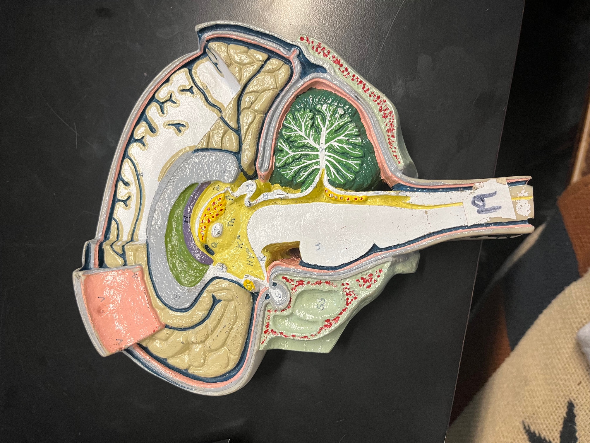
V
arachnoid villus, reabsorbs CSF into bloodstream
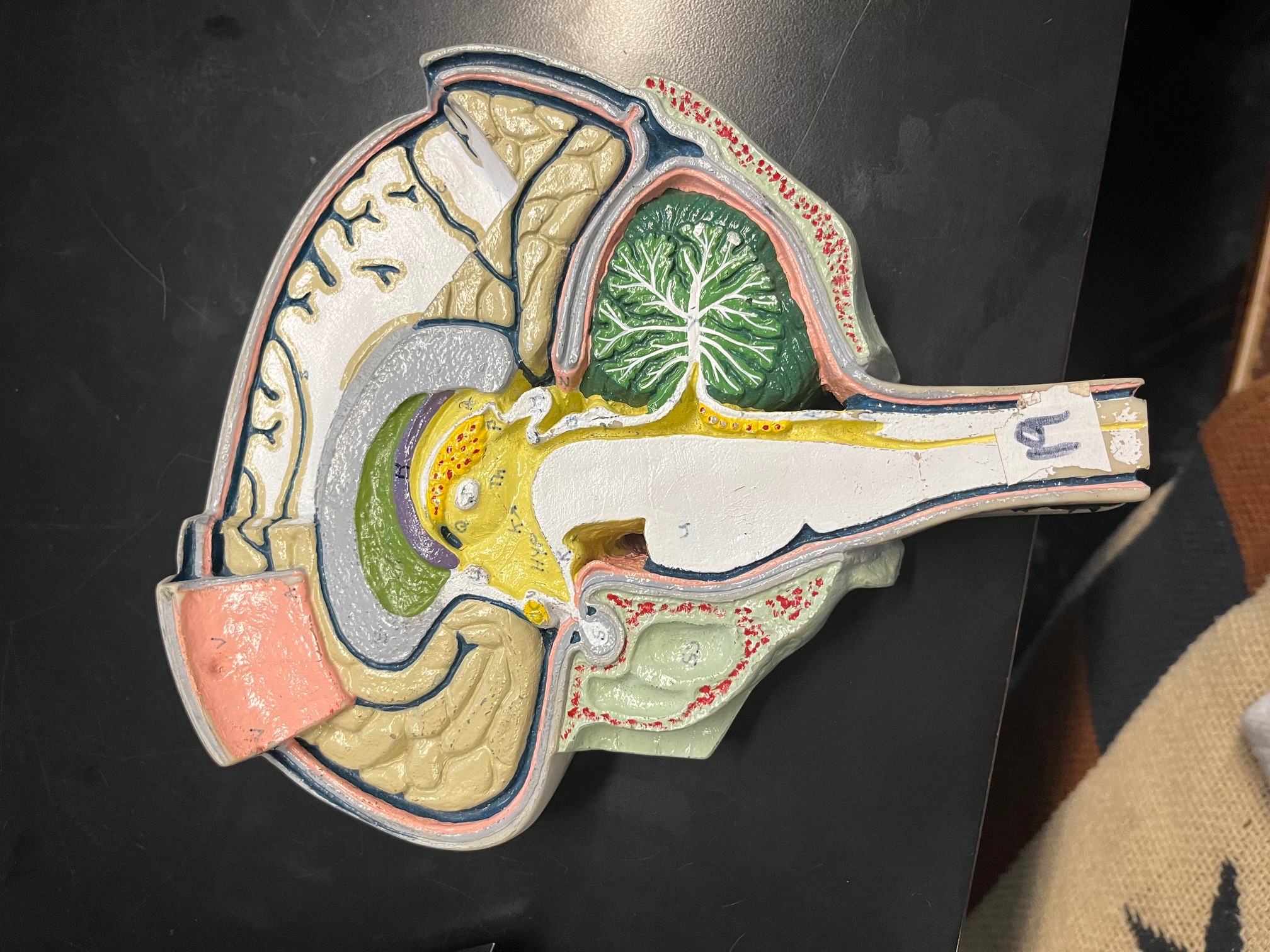
A
arachnoid mater on falx cerebri
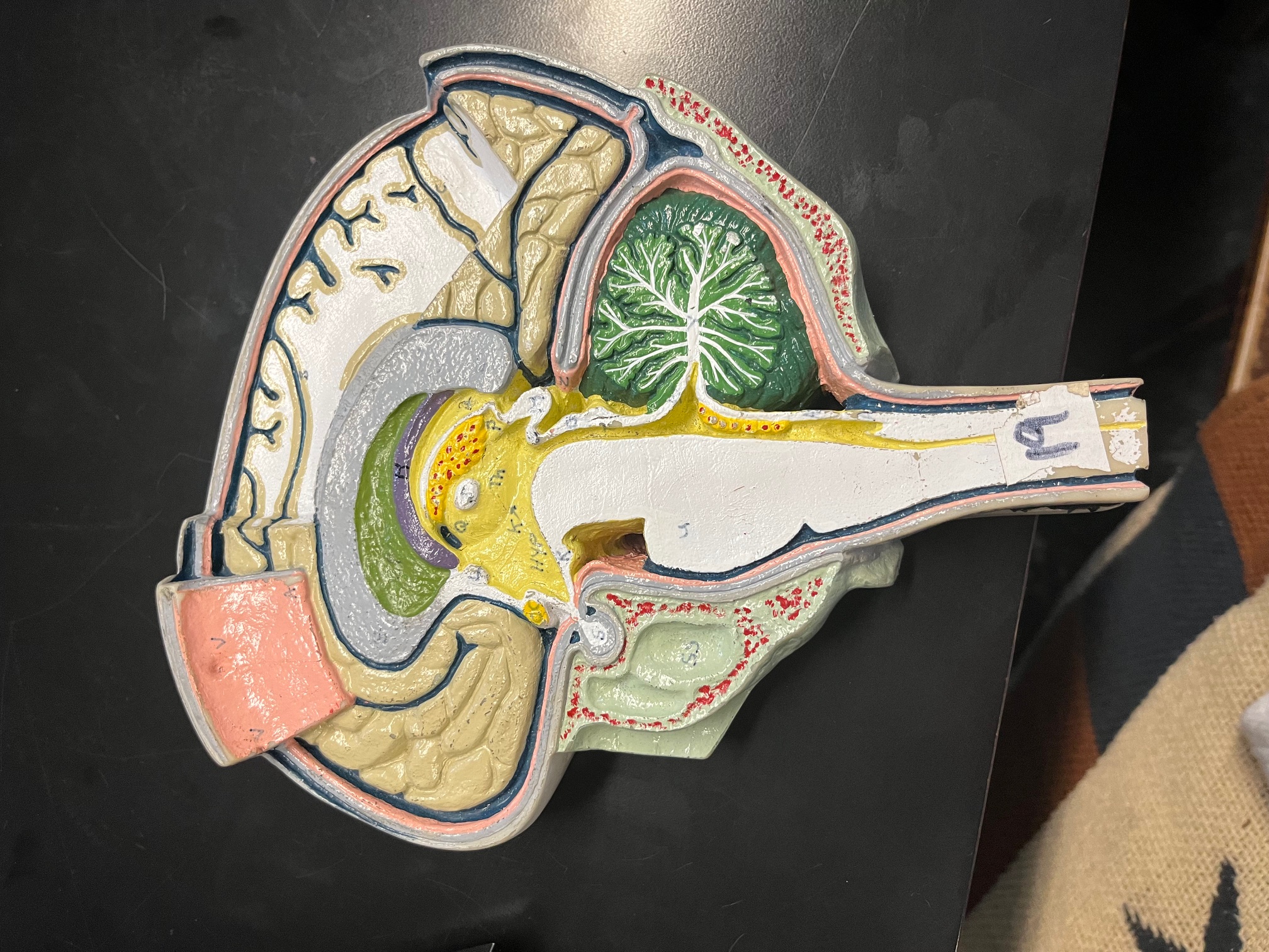
C
parieto-occipital suclus, separates parietal lobe from occipital lobe
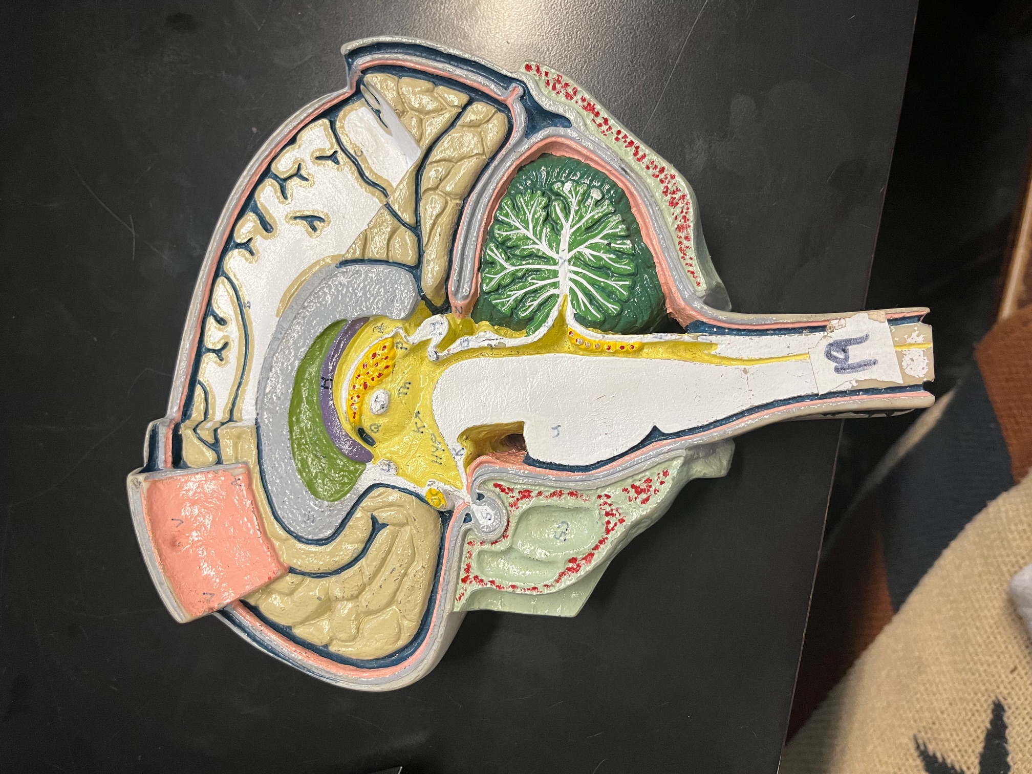
E (grey curved part)
corpus callosum, allows for communication between cerebral hemispheres
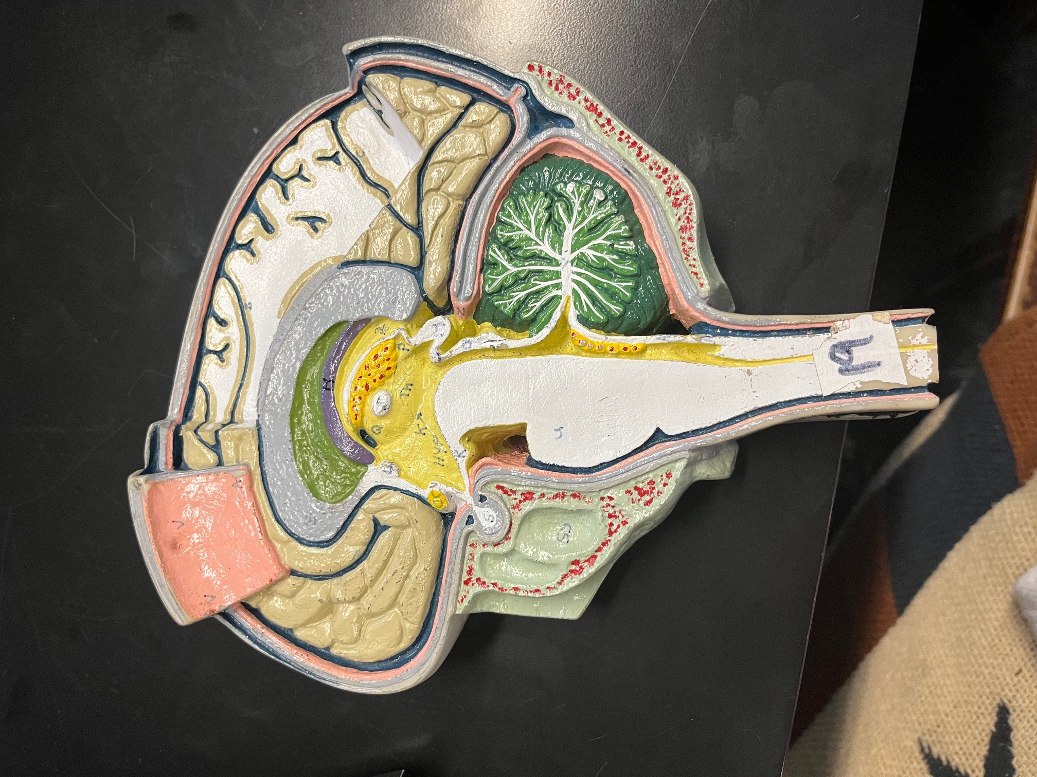
dark blue line above cerebrum
superior sagittal sinus, collects brain draining from brain tissue
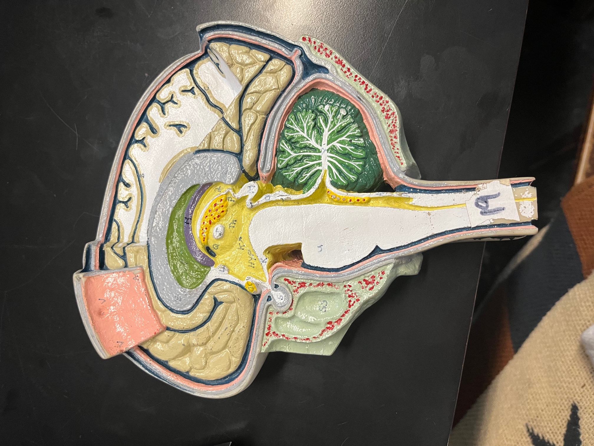
grey line above cerebrum
periosteal and meningeal layer of dura mater
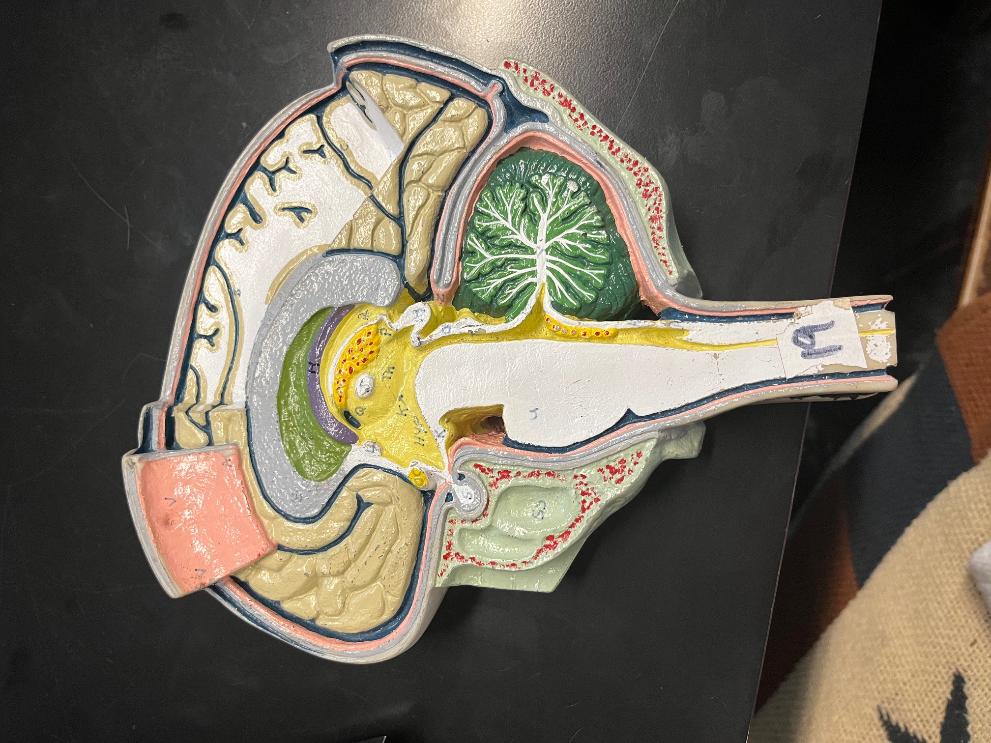
pink line
arachnoid mater, protects the brain by housing cushioning CSF
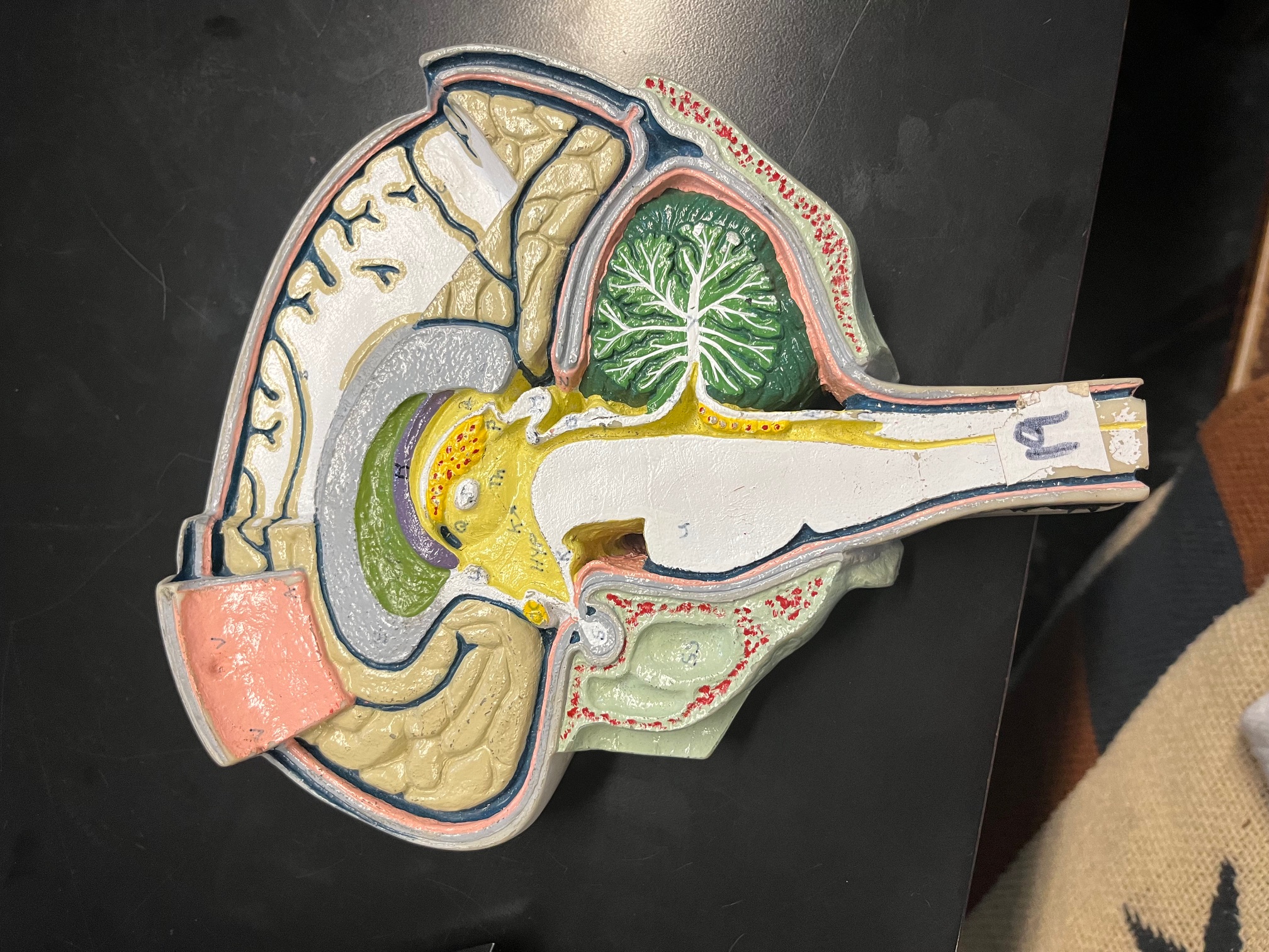
navy blue line under pink line
subarachnoid space
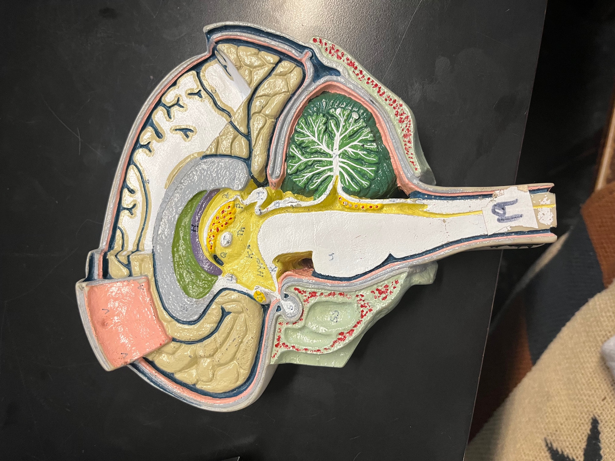
OC (yellow dot)
optic chiasma, area where medial portion of optic nerves cross
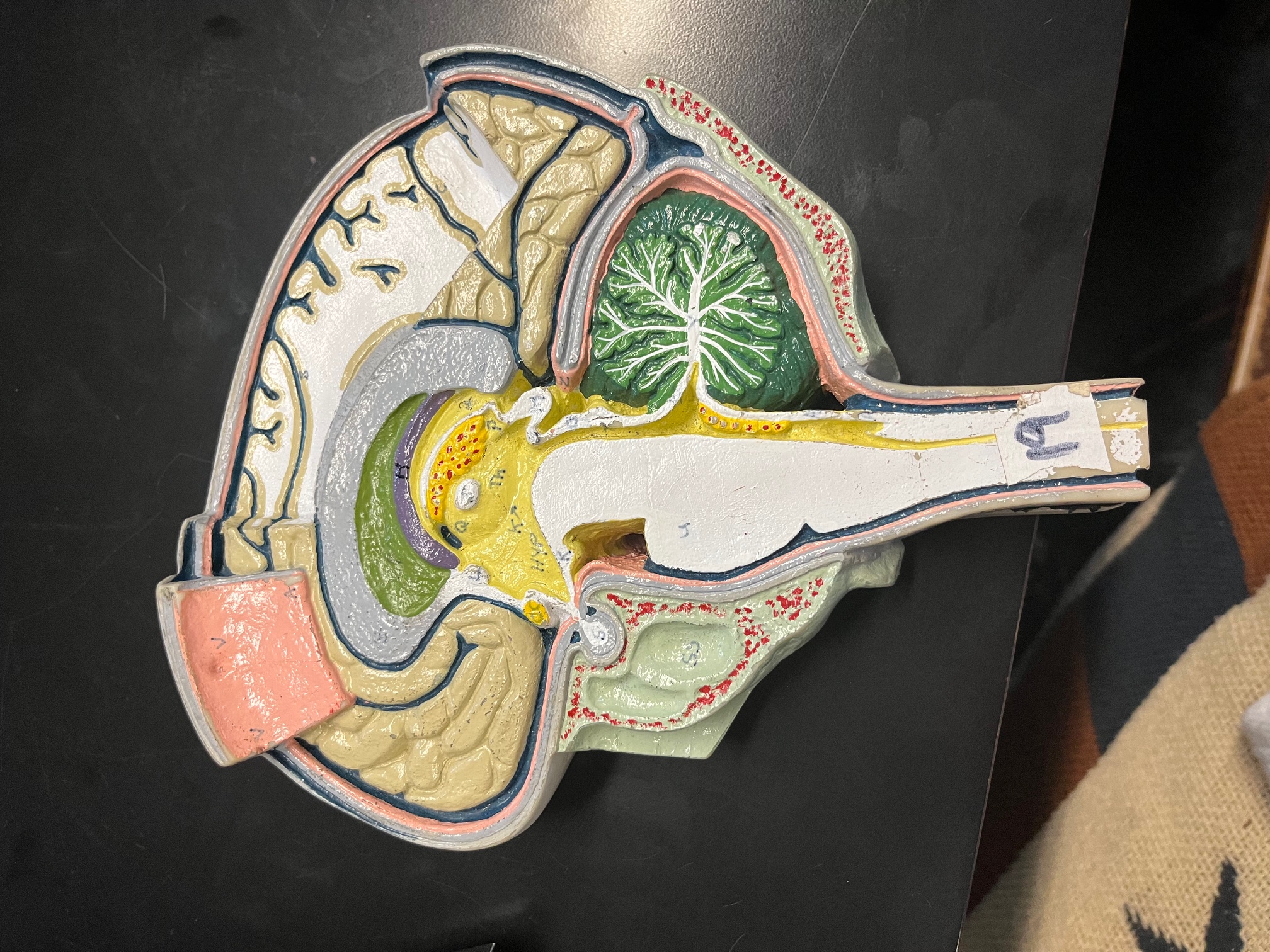
yellow with red dots
choroid plexus, makes CSF
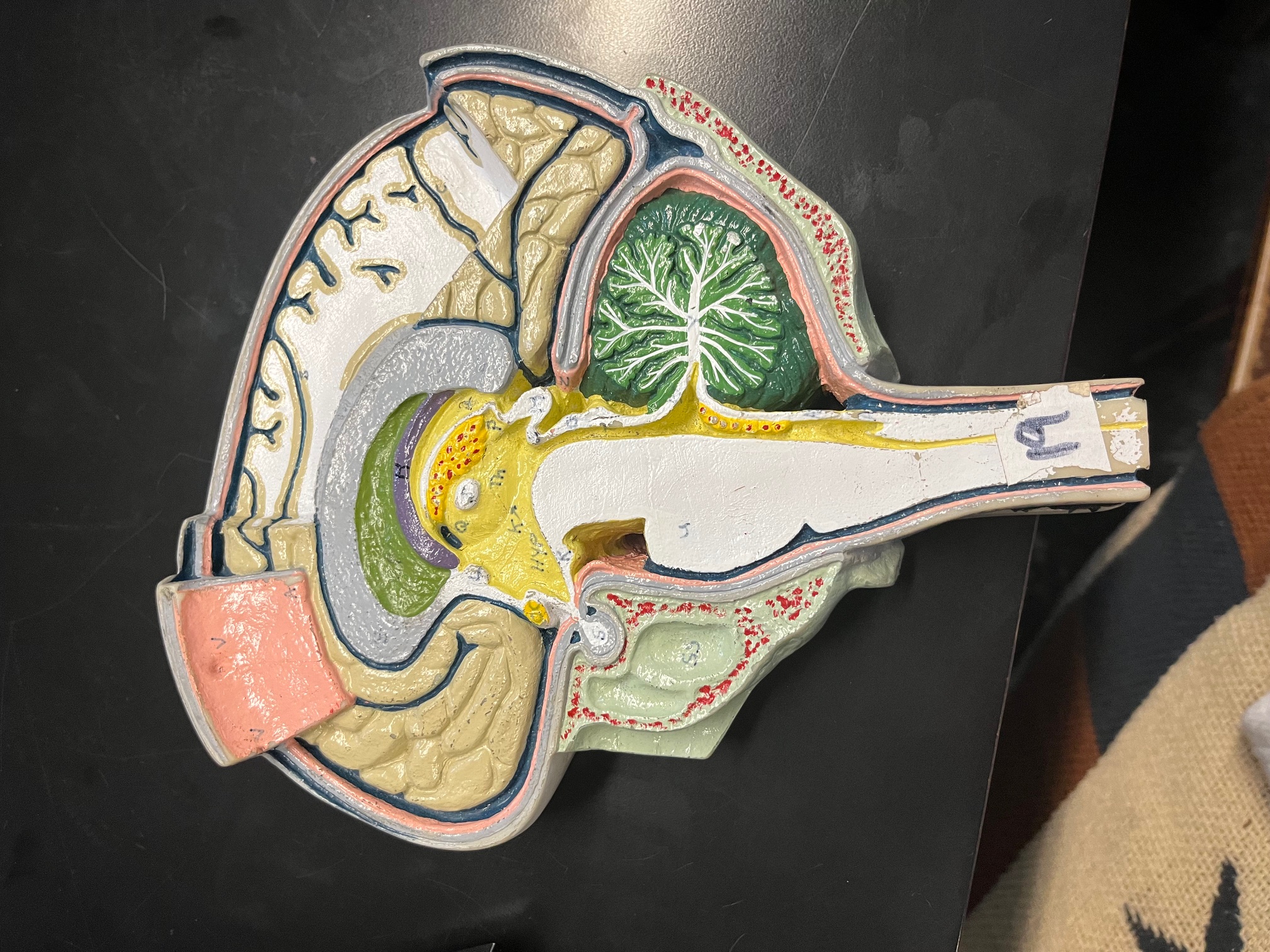
green space under grey curve
septum pellucidum, separates lateral ventricles
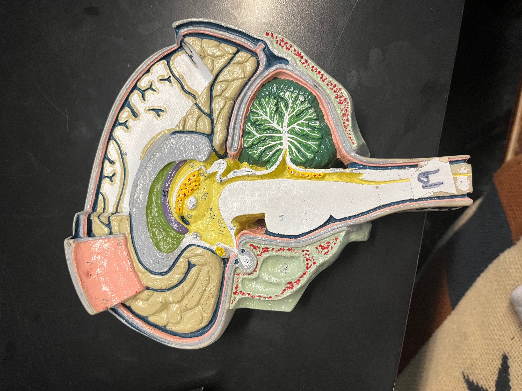
H
fornix, concerned with olfaction
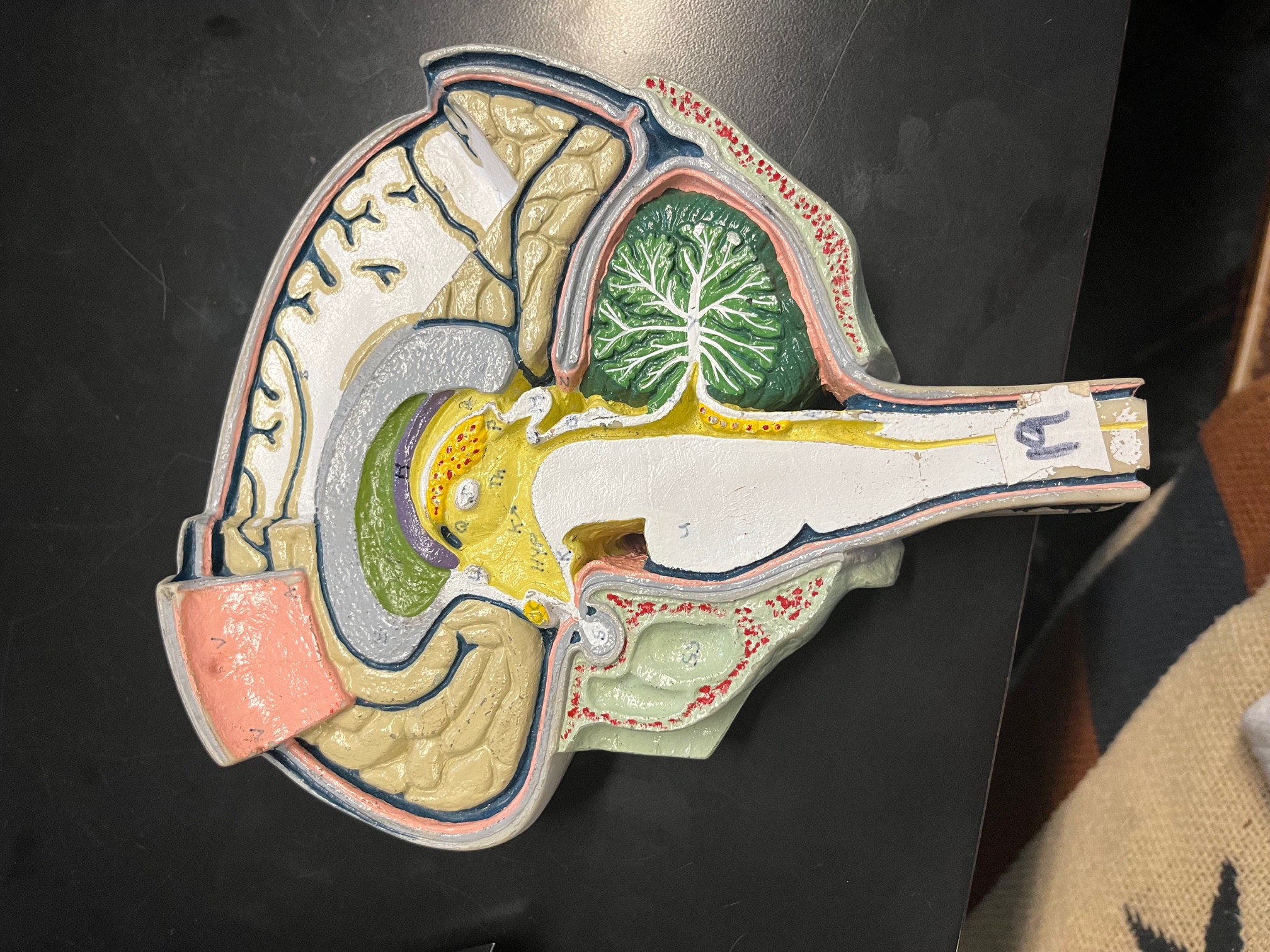
Th
thalamus, relay station for sensation except smell
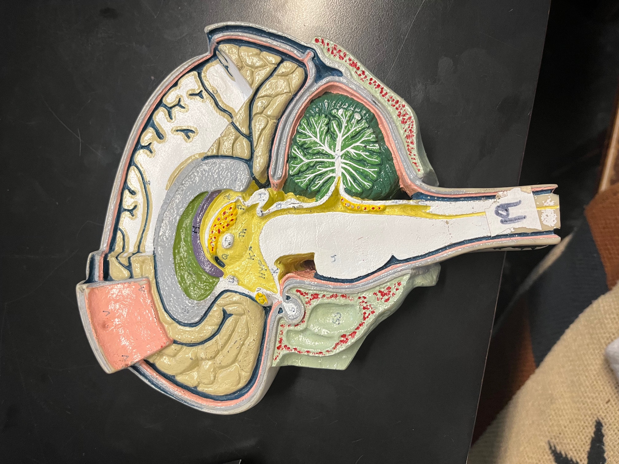
J (white dot in middle of yellow)
intermediate mass of thalamus, connects thalamic lobes
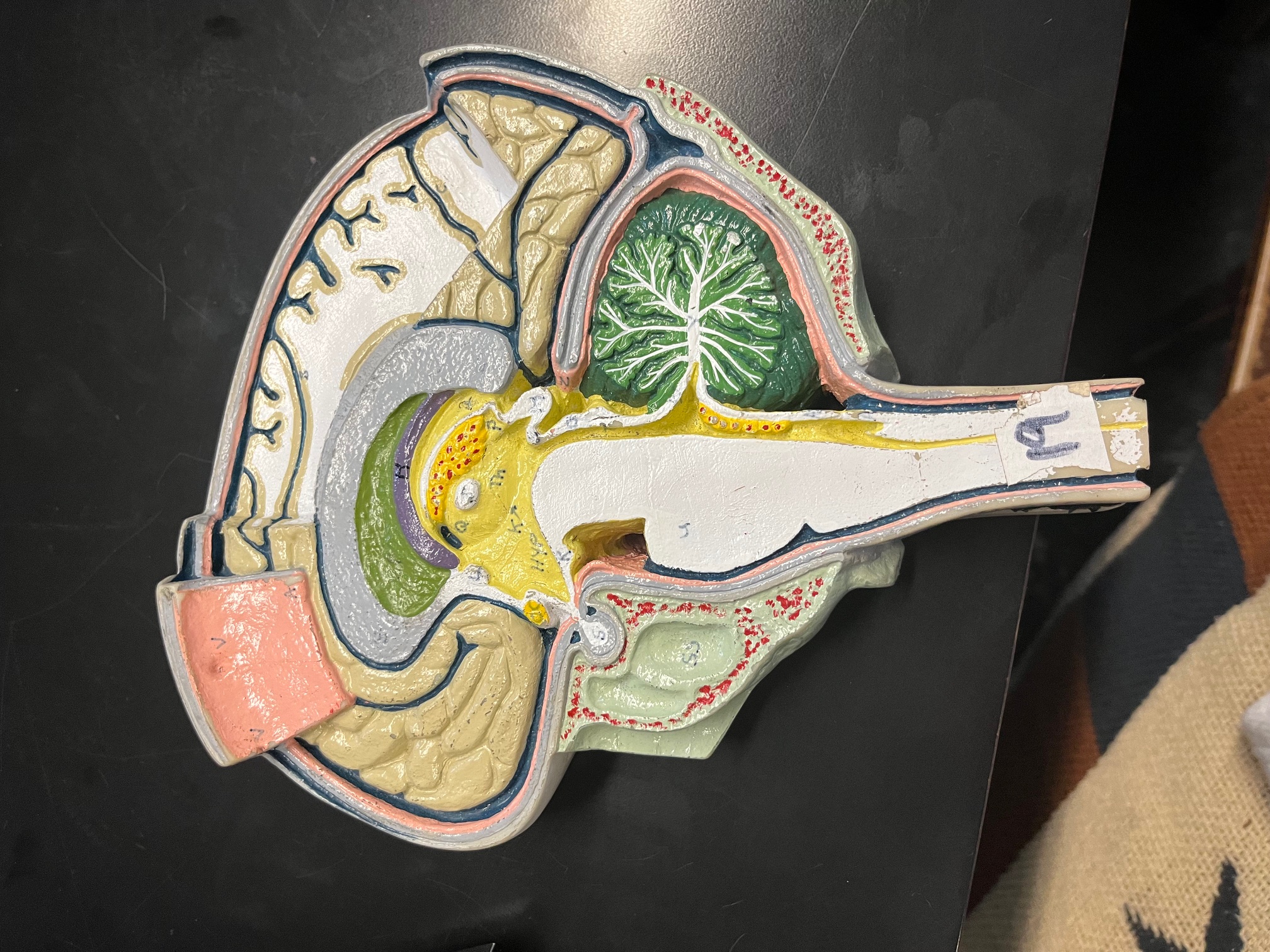
HYP
hypothalamus, drives (sex, hunger, thirst)
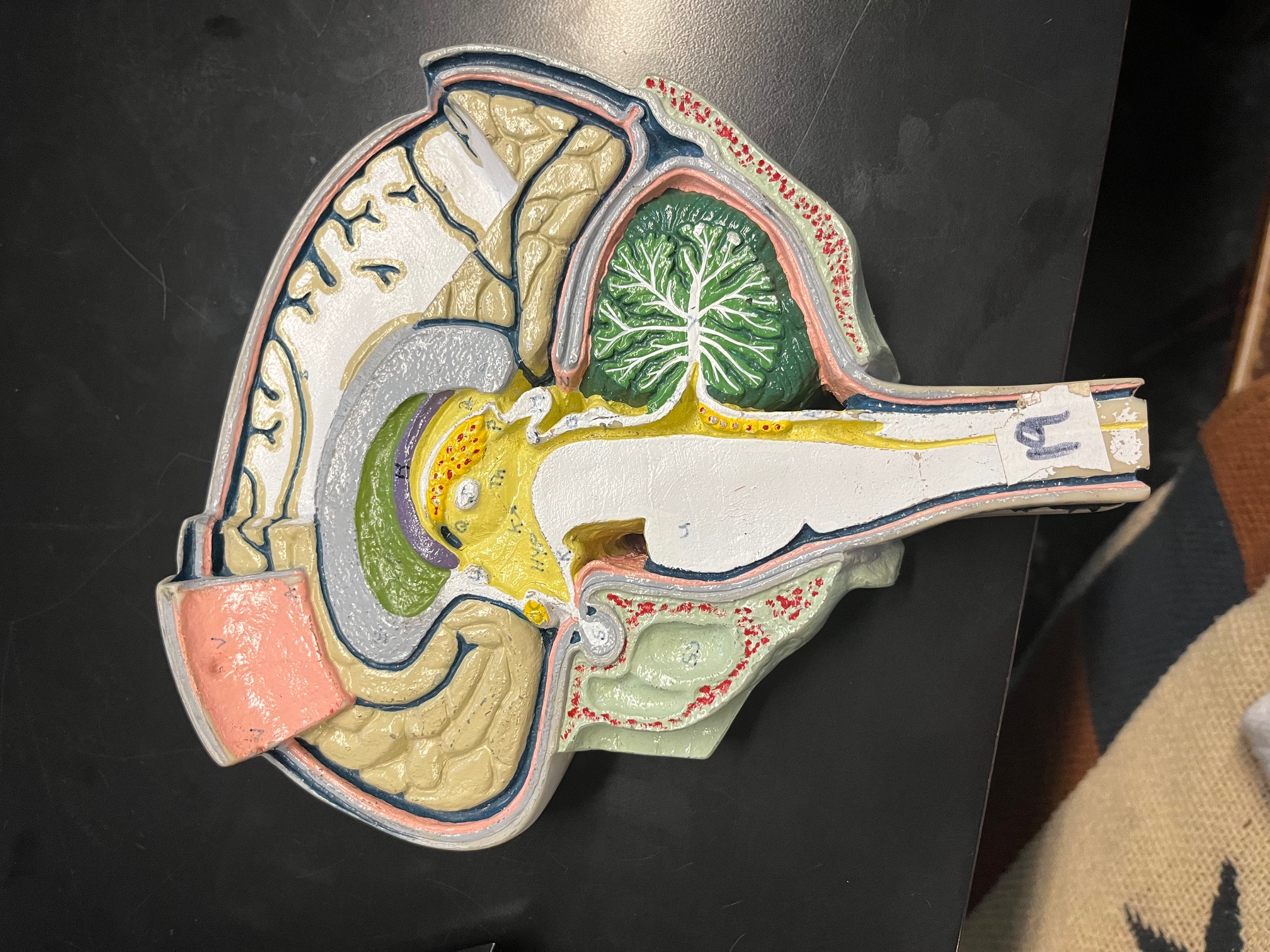
L (white dot on left of yellow)
anterior commissure, allows communication between cerebral hemispheres
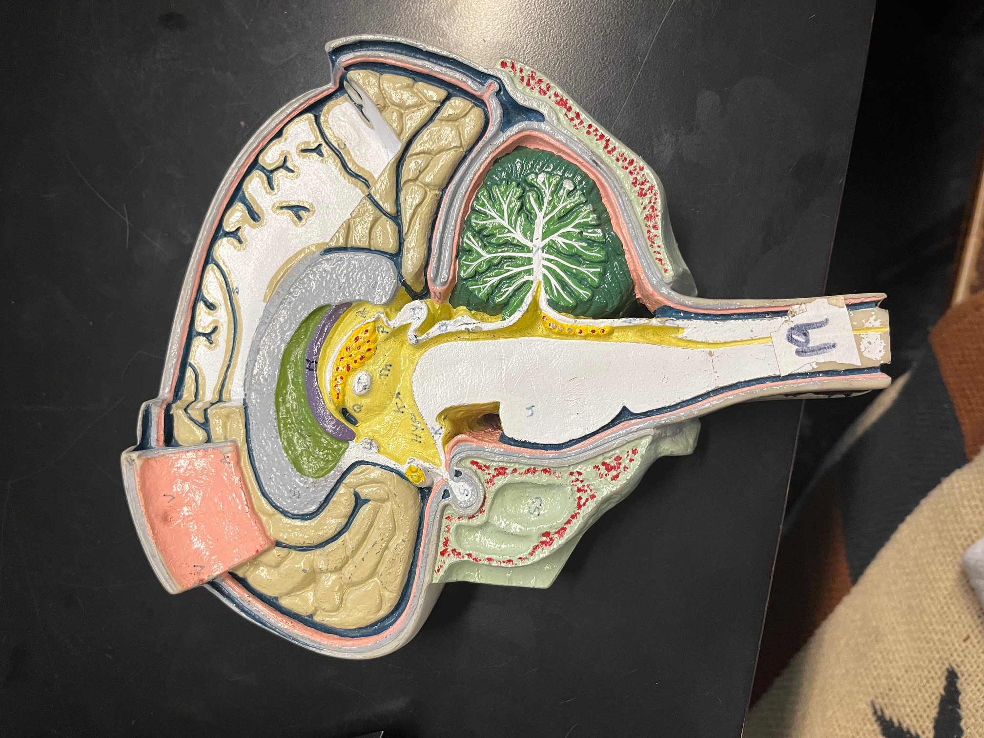
M (white dot on right of yellow)
pineal gland, produces melatonin
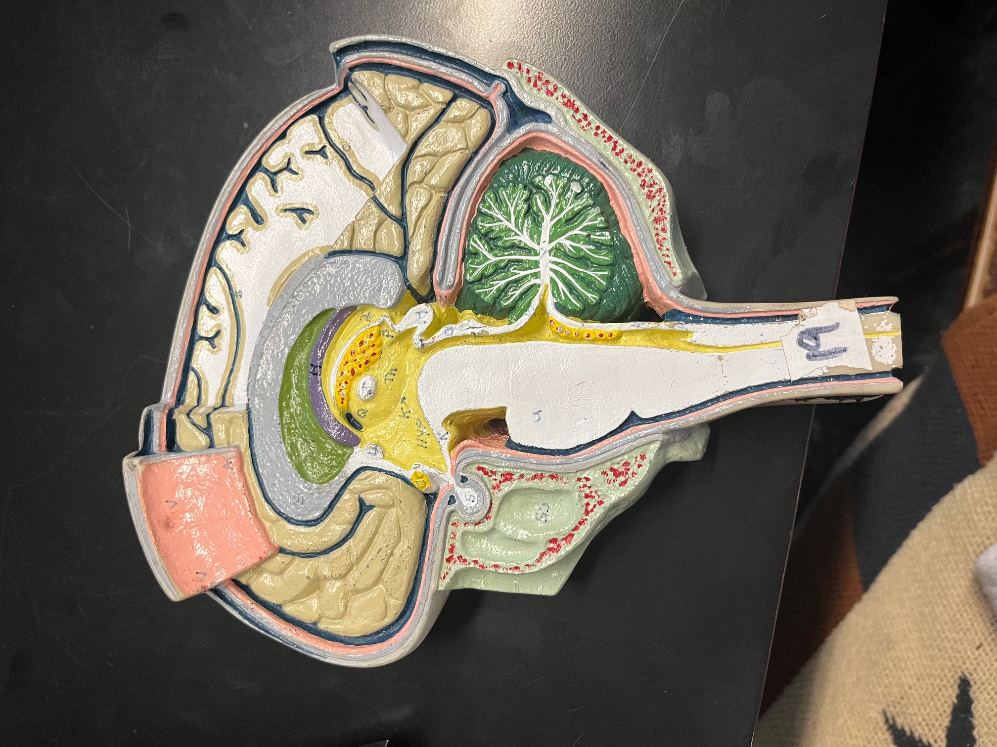
S
pituitary gland, produces hormones
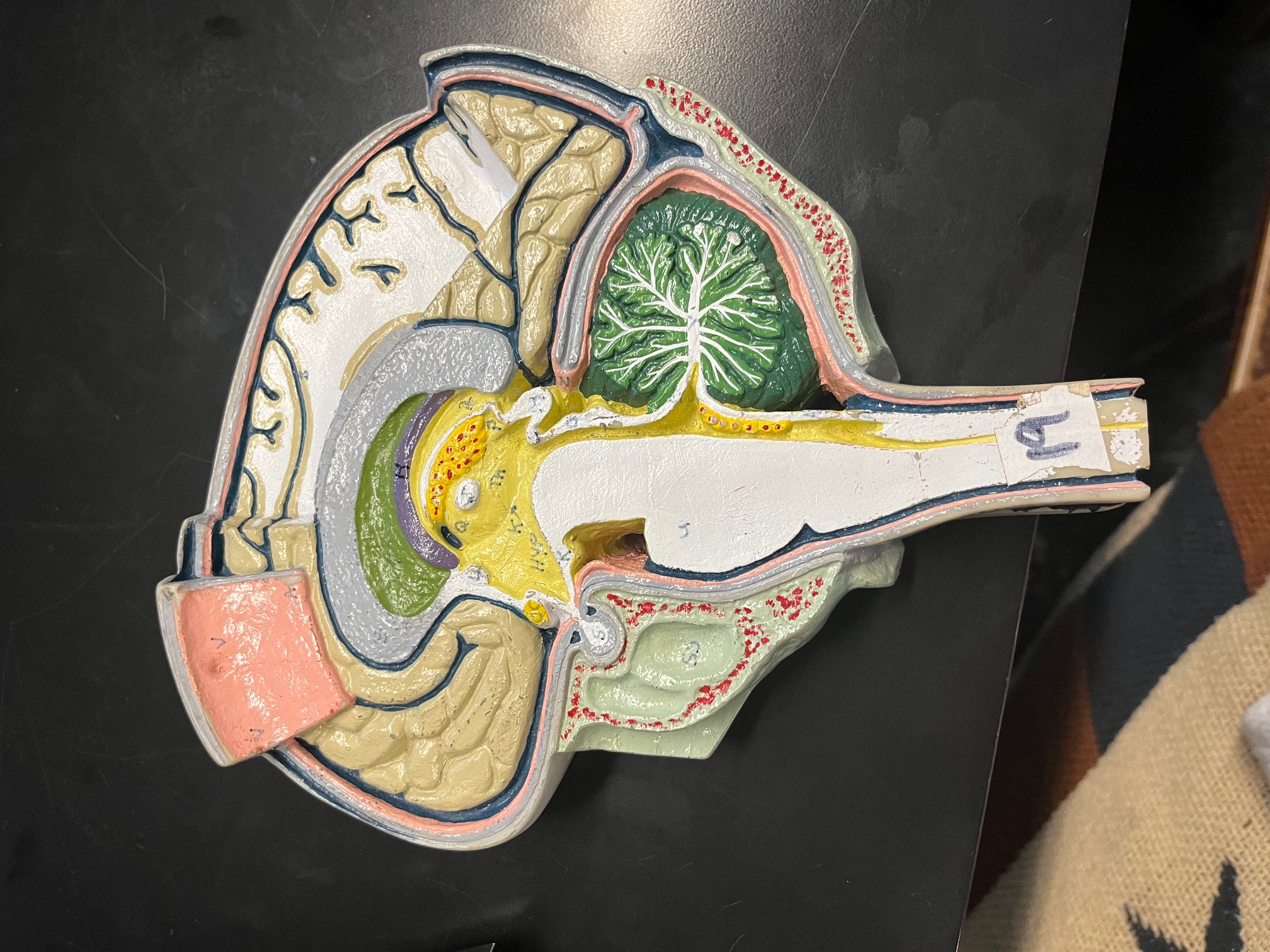
O
corpora quadrigemina, contains visual and auditory reflex center
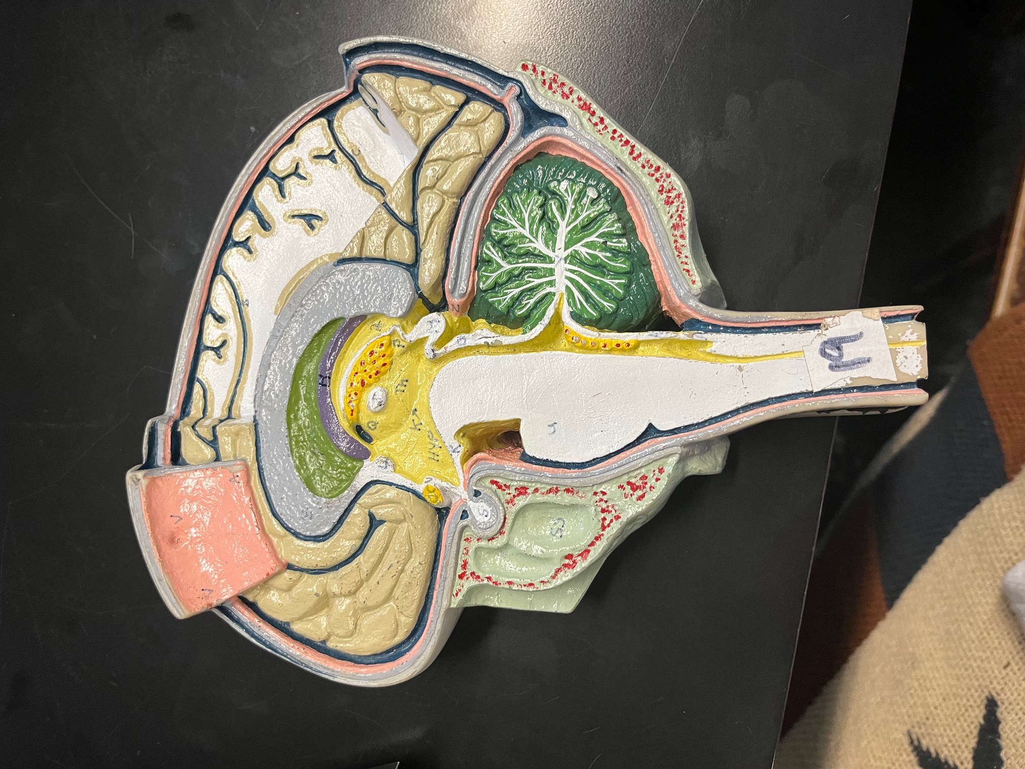
Q
interventricular foramen, takes CSF from lateral ventricles to third ventricle
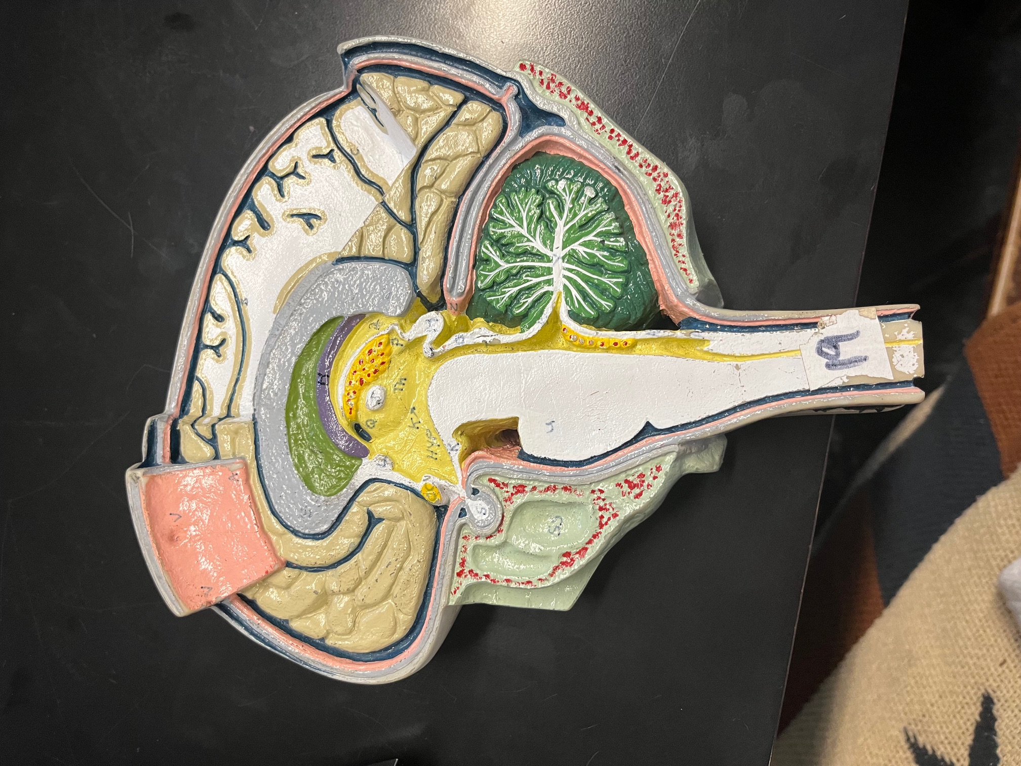
R (under HYP)
mammillary body, relay station for olfaction
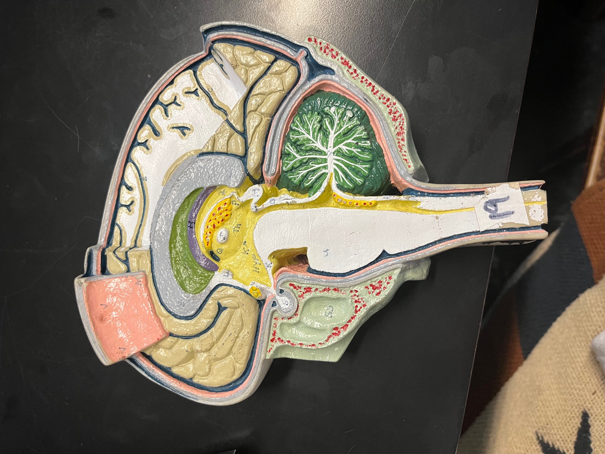
T (below O)
cerebral aqueduct, takes CSF from third to fourth ventricle
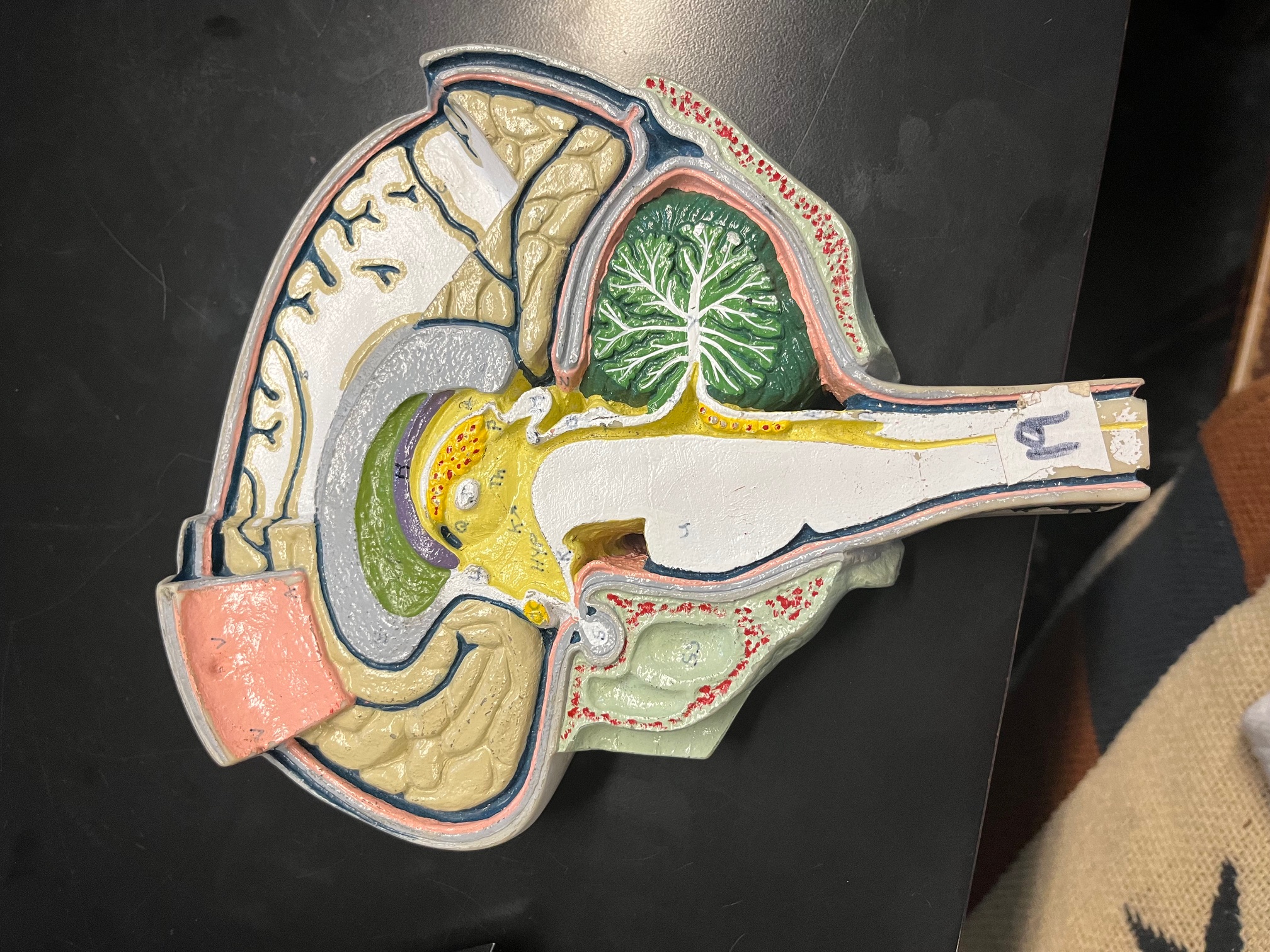
Z
straight sinus, involved in CSF flow
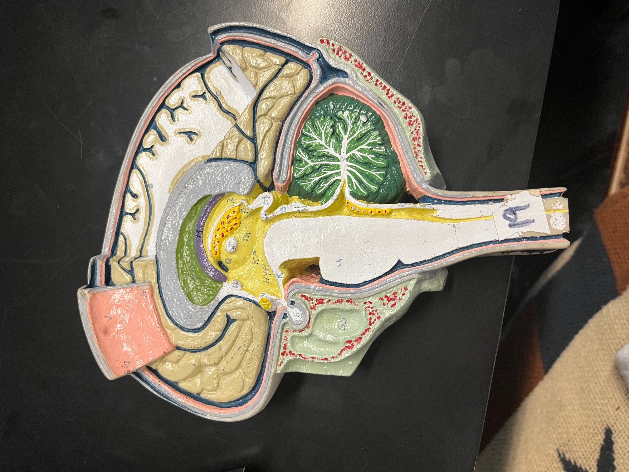
grey line between cerebrum and cerebellum
tentorium cerebelli, separates cerebrum from cerebellum
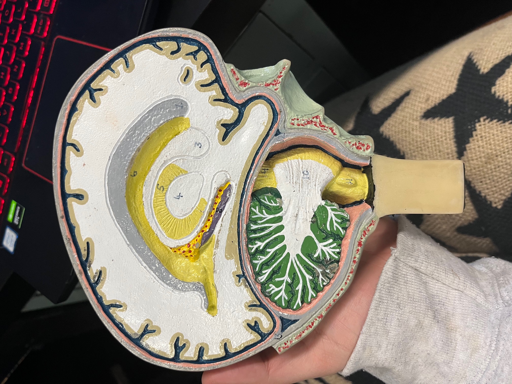
9
cerebral peduncle, connects cerebrum to pons
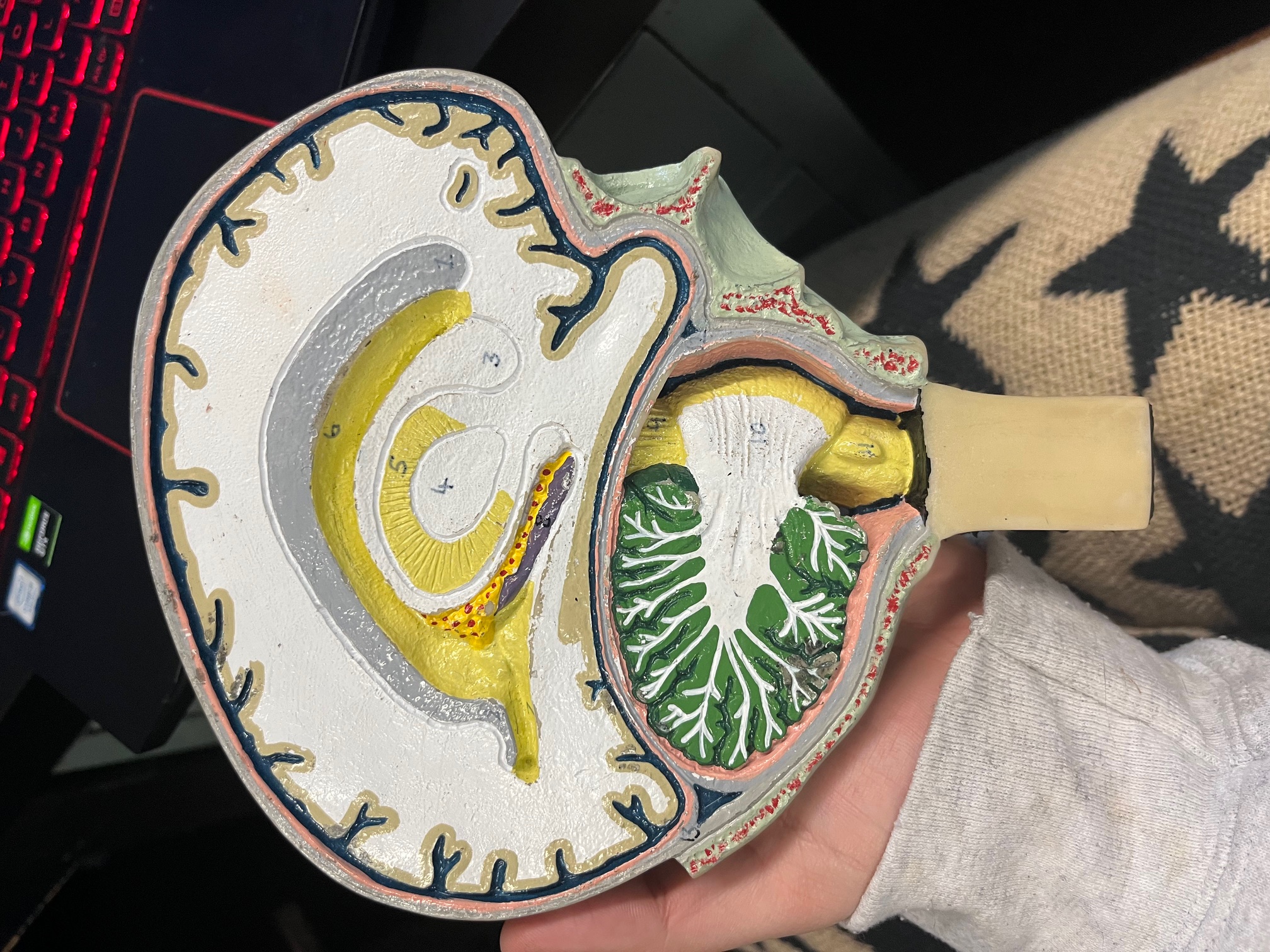
10
pons, connects brain with lower CNS centers
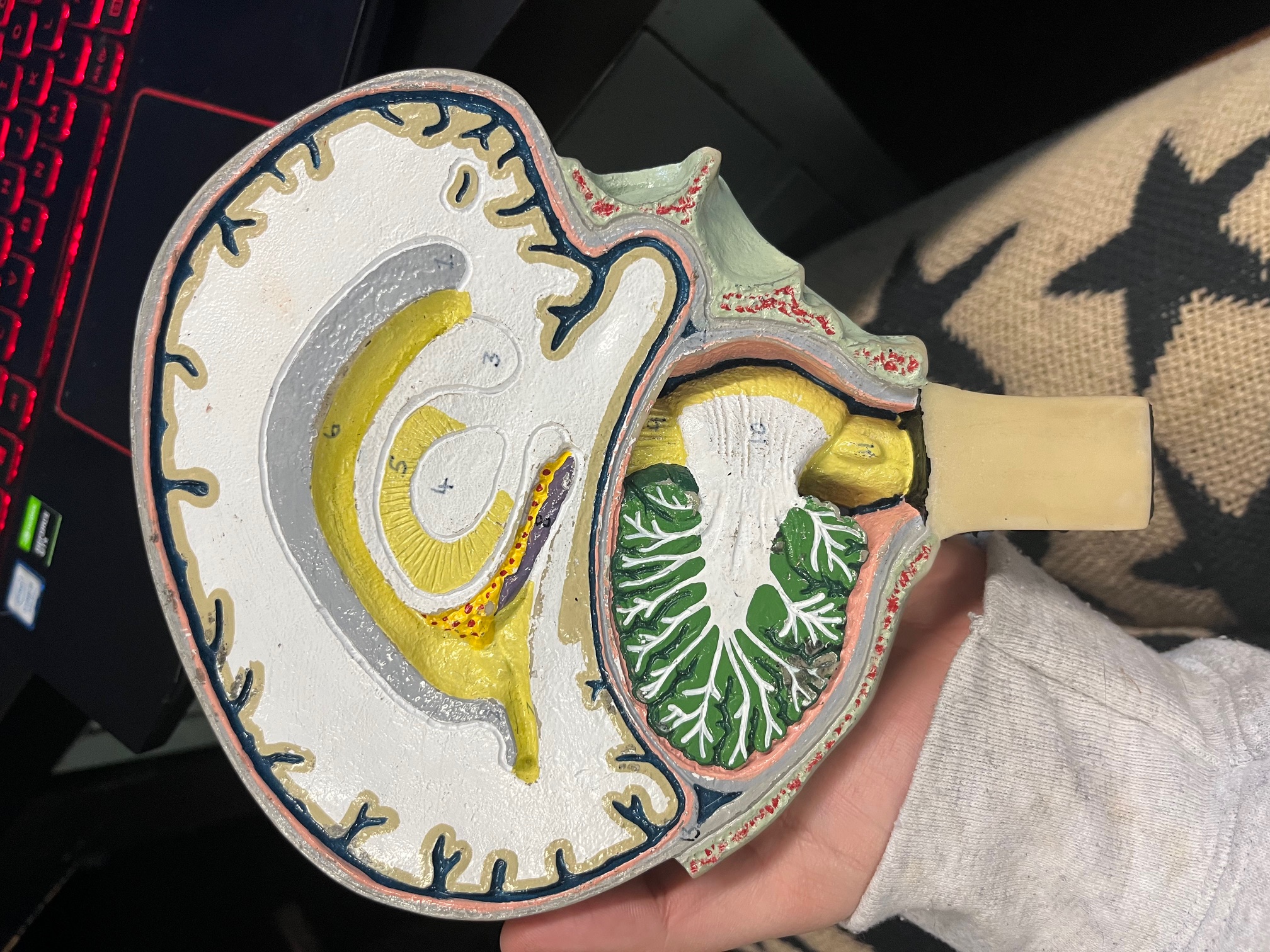
13
tranverse sinus, drains venous blood from brain
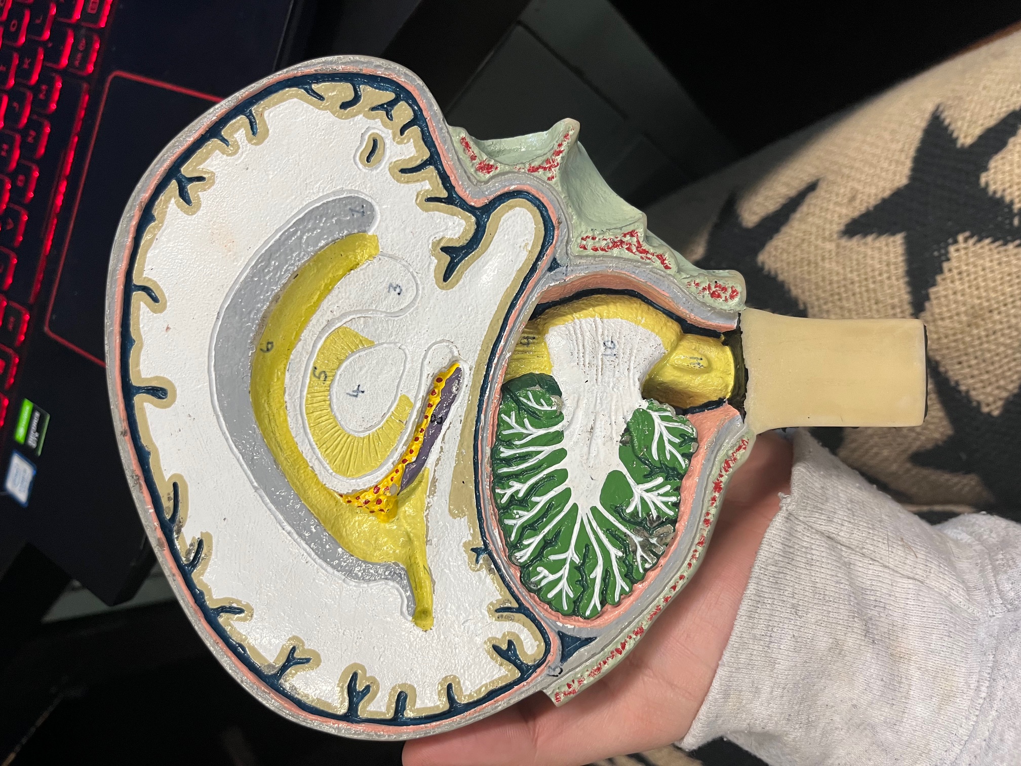
6
right lateral ventricle, takes CSF through interventricular foramen into third ventricle
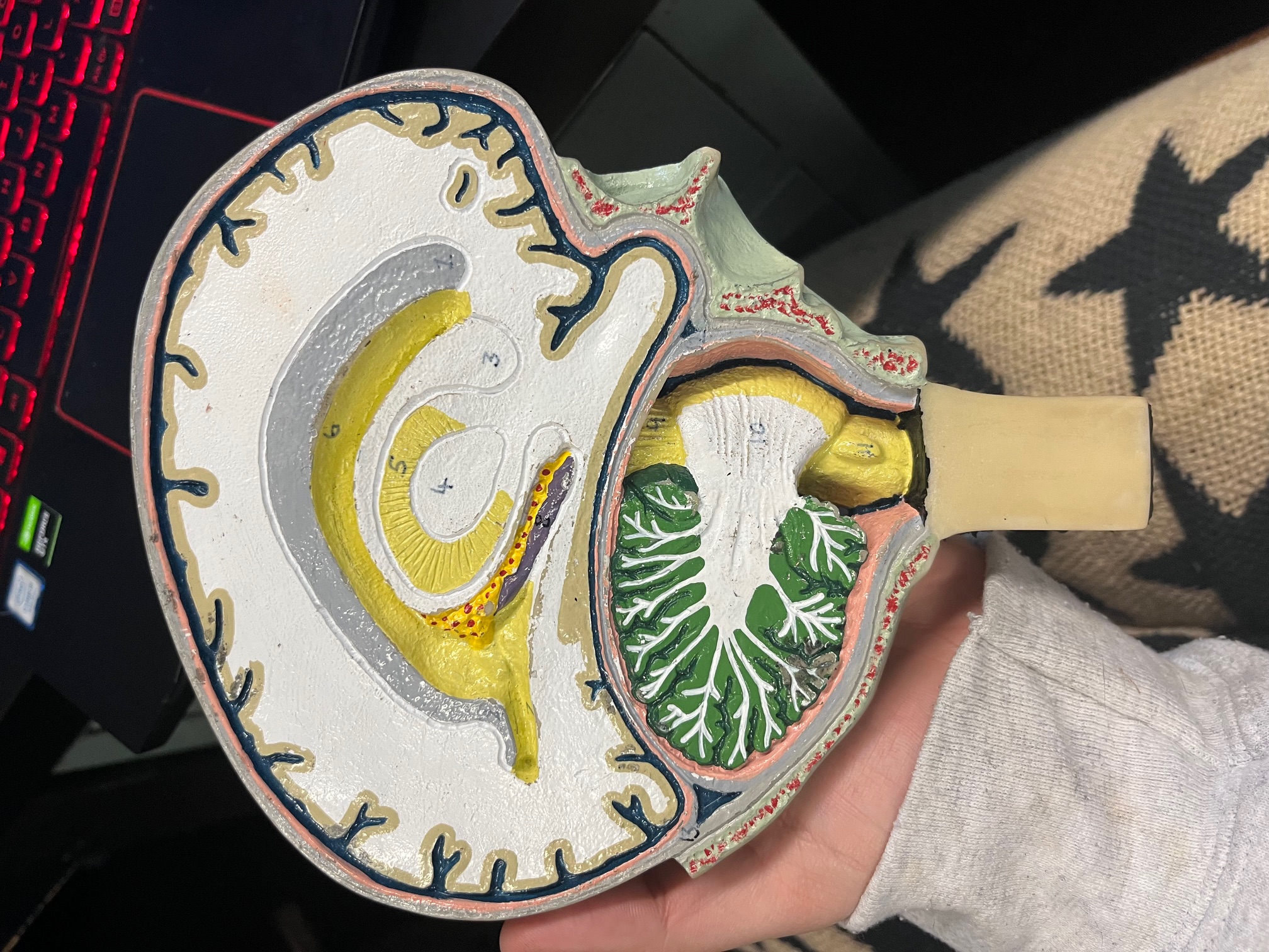
7 (name only)
posterior horn of lateral ventricle
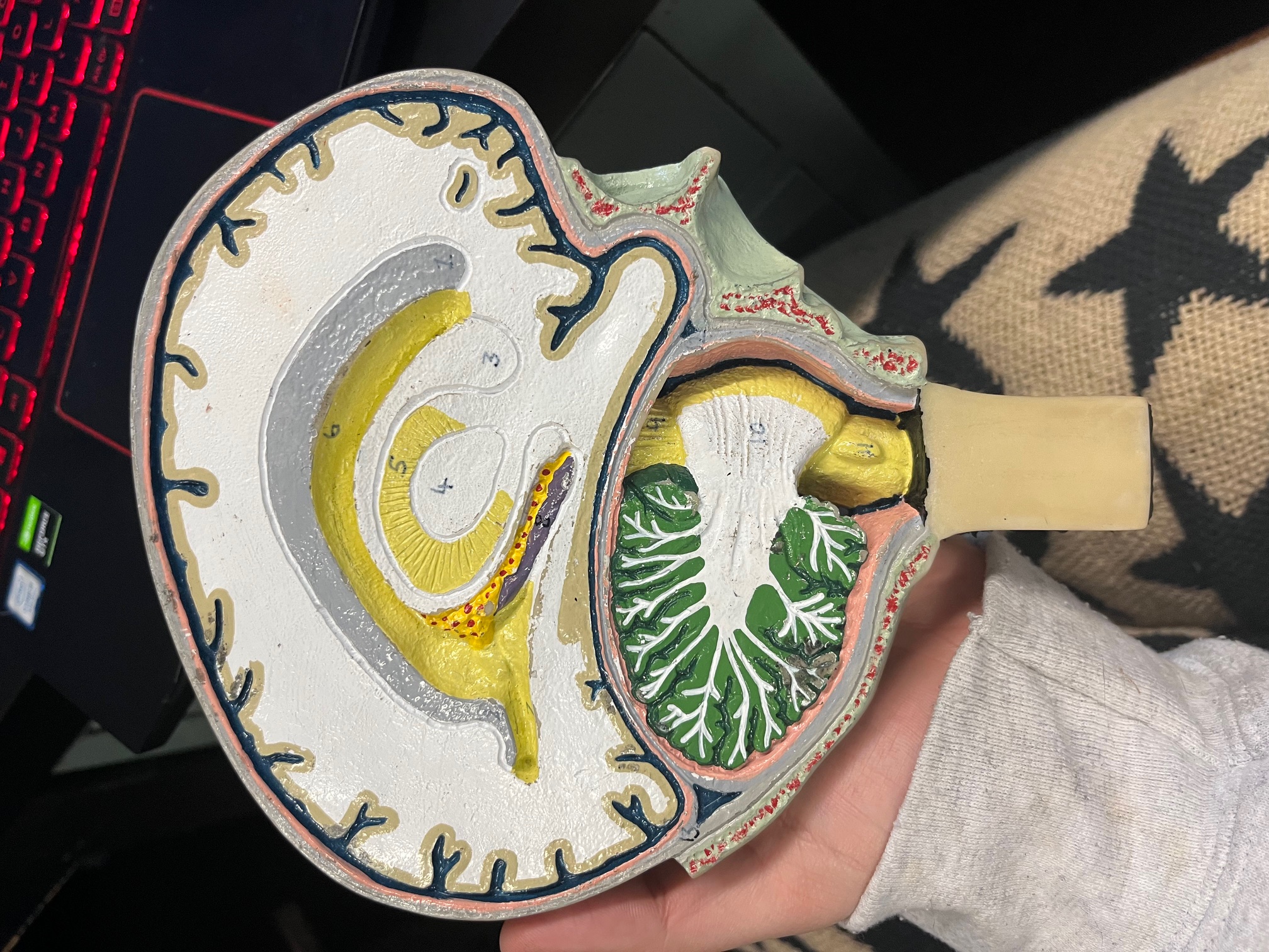
8 (yellow space in between 1 and 3) (name only)
inferior horn of lateral ventricle