eso + pancreatic + endo
 Call Kai
 Learn
 Practice Test
 Spaced Repetition
 Match
 Flashcards
 Knowt Play
1/162

There's no tags or description
Looks like no tags are added yet.
163 Terms
incidence of esophageal cancer
1% of all cancers
2026 ⇒ estimates 2,800 diagnosed // 2,500 deaths
mortality of esophageal cancer
HIGH
2026 ⇒ estimates 2,800 diagnosed // 2,500 deaths
gender affected by esophgeal cancer
men>women (3-4:1)
age affected by esophageal cancer
55-85yo
geography of esophgeal cancer
northern china, northern iran, south africa
race affected of esophageal cancer
Caucasian ⇒ adenocarc
Black person ⇒ SCC
cause/risk factors of adenocarcinoma (esophgaeal cancer)
Barrett's esophagus
Gastroesophageal reflex disease (GERD)
Obesity (by ↑ risk of GERD)
cause/risk factors of squamous cell carcinoma
Smoking
Alcohol
Dietary and environmental factor that cause chronic irritation and inflammation of esophageal mucosa
Tylosis, achalasia, esophageal diverticula and webs, plummer-winson syndrome, HPV infection
barretts esophagus
normal flat, pink lining (squamous epithelium) of the lower esophagus changes into a thicker, red, intestinal-type lining (columnar epithelium with goblet cells)
Associated with GERD
GERD
gastroesophageal reflex
chronic digestive condition where stomach acid and contents frequently flow back up into the esophagus (food pipe), irritating its lining
how is GERD related to esophageal cancer
If regular stomach acid and bile splashes up into the lower esophagus, it irritates and injuries the unprotected lining ⇒ may lead to barretts esophagus ⇒ dysplasia
achalasia
disorder when the lower ⅔ of esophagus loses normal peristaltic activities and becomes dilated (“megaesophagus”) and the esophagogastric junction sphincter fails to relax – ∴ prohibits passage of food into stomach
Pts w achalasia have a 5-20% risk of developing squamous cell cancer of esophagus
what vertebral does esophagus from/to
C6 to T11
begins inferior to the laryngopharynx at the level of the cricoid cartilage (C6) at the upper esophageal sphincter to stomach (T11)
relational anatomy of esophagus
posterior to the trachea, anterior to the vertebral bodies. Lateral and left of esophagus is the aortic arch
which is more ANT/POST: trachea vs esophagus
trachea = more ANT
esophagus = more POST
muscle composition of esophagus
deep to superficial
Lamina propria, Mucosa, muscularis mucosa, submucosa, circular muscular layer, longitudinal muscle, with interconnecting lymphatics
⤷ lacks a serosal layer
significance of lacking serosal layer in esophagus
esophagus structurally weaker, more prone to tearing, and lacking a physical barrier to stop the spread of cancer and infection into the chest cavity
regions of esophagus
upper thoracic, middle thoracic, lower thoracic/GE junction
why are esophagus references from upper incisors (front teeth)
fixed and identifiable landmark for endoscopy etc.
longitudinal or radial growth of cancer in esophagus
longitudinally (up/down)
direct invasion of esophagus
Longitudinal growth first (up/down)
Tumours can grow large before obstruction
Esophagus = NO serosal layer ⇒ ∴ less resistance against local spread
Fistula with bronchus or trachea possible
lymphatics spread of esophagus
NOT CONTIGUOUS
early lymphatics invasion + skip lesions distant from primary site
drainage = unpredictable
Upper ⅓ (cervical area) ⇒ drains into…
Internal jugular, cervical, paraesophageal, supraclavicular LNs
Upper/middle thoracic portion drain into…
Paratracheal, hilar, subcarinal, paraesophgeal, paracardial LNs
Distal/lower ⅓ of esophagus drains…
Celiac axis, left gastric nodes, nodes of the lesser curvature of the stomach

significances of esophagus having a rich lymphatic network
cancer can spread through LN far away from orginal tumour (∴ lymphatic spread if unpredictable)
where do LNs from the upper 1/3 of cervical area drain into
Internal jugular
cervical
paraesophageal
supraclavicular LNs
where do LNs from middle thoracic esophagus drain into
paratracheal
hilar
subcarinal
paraesophageal
paracardinal
where do LNs from distal/lower 1/3 of esophagus drain
celiac axis
left gastric nodes
nodes of the lesser curvature of the stomach
what is unqiue about esophagus LN
longitudinal flow (bidirectional spread) + skip metastases (able to drain into any adjacent draining nodal bed)
possible mets to esophageal cancer (what are most common)
*liver, *lung, bone, CNS
Most patients present with advanced diseases
does esophageal cancer have early or late stage presentation? + why?
**early-stage = Ū ASYMPTOMATIC/nonspecific
∴ esophgeal cancer has late-stage presentation (when symptoms finally show = ū alr advanced)
Why? Esophagus is used to having food into (allow things to pass/flexible), hard to notice tumour in there
early stage SS clinical presentation of esophageal cancer
90%: dysphagia (difficulty swallowing), weight loss
others
coughing and regurgitation
50%: odynophagia (painful swallowing)
upper GI bleeding
regional invasion SS esophageal cancer
respiratory fistula (hole between trachea/esophagus)
hoarseness from recurrent laryngeal nerve invasion
pain caused by local spread
advanced distant stage SS for esophageal cancer
Hypercalcemia (but no brain mets = paraneoplastic syndrome bc of parathyroid production protein)
Mets to lungs, liver, central nervous system
Hematemesis: vomiting blood
what paraneoplastic syndrome is related to esophagus cancer
hypercalcemia
are pts w esophageal cancer diagnosed in early or late stage
late stage (ū locally advanced/metastatic) bc early-stage ū asymptomatic/subtle
esophagus also has no serosal layer… ∴ able to grow/infiltrate deep muscle and lymph nodes before obstruction occur
SS related to esophagus
dysphagia, odynophagia, regurgitation + mucus, esophagus bleeding, anorexia + weight loss bc of inability to eat
SS related to surrounding anatomy of esophgeal cancer
chest pain, hoarseness, chronic cough, dyspnea (SOB), bone/back pain
tracheoesophageal fistula
abnormal tract connecting the esophagus to the respiratory tract (trachea)
⤷ easily occurs in esophagus bc of no serosal layer
imaging that can be done for esophagus cancer
barium swallow contrast w xray
Endosocpic US
bronchoscopy
CT chest/upper abdomen
PET-FDG for mets
which imaging technique is more accurate for staging primary/LNs of esophgeal cancer
endoscopic US — assess depth of tumour, LN assessment, biopsy
barium contrast swallow w xray
drink chalky, liquid barium sulfate mixture that coats the inner lining of your upper digestive tract so tumour shows up bright white on X-rays (highlight)
histopathology for esophagus
Upper and middle esophagus ⇒ squamous cell carcinoma
Lower esophagus/gastroesophageal (GE) junction ⇒ adenocarcinoma
Other rare histology
Adenoid cystic carcinoma
Mucoepidermoid carcinoma
Adenosquamous carcinoma
Undifferentiated carcinoma
what histology for upper/middle esophagus
SCC
what histology for lower/GE junction
adenocarcinoma
adverse prognostic factor of esophgeal cancer
Size of primary tumour >5cm
Weight loss 10%
Poor performance status (impact their ability to tolerate treatment)
Age >65yo
5-year survival of esophgeal cancer
5-30% overall (assuming pt is amendable)
Localized (confined to esophagus) – 43%
Regional – 23%
Distant – 5%
goal of treatment for esophageal cancer
Goal of treatment… relieve dysphagia and provide chance for cure
⤷ esophageal cancer is treatable but rarely curable as most patients have locally advanced/metastatic disease at diagnosis
radical intent treatment for esphageal cancer
1) Chemoradiation therapy (*standard treatment*)
2) Neoadjuvant pre-op chemo radiation
Chemoradiation = choice of treatment (incl. brachy)
Surgery requires extremely extensive procedure
palliative intent tx for esophageal cancer
Symptom management = PRIMARY GOAL [pain control, improvement in swallowing, quality of life]
Various combinations ⇒ surgery, chemotherapy, radiation therapy, stent, photodynamic therapy, endoscopic laser therapy
total esophagectomy
removes all of esophagus + LN + portion of upper stomach
Ū stomach is pulled up into the chest/neck and connected to the remaining upper esophagus/pharynx
Depends on location of lesion and extent of disease
Transthoracic resection (TTE) (ivor lewis procedure
removes all of esophagus, LN, upper portion of stomach
Laparotomy + right thoracotomy to remove esophagus and pull stomach up into thoracic cavity
transhiatal approach
esophagus is removed through abdomen and neck… without opening chest (works through esophageal hiatus: opening in diaphragm)
laparotomy
large abdominal incision
right thoracotomy
surgical incision on right side of the chest (ū bc esophagus is easier to access from right side)
bc left side has aortic arch
cons of surgery for esophageal cancer
high morbitdity and mortality rate
which part of the esophagus cannot be surgically operated on
Challenging to operate in upper ⅓ near cervicals → ū use chemoradiation in that area
high surgical risk (high morality/morbitity)
tight anatomical space with nearby organs (larynx, trachea, thyroid, BV, nerves etc)
chemotherapy as tx for esophagus process
Three-step approach:
1) induction chemotherapy – ū to shrink before chemotherapy ?
2) pre-op chemoradiation
3) surgery
continous infusion of 5FU and cisplatin = standard (concurrent w RT) — but a lot of side effects (other regiments are being studied)
3-field approach of RT
cannot have beam from direct posterior field (hitting spinal cord)
Instead = have 2 oblique posterior fields (avoids spinal cord) + anterior field
compare dose of RT and chemoradiation
dose of RT alone = higher than chemoradiation
lower dose bc chemotherapy makes cells more sensitive to radiation… so don’t need that high of a dose to have the same effect
RT alone = 6000-6500cGy
Chemoradiation = 5040 cGy
organs at risk/sensitive to radiaiton damage
lung, heart, spinal cord, (stoamch)
which structures/organs are the most radiosensitive
lung ??
non-surgical tx of choice for esophagus cancer
RT + concurrent chemo
screening for esophgeal cancer
For public/people with GERD ⇒ no regular screening test bc no evidence that show more benefits than risks
For high-risk people (has Barrett’s esophagus, inherited syndromes) ⇒ carefully monitored for early/precancer + regular upper endoscopies
prevention of esophagus cancer
Quit smoking
limit/avoid alcohol!
Maintain healthy weight
Eat a diet rich of fruits/veggies
why are long radiation fields used in esophagus cancer
bc of esophag’s longitudinal spread + extensive lymphatic drainage + skip metastasis
to ↓ risk of recurrence, margin size is 3-5cm above and below tumour margins to also target more longitudinally and possible skip metastasis
incidence of pancreatic
2% of all cancers all cancers in US
mortality of pancreatic
4th leading cause of cancer in US, one of the deadliest malignancies
age affect of pancreatic
50-80yo, most >70yos
gender affected of pancreatic
males > females
race affected of pancreatic
african american > white
known risk factors
Tobacco
Obesity
Diabetes
Family history of pancreatic cancer
Genetic conditions
Occupational exposure to chemicals
Chronic pancreatitis
possible risk factors
Alcohol
Helicobacter pylon (bacteria)
Hepatitis B virus
Exposure to pesticides
lower risk
Diet high in fruits/veggie
location of pancreas
between aorta and IVC, covered by stomach and liver
Internal vertebral relation to pancreas – pancreas is located retroperitoneally to T12-L2
what vertebrae is associated with pancreas
T12-L2
3 anatomical portions of pancreas
Head – surrounded by duodenum
Body
Tail – in the hilum of the spleen
function of pancreas
exocrine ⟹ Make digestive enzymes that are collected in main pancreatic duct and emptied into the duodenum (digest fats, sugar, proteins)
endocrine ⟹ make hormones — Islets of langerhans: produce hormones (eg. insulin) that are released into bloodstream
why is tumour at head of pancreas bad
Head of pancreas has the pancreatic due emptying enzymes into the duodenum + bile duct deliver bile from liver and gallbladder. Tumour ū block both pancreatic and bile duct (∴ also cause jaundice)
bile
yellow-green fluid produced by liver, stored in gallbladder that breaks down dietary fats, absorbs fat-soluble vitamins, neutralizes stomach acids, remove waste
early stage of pancreatic cancer symptoms
asymptomatic in early — “silent disease“ (∴ diagnosed ū at later stage)
direct extension of pancreatic (what structures does it spread to)
spread to stomach, duodenum, colon, spleen
IF spread to mesenteric artery, portal vein, celiac axis artery, nerve involvement ⟹ ∴ UNRESECTABLE
local extension, if spread to these structures, will mean unresectable
IF spread to mesenteric artery, portal vein, celiac axis artery, nerve involvement, ⇒ UNRESECTABLE
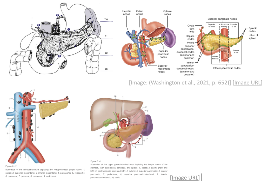
lymphatics spread to pancreatic
Left side ⇒ drains along tail into splenic hilar nodes
Superior pancreatic LNs and celiac axis
Inferior pancreatic, superior mesenteric, and left paraaortic nodes
Right side ⇒ drains to anterior and posterior pancreaticoduodenal nodes + right para-aortic node
how much % of pancreatic cancer have mets at the time of presentation?
50%
where does metastatsis spread to for pancreatic cancer
spread to liver (via portal vein), lungs, peritoneal seeding into perionteal cavity, bone (less common but possible for advanced stage)
percentage of tumour found in the location segments of pancreas
Head/neck = 50-60%
Body = 25%
Tail = 25%
ealry stage clinical presentaiton of pancreatic cancer
asymptomatic in early stage (“silent disease”)
main S/S of pancreatic cancer
Jaundice (+itching): yellowing of skin and sclera of eye
tumor blocks the common bile duct, preventing bile and the yellow pigment bilirubin from flowing from the liver into the small intestine
Abdominal pain
Weight loss
Anorexia
other S/S of pancreatic cancer
Jaundice (+itching): yellowing of skin and sclera of eye
tumor blocks the common bile duct, preventing bile and the yellow pigment bilirubin from flowing from the liver into the small intestine
Abdominal pain
Weight loss
Anorexia
Back pain
Digestive problems
Diabetes mellitus (type 2 adult onset)
Dark urine
Light stools
Enlarged pancreas, gallbladder, or liver possibly
why is pancreatic cancer difficulty to detect and diagnoses
No noticeable signs/symptoms in early stages of pancreatic cancer
The signs of pancreatic cancer, when present, are S/S of other illnesses (eg. pancreatitis, ulcer)
The pancreas is obscured by other organs in abdomen and is hard to visualize on imaging
what lab tests are done for pancreatic cancer
Lab testing
CBC + blood chemistry test
Liver enzyme test — see liver (bc one of main met sources)
Creatinine — tests how well kidney filter for possible contrast/dye injected in body/ducts
Blood urea nitrogen (BUN): measures waste product formed when liver breaks down protein — liver break down ⟹ relates to liver
glucose — pancreas aids in blood sugar levels (insulin)
what imaging done for pancreatic cancer
CT — common!
MRI
endoscopic retrograde Cholangiopancreatography (ERCP): see bile ducts, gallbladder
ERCP = Using fluoroscopy (live xray) + use contrast to visualize anatomy of pancreatobiliary ductals (whats going on in pancreas and bile duct)
endorectal ultrasound (ERUS) or AKA transrectal US
diagnostic tests that can be done to test for pancreatic cancer
lab tests,
imaging (CT/MRI/ERCP/ERUS)
FNA (needle to collect sample — biopsy)
Cholangiography: inject dye into bile duct + branches to see obstruction
Visceral angiography to determine vascular invasion
histology of pancreatic
80% adenocarcinoma
types of pancreatic cancer
Exocrine cancer: 95% of cases, and arise from cells of the pancreatic duct
Functions to produce digestive enzymes
Endocrine cancer: 5% of cancers
Arise in cells that produce hormones
which type of pancreatic cancer is more common
exocrine (95% of cases) adenocarc
staging of pancreatic cancer
TNM
T-stage ⇒ based on tumour diameter size
T1 = <2cm
T2 = 2-4cm
T3 = >4cm
T4 = growing outside of pancreas and into nearby major blood vessels
N-stage ⇒ # of nodes affected
N1 = 1-3 LNs
N2 = 4+ LNs
good prognostic factors of pancreatic cancer
Good prognostic factors
Clear surgical margins
Negative lymph nodes
Reduced perioperative mortality (death bc of another surgical procedure
5-year survival
Overall – ~5%
Local disease = 19%
Regional disease = 8%
Distant disease = 2%
radical surgery for pancreatic cancer
surgery = treatment of chocie
Distal pancreatectomy: remove only body + tail of pancreas + maybe spleen and LNs
Total pancreatectomy: remove ENTIRE pancreas + surrounding organs
Pancreaticoduodenectomy (whipple procedure): remove head of pancreas + first part of SI + gallbladder + bile duct ⇒ and reconnect to allow for flow between
Ū for tumours that have NOT spread beyond pancreas (still resectable)
Anatomy anastomosed (reconnected) ⇒ remaining pancreatic tail to jejunum//remaining common bile duct to jejunum//remaining stomach to jejunum
⤷ surgery may be done laparoscopically
distal pancreatectomy
remove only body + tail of pancreas + maybe spleen and LNs