NEUR30080 L3 SQ action potential
 Call Kai
 Learn
Learn Practice Test
 Spaced Repetition
 Match
Match Flashcards
 Knowt Play
1/42

There's no tags or description
Looks like no tags are added yet.
Name | Mastery | Learn | Test | Matching | Spaced | Call with Kai | Chat |
|---|
No analytics yet
Send a link to your students to track their progress
43 Terms
learning objectives
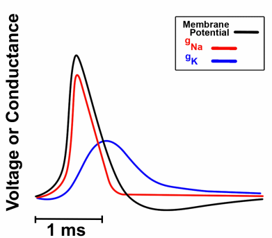
the waveform: one spike, five phases
rest (-65 mV): K+ leak channels dominate, holding Vm near Ek
threshold (-55 mV): the point where the feedback runs away
upstroke and overshoot (+30 mV): Na+ enters in and briefly flips the polarity
repolarisation: Na+ channels inactivate and K+ efflux pulls Vm back down
after-hyperpolarisation (-80 mV): K+ channels close late, so Vm dips past rest
waveform image

all-or-nothing
sub-threshold input: elicits only passive electrotonic decay (the Lecture 2 membrane)
supra-threshold input: triggers an identical, full-amplitude AP regardless of stimulus excess
amplitude is uninformative: fixed size of the spike means individual APs carry no graded information
rate coding: stimulus intensity is encoded entirely by spike frequency (Hz), never by amplitude
this is why a pinprick and a hammer blow produce the same size of AP, and very different firing rates
all-or-nothing image

recording the spike two ways
intracellular recording: the electrode penetrates the cell, directly measuring the full 100 mV transmembrane voltage (Vm)
extracellular recording: an external electrode detects minute local current sinks and sources in the EC fluid
signal attenuation: the recorded wavelength is tiny (100 uV), biphasic
non-destructive: allows for stable, LT in vivo recording without compromising the cell memb

one channel, two gates
activation gate:
closed at rest
opens rapidly upon depol via the S4 voltage sensor
inactivation gate:
open at rest
closes slowly upon depol
steric occlusion via a tethered ‘ball-and-chain’ mechanism
closed different to inactivated

inactivation and activation gates: kinetic disparity
both gates respond to depolarisation, but kinetic delay between activation and inactivation creates the transient Na+ current
inactivation and activation gates: voltage-dependent reset
repolarisation is an absolute requirement to dislodge the inactivation gate
threshold is where the feedback runs away
The Hodgkin Cycle: Initial depolarisation opens Nav channels → Na+ influx → further depolarisation → more Nav open
Positive Feedback: This self-amplifying loop rapidly drives the membrane toward ENa
Sub-threshold Damping: Below threshold, K+ efflux exceeds Na+ influx, damping the cycle and restoring the resting potential
Threshold Crossing: The critical voltage where inward Na+ current irreversibly exceeds outward K+ current
threshold is not s fixed channel property, it is the precise voltage where inward Na+ current irreversibly exceeds outward K+ current

why the peak stops short
The Thermodynamic Limit: ENa is +58 mV, but the peak arrests at ~+30 mV.
Nav Inactivation: The slow inactivation gate sterically occludes the pore, terminating Na+ influx
Delayed Rectifier Activation: Voltage-gated K+ channels (Kv) finally open, introducing a massive outward K+ current
Continuous Leak: Resting K+ leak channels continuously oppose depolarisation
the AP is a kinetic race between an inward current that inactivates early and an outward current that activates late

repolarisation and the undershoot
Dual Mechanism Repolarisation: Nav inactivation removes inward current, while Kv activation provides strong outward current
Kinetic Delay (Kv): Delayed rectifier channels open late, allowing the initial rapid upstroke.
Prolonged gK: Kv channels are slow to deactivate, maintaining high K+ permeability as Vm drops
The Undershoot (AHP): Elevated PK transiently drives Vm closer to EK (~ −80 mV) than at rest
the undershoot is not a separate mechanism, it is just the K+ channels being late to leave
phases of the spike table

refractory periods due to inactivation
absolute refractory
relative refractory
frequency cap
complete reset
absolute refractory period
infinite threshold
Nav channels are physically inactivated (sterically occluded)
a channel state, not a shortage of ions
relative refractory period
elevated threshold
a subset of Nav channels have reset, but residual gk strongly opposes depolarisation
frequency cap
together, the refractory periods restrict maximum firing rates to a few 100 Hz
complete reset
once fully repolarised, the channels fully reset
gradients are functionally unconsumed
refractory periods due to inactivation image

research using giant squid axon
Hodgkin and Huxley (1952): Utilised the squid giant axon and voltage clamp technique
Mathematical Inference: Separated ionic currents and mathematically predicted the kinetic behaviour of distinct 'gates
Predictive Power: The model accurately predicted gating charge movements, physically verified decades later
Predictive Power: The model accurately predicted gating charge movements, physically verified decades late


Q:

the trigger zone
the axon hillock segment
a highly specialised 20-60 um unmyelinated domain directly proximal to the soma
Ankyrin-G and betaIV-spectrin physically anchor the channels into this high-density matrix
low threshold Nav1.6 is clustered distally (where the spike initiates), while Nav1.2 sits proximally

ankyrin-G significance
lose ankyrin-G and the axon will grow spines and start behaving like a dendrite
the axon initial segment (AIS) defines neuronal polarity
channels are localised
Nav clustering: strictly localised to the AIS and the Nodes of Ranvier - the exclusive sites of active regeneration
Kv1 localisation: sequestered at juxtaparanodes beneath the myelin to damp pathological repetitive firing
Kv7 (M-current): positioned at the AIS and nodes to regulate threshold dynamics and resting excitability
structural plasticity: the AIS is dynamic, chronic depol physically shifts the structure distally to reduce excitability
channels are localised image
a neuron does not have one uniform excitability
different subcellular patches express completely discrete channel populations

mechanism of spread: local circuits
the actively firing membrane patch acts as a massive current source due to local Na+ influx
electrotonic spread: current spreads passively down the axon, dictated entirely by the length constant (lambda)
the advancing passive depol triggers durther depol in the adjacent memb patch
nothing physically ‘travels’, each discrete patch ignites the next, analogous to a burning fuse
passive electrotonic spread drives current forward and active regeneration restores the amplitude
both essential for long-distance propagation
mechanism of spread image

direction
only goes forwards because intracellular current spreads bidirectionally
the immediately preceding membrane patch is in its absolute refractory period (Nav channels inactivated)
consequently, only the ‘resting’ membrane ahead can successfully depolarise
stimulate mid-axon: initiates bidirectional propagation (orthodromic and antidromic) because both adjacent patches are resting
direction enforced by refractoriness, not by structure
direction image

conduction velocity

increasing diameter of axons
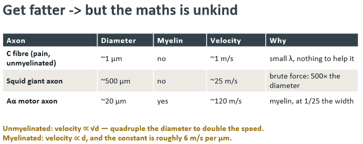
myelin: optimises by changing both constants at once
maximising lambda: multiple lipid layers in series massively increase transmemb resistance (Rm), essentially eliminating passive leak
minimising tau: increasing the dielectric thickness drastically reduces total membrane capacitance (Cm), allowing near-instantaneous charging
lambda and tau up and down together: myelin successfully optimises both biophysical constraints simultaneously
internodes about 100x the axon diameter
myelinated axon image

oligodendrocytes
myelinate many axons each in the CNS
schwann cells
make exactly one internode each in the PNS
Nodes of Ranvier
one bare membrane, every 2 mm
Nodes of Ranvier (1 um) contain the highest Nav channel density in the nervous system.
Under the myelin, the signal spreads purely via fast, passive electrotonic decay. Nothing physically 'jumps'
At the node, the arriving passive voltage triggers the depolarisation, rebuilding a fresh action potential from scratch
The Safety Factor (~3-5x): The passive current arriving at a node is safely several times larger than the required threshold current
saltatory conduction misnomer
implies leaping, but this is misnomer
the signal is passively carried and actively rebuilt
demyelination
the safety factor falls below one
Demyelination causes Rm to plummet and Cm to spike; lambda shortens catastrophically while tau lengthens
Passive current decays completely before reaching the next node
Conduction Block: The safety factor drops below 1.0, causing the propagating signal to permanently stall and die
Increased temperature accelerates channel kinetics, prematurely terminating Nav influx and further reducing the safety factor
demyelination image

Uthoff’s phenomenon
MS symptoms worsen with a hot bath, exercise or fever
how you take the spike apart table


Q:

learning outcomes
