Management of Odontogenic Cysts & Tumors
 Call Kai
 Learn
 Practice Test
 Spaced Repetition
 Match
 Flashcards
 Knowt Play
1/35

There's no tags or description
Looks like no tags are added yet.
36 Terms
origins of odontogenic cysts/tumors
-residual tooth-forming tissue is source
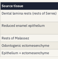
source tissue, where it sits, what it can become

cyst v. tumor
-cyst: epithelium-lined cavity with fluid or keratin, grows by internal pressure and pushing bone ahead of it, usually separates cleanly from bone, shrinks when drained → enucleation or decompression
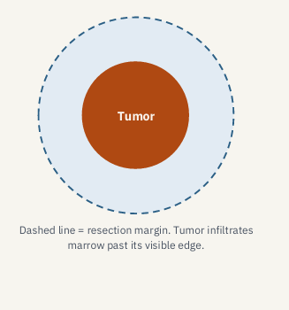
-tumor: true neoplasm with autonomous growth, infiltrative types extend into marrow beyond the visible edge, encapsulated types shell out, draining does not cure it → enucleation (encapsulated) or resection with margins (infiltrative)
classification of cysts, benign tumors, malignant

describing the lesion- locularity, relation to teeth, borders, neighbors, contents

pericoronal differential
-dentigerous cyst
-OKC
-unicystic ameloblastoma
-AOT (anterior, canine)
-ameloblastic fibroma
-CEOT, COC
periapical/lateral root differential
-PAR/radicular cyst
-lateral periodontal cyst
-early COD
-cementoblastoma
-traumatic bone cyst
multilocular differential
-OKC
-ameloblastoma
-myxoma
-CGCG
-AV malformation
-aneurysmal bone cyst
-intraosseous carcinoma
multifocal differential
-NBCCS (multiple OKCs)
-cherubism
-langerhans cell histiocytosis
-hyperparathyroidism (brown tumors)
-early COD
-multiple myeloma
multilocular radiolucency? think…
MACHO
-myxoma: young adults; straight “tennis racket” septa
-ameloblastoma: 30-70; posterior mandible; soap bubble/honeycomb appearance
-central giant cell granuloma: younger; anterior mandible
-hemangioma: vascular lesion
-OKC: 2nd-3rd decade; little expansion
treatment for MACHO differential


aspirate returns, think, next

treatment ladder
-decompression/marsupialization (shrink it first, drain placement following incisional biopsy)
-enucleation with/without curettage
-enucleation + adjunct (peripheral ostectomy, Carnoy’s 5-FU, cryotherapy)
-marginal resection (inferior border kept; continuity preserved)
-segmental resection + reconstruction (continuity defect; bone graft or free flap), for infiltrative lesions
-up the ladder: more morbidity, lower recurrence; pick the rung by diagnosis, size, location, age, nearby vital structures, recurrence risk, the patient
traumatic bone cyst treatment
-exploring it is the treatment

decompression
-lesion shrinks, bone backfills
-cyst wall thickens, so later enucleation is easier
-lining histology changes
-choose when large lesion near the IAN, sinus, or roots, child with developing teeth that may still erupt, patient who can’t tolerate a large surgery
-considerations: months of irrigation, compliance matters, usually a second surgery to enucleate
-biopsy before or at drain placement, while the lining is still diagnostic
dentigerous cyst
-about 1/3 of odontogenic cysts
-fluid between the reduced enamel epithelium and the crown; attached at the CEJ
-most often: mandibular 3rd molar > maxillary canine > maxillary 3rd molar > mandibular 2nd premolar
-central (most common), lateral, or circumferential
-unilocular and corticated; can push a tooth far from its site and resorb roots
-a pericoronal (follicular) space wider than about 3-5mm on radiograph is the conventional threshold that should raise suspicion for a dentigerous cyst rather than a normal dental follicle
-treatment: small- enucleation with the tooth; large- marsupialization, then enucleation once it has decompressed; recurrence is uncommon, always send to pathology
cyst, key features, treatment: radicular, residual, lateral periodontal, glandular, calcifying (Gorlin)
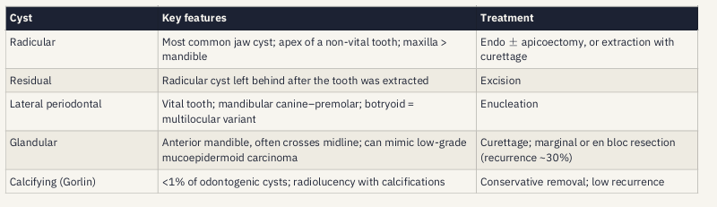
the most common radiolucency at a root apex
-periapical lesion of endodontic origin
-test vitality before anything else
-non-vital tooth: treat the tooth first
-persists after endo: biopsy (+ apicoectomy)
-vital tooth: think beyond endo
OKC
-cyst that behaves like a tumor
-2nd-3rd decade; slight male predilection
-posterior mandible (3rd molar/ramus) most common
-from dental lamina rests; PTCH tumor-suppressor gene
-tunnels through marrow with little expansion; displaces the IAN, resorbs roots
-thin, friable lining → hard to remove in one piece
-recurs, mostly within 5 years

nevoid basal cell carcinoma syndrome (Gorlin)
-multiple OKCs (~90%)
-multiple basal cell carcinomas (~90%), can start in childhood
-palmar and plantar pits
-calcified falx cerebri
-bifid or fused ribs, spina bifida
-frontal bossing, macrocephaly, hypertelorism
-medulloblastoma, ovarian fibromas
-cleft lip/palate (~5%)
-genetics: autosomal dominant, PTCH1 (9q22.3) in most, also PTCH2/SUFU, hedgehog signaling pathway, ~40% arise de novo, ~1 in 57,000 (estimates vary)
-care: lifelong jaw surveillance, dermatology for skin cancer checks, genetics referral (screen the family), avoid radiotherapy, minimize ionizing radiation
OKC recurrence rates with treatment options


OKC- adjuncts for bone cavity, how it works, watch out for

treatment of biopsy-proven OKC based on size/aggressiveness
-small, accessible: enucleation + peripheral ostectomy + adjunct (5-FU or Carnoy’s)
-large, near IAN or teeth, child: decompress first, then enucleate + adjunct once smaller
-repeated recurrence, aggressive: resection with ~1cm margins
-for everyone: radiographic follow-up for at least 5 years- most recurrences show up by then, NBCCS: lifelong
ameloblastoma
-most common clinically significant epithelial odontogenic tumor
-age 30-70; mandibular molar/ramus
-slow, painless expansion
-uni- or multilocular: “soap bubble” (large locules), “honeycomb” (small)
-root resorption; can mimic a dentigerous cyst
-variants: conventional, unicystic, peripheral, malignant
pattern and histology of ameloblastoma- follicular, plexiform, acanthomatous, granular cell, desmoplastic
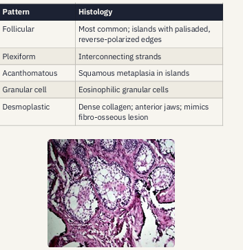
why we resect a benign tumor
-70-85% recur after enculeation and curettage, usually within 5 years; palliative only
-1.0-1.5cm bony margins plus one uninvolved anatomic barrier: curative marginal or segmental resection
unicystic ameloblastoma
-type drives management
-13-21% of ameloblastomas, younger pts, mandibular 3rd molar, looks like a dentigerous cyst

ameloblastoma- less common variants (variant, behavior, treatment)


odontogenic myxoma
-ages ~10-40, mean ~30
-no sex predilection
-mandible more often than maxilla
-from odontogenic ectomesenchyme; gelatinous, with no capsule
-film: multilocular, thin straight “tennis racket” septa; soap bubble or honeycomb
-histology: loose myxoid stroma with spindle- and stellate-shaped cells
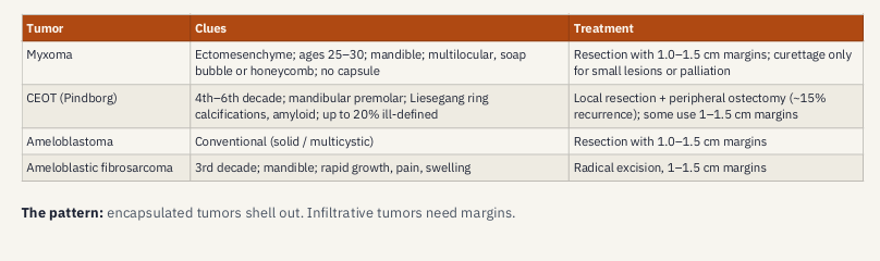
-treatment: resection, 1.0-1.5cm margins + one uninvolved anatomic margin, curettage only for small lesions or palliation, no capsule: curettage leaves tumor in marrow
central giant cell granuloma
-peak ages 5-15; female 2:1
-anterior mandible; uni-or multilocular
-same histology as a brown tumor: rule out hyperparathyroidism when the picture fits
-aggressive if: 1 major (>5cm or recurrence) or 3 minor (rapid growth, tooth displacement or loosening, cortical thinning or perforation, root resorption)
-treatment: enculeation + peripheral ostectomy (~20% recurrence), recurrent- resection, 1cm margins; large- intralesional steroids, weekly x6 weeks, other adjunct medications
cherubism
-child
-bilateral
-symmetric multilocular jaws
-autosomal dominant
-regresses after puberty, observe
AV malformation
-any multilocular lesion could be one
-aspirate first: bleeding can be fatal
COD
-vital teeth
-predominantly seen in black women
-4th-5th decade
-early lesions radiolucent
-don’t biopsy or extract- risk of osteomyelitis
nasopalatine duct cyst
-midline anterior maxilla
-vital incisors
-non-odontogenic
-enucleation
tumors that don’t require margins

tumors that need margins