Chapter 8.4, 10.4, 12.3, 12,4.
 Call Kai
 Learn
 Practice Test
 Spaced Repetition
 Match
 Flashcards
 Knowt Play
1/49

There's no tags or description
Looks like no tags are added yet.
50 Terms
A DNA molecule 300 bp long has 20 complete rotations. This DNA molecule is:
a) positively supercoiled
b) negatively supercoiled
c) relaxed
b
How does bacterial DNA differ from eukaryotic DNA?
Bacterial DNA is not complexed to histone proteins and its circular
The percentage of cytosine in a double-stranded DNA molecule is 40%. What is the percentage of thymine?
In double-stranded DNA, A pairs with T, whereas G pairs with C; so the percentage of A equals the percentage of T, and the percentage of G equals the percentage of C. If C 40%, then G also must be 40%. The total percentage of C G is therefore 40% 40% 80%. All the remaining bases must be either A or T; so the total percentage of A T 100% – 80% 20%; because the percentage of A equals the percentage of T, the percentage of T is 20%/2 10%.
How does supercoiling arise? What is the difference between positive and negative supercoiling?
Supercoiling arises from the overwinding (positive supercoiling) or underwinding (negative supercoiling) of the DNA double helix; from a lack of free ends, as in circular DNA molecules; when the ends of the DNA molecule are bound to proteins that prevent the ends from rotating about each other.
What functions does supercoiling serve for the cell?
Overrotation or underrotation of a DNA double helix places strain on the molecule, causing it to supercoil. Supercoiling is controlled by topoisomerase enzymes. Most cellular DNA is negatively supercoiled, which eases the separation of nucleotide strands during replication and transcription and allows DNA to be packed into small spaces
Which region of bacterial mRNA contains the Shine-Dalgarno sequence?
5' untranslated region
If splice site were mutated so that splicing did not take place, what would the effect be on the protein encoded by the mRNA?
It would be longer than normal
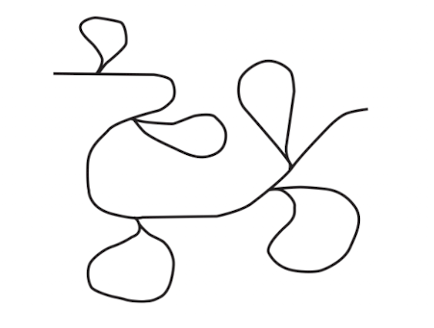
DNA from a eukaryotic gene was isolated, denatured, and hybridized to the mRNA transcribed from the gene; the hybridized structure was then observed with the use of an electron microscope. The following structure was observed.
a. How many introns and exons are there in this gene? Explain your answer.
b. Identify the exons and introns in this hybridized structure (picture).
a. Each of the loops represents a region in which sequences in the DNA do not have corresponding sequences in the RNA; these regions are introns. There are five loops in the hybridized structure; so there must be five introns in the DNA and six exons.
b. (picture)

Draw a typical bacterial mRNA and the gene from which it was transcribed. Identify the 5 and 3 ends of the RNA and DNA molecules, as well as the following regions or sequences:
a. Promoter
b. 5 untranslated region
c. 3 untranslated region
d. Protein-coding sequence
e. Transcription start site
f. Terminator
g. Shine–Dalgarno sequence
h. Start and stop codons

A test-tube splicing system contains all the components (snRNAs, proteins, splicing factors) necessary for the splicing of nuclear genes. When a piece of RNA containing an intron and two exons is added to the system, the intron is removed as a lariat and the exons are spliced together. If the RNA molecule added to the system has the following mutations, what intermediate products of the splicing reactions will accumulate? Explain your answer.
a. GT at the 5 splice site is deleted.
b. A at the branch point is deleted.
c. AG at the 3 splice site is deleted.
a. The GT sequence at the 5 splice site is required for the attachment of the spliceosome and the first cleavage reaction. If this sequence is mutated, cleavage will not take place. Thus, the original pre-mRNA with the intron will accumulate.
b. After cleavage at the 5 splice site, the 5 end of the intron attaches to the A at the branch point in a transesterification reaction. If the A at the branch point is deleted, no lariat structure will form. The separated first exon and the intron attached to the second exon will accumulate as intermediate products.
c. The AG sequence at the 3 splice site is required for cleavage at the 3 splice site. If this sequence is mutated, accumulated intermediate products will be: (1) the separated first exon and (2) the intron attached to the second exon, with the 5 end of the intron attached to the branch point to form a lariat structure.
What is the function of Shine-Dalgarno consensus sequence
The Shine-Dalgarno sequence is a ribosome-binding site found in prokaryotic mRNA, located a few nucleotides upstream of the start codon (AUG).
Its main functions are:
Helps recruit the small (30S) ribosomal subunit to the mRNA.
Base-pairs with a complementary sequence in the 16S rRNA.
Correctly positions the ribosome at the start codon to initiate translation.
In short: The Shine-Dalgarno sequence ensures proper ribosome binding and accurate initiation of protein synthesis in prokaryotes.
What is the 5 cap?
The 5’ end of eukaryotic mRNA is modified by the addition of the 5’ cap. The cap consists of an extra guanine nucleotide (linked 5’ to 5’ to the mRNA molecule) that is methylated at position 7 of the base and at adjacent bases whose sugars are methylated at the 2’ -OH group.
What is the function of the spliceosome?
The spliceosome consists of five snRNPs. The splicing of pre- mRNA nuclear introns takes place within the spliceosome.
What is alternative splicing? How does it lead to the production of multiple proteins from a single gene?
Alternative splicing is a process in which a single pre-mRNA molecule is spliced in different ways, allowing different combinations of exons to be included in the mature mRNA.
During this process:
Introns are removed from pre-mRNA.
Exons can be included or excluded in different combinations.
Different mature mRNA molecules are produced from the same gene.
These mRNAs are translated into different protein isoforms, which may have different structures and functions.
Alternative splicing enables exons to be spliced together in different combinations to yield mRNAs that encode different proteins.

Summarize the different types of processing that can take place in pre-mRNA.
The different types of processing are: addition of the 5 cap to the 5 end of the pre-mRNA; cleavage of the 3 end of a site downstream of the AAUAAA consensus sequence of the last exon; addition of the poly(A) tail to the 3 end of the mRNA immediately after cleavage; and removal of the introns (splicing).
Briefly describe the structure of tRNAs.
short answer: All tRNAs are similar in size and have a common secondary structure known as the cloverleaf. Transfer RNAs contain modified bases and are extensively processed after transcription in both bacterial and eukaryotic cells.
textbook answer: A unique feature of tRNA is the presence of rare modified bases. All RNAs have the four standard bases (adenine, cytosine, guanine, and uracil) specified by DNA, but tRNAs have additional bases, including ribothymine, pseudouridine (which is also occasionally present in snRNAs and rRNA), and dozens of others. The structures of all tRNAs are similar, a feature critical to tRNA function. Some of the nucleotides in a tRNA are complementary to each other and form intramolecular hydrogen bonds. As a result, each tRNA has a cloverleaf structure (Figure 10.22). All tRNAs have the same sequence (CCA) at the 3 end, where the amino acid attaches to the tRNA. At the other end of the tRNA is a set of three nucleotides that make up the anticodon, which pairs with the corresponding codon on the mRNA during protein syn- thesis to ensure that the amino acids link in the correct order. Although each tRNA molecule folds into a cloverleaf owing to the complementary pairing of bases, the cloverleaf is not the three-dimensional (tertiary) structure of tRNAs found in the cell. The results of X-ray crystallographic studies have shown that the cloverleaf folds on itself to form an L-shaped structure, as illustrated by the space-filling and ribbon models .
chatGPT answer: Transfer RNAs (tRNAs) are small, single-stranded RNA molecules, typically 70–90 nucleotides long, that fold into a cloverleaf-shaped secondary structure and an L-shaped 3D structure.
Their main structural features include:
Anticodon loop: Contains three nucleotides that recognize a complementary codon on mRNA.
Amino acid attachment site: Located at the 3′ end (CCA sequence), where a specific amino acid binds.
D loop and TΨC loop: Help maintain the tRNA structure and facilitate interactions with enzymes and ribosomes.
Acceptor stem: Formed by base pairing between the 5′ and 3′ ends.
In short: tRNAs act as adaptor molecules, carrying specific amino acids to the ribosome during protein synthesis.

What is the origin of small interfering RNAs and microRNAs? What do these RNA molecules do in the cell?
Small interfering RNAs (siRNAs) originate from long double-stranded RNA molecules, often produced by viruses, transposons, or cellular processes. They are processed by the enzyme Dicer into short RNA fragments.
MicroRNAs (miRNAs) originate from genes in the cell's own genome. They are transcribed into RNA precursors that form hairpin structures and are processed by Drosha and Dicer.
Functions in the cell:
siRNAs: Usually bind to complementary mRNA molecules and trigger their degradation, preventing protein production.
miRNAs: Usually bind to partially complementary mRNA molecules, inhibiting translation or promoting mRNA degradation.
In short: Both siRNAs and miRNAs regulate gene expression through RNA interference (RNAi), reducing protein production by targeting specific mRNAs.
What is the difference between a structural gene and a regulator gene
Structural genes encode proteins; regulator genes control the transcription of structural genes
Why is transcription a particularly important level of gene regulation in both bacteria and eukaryotes?
Because it is the first step in the process of information transfer from DNA to proteins. For cellular efficiency, gene expression is often regulated early in the process of protein production.
What is a constitutive gene?
A constitutive gene is not regulated and is expressed continually
In a negative repressible operon, the regulator protein is synthetized as
an inactive repressor
The lac Operon of E. coli
The lac operon of E. coli controls the transcription of three genes needed in lactose metabolism: the lacZ gene, which encodes B-galactosidase; the lacY gene, which encodes permease; and the lacA gene, which encodes thiogalactoside transacetylase. The lac operon is negative inducible: a regulator gene produces a repressor that binds to the operator site and prevents the transcription of the structural genes. The presence of allolactose inactivates the repressor and allows the transcription of the lac operon.

In the presence of allolactose, the lac repressor
cannot bind to the operator
Have you spotted the flaw in the explanation just given for the induction of the lac proteins? You might recall that permease is required to transport lactose into the cell. If the lac operon is repressed and no permease is being produced, how does lactose get into the cell to inactivate the repressor and turn on transcription? Furthermore, the inducer is actually allolactose, which must be produced from lactose by B-galactosidase. If B-galactosidase production is repressed, how can lactose metabolism be induced?
The answer is that repression never completely shuts down transcription of the lac operon. Even with active repressor bound to the operator, there is a low level of transcription and a few molecules of -galactosidase, permease, and transacetylase are synthesized. When lactose appears in the medium, the permease that is present transports a small amount of lactose into the cell. There, the few molecules of B-galactosidase that are present convert some of the lactose into allolactose, which then induces transcription.
For E. coli strains with the following lac genotypes, use a plus sign (+) to indicate the synthesis of B-galactosidase and permease and a minus sign (-) to indicate no synthesis of the enzymes when lactose is absent and when it is present.
Genotype of strain
a. lacI+ lacP+ lacO+ lacZ+ lacY+
b. lacI+ lacP+ lacOc lacZ- lacY+
c. lacI+ lacP- lacO+ lacZ+ lacY-
d. lacI+ lacP+ lacO+ lacZ- lacY- /lacI- lacP+ lacO+ lacZ+ lacY+
a. All the genes possess normal sequences, and so the lac operon functions normally: when lactose is absent, the regulator protein binds to the operator and inhibits the transcription of the structural genes, and so B-galactosidase and permease are not produced. When lactose is present, some of it is converted into allolactose, which binds to the repressor and makes it inactive; the repressor does not bind to the operator, and so the structural genes are transcribed, and B-galactosidase and permease are produced.
b. The structural lacZ gene is mutated; so B-galactosidase will not be produced under any conditions. The lacO gene has a constitutive mutation, which means that the repressor is unable to bind to lacO, and so transcription takes place at all times. Therefore, permease will be produced in both the presence and the absence of lactose.
c. In this strain, the promoter is mutated, and so RNA polymerase is unable to bind and transcription does not take place. Therefore, B-galactosidase and permease are not produced under any conditions.
d. This strain is a partial diploid, which consists of two copies of the lac operon—one on the bacterial chromosome and the other on a plasmid. The lac operon represented in the upper part of the genotype has mutations in both the lacZ and the lacY genes, and so it is not capable of encoding -galactosidase or permease under any conditions. The lac operon in the lower part of the genotype has a defective regulator gene, but the normal regulator gene in the upper operon produces a diffusible repressor (trans acting) that binds to the lower operon in the absence of lactose and inhibits transcription. Therefore, no -galactosidase or permease is produced when lactose is absent. In the presence of lactose, the repressor cannot bind to the operator, and so the lower operon is transcribed and B-galactosidase and permease are produced.

Trp Operon of E.coli
The trp operon is a negative repressible operon that controls the biosynthesis of tryptophan. In a repressible operon, transcription is normally turned on and must be repressed. Repression is accomplished through the binding of tryptophan to the repressor, which renders the repressor active. The active repressor binds to the operator and prevents RNA polymerase from transcribing the structural genes.

In the trp operon, what happens to the trp repressor in the absence of tryptophan?
It cannot bind to the operator and transcription takes place
Most transcriptional activator proteins affect transcription by
the basal transcription apparatus
how does the binding of transcriptional activator proteins to enhancers affect transcription at genes that are thousands of base pairs away?
The DNA between the enhancer and the promoter loops out, and so transcription activators bound to the enhancer are able to interact directly with the transcription apparatus
How does the poly(A) tail affect stability?
The poly (A) tail stabilizes the 5’ cap, which must be removed before the mRNA molecule can be degraded from the 5’ end, because the endonucleases are busy chewing the poly (A) tail
In RNA silencing, siRNAs and miRNAs usually bind to which part of the mRNA molecules that they control?
3’ UTR
A comparison of Bacterial and Eukaryotic Gene Control
1. Most gene regulation in bacterial cells is at the level of transcription (although it does exist at other levels). Gene regulation in eukaryotic cells most often takes place at multiple levels, including chromatin structure, transcription, mRNA processing, RNA stability, RNA interference, translation, and posttranslational control.
2. Complex biochemical and developmental events in bacterial and eukaryotic cells may require a cascade of gene regulation, in which the activation of one set of genes stimulates the activation of another set.
3. Much of gene regulation in both bacterial and eukaryotic cells is accomplished through proteins that bind to specific sequences in DNA.
4. Chromatin structure plays a role in eukaryotic (but not bacterial) gene regulation. In general, condensed chromatin represses gene expression; chromatin structure must be altered before transcription can take place. Chromatin structure is altered by the modification of histones, chromatin-remodeling proteins, and DNA methylation.
5. In bacterial cells, genes are often clustered in operons and are coordinately expressed by transcription into a single mRNA molecule. In contrast, each eukaryotic gene typically has its own promoter and is transcribed independently. Coordinate regulation in eukaryotic cells takes place through common response elements, present in the promoters and enhancers of the genes. Different genes that have the same response element in common are influenced by the same regulatory protein.
6. Regulatory proteins that affect transcription exhibit two basic types of control: repressors inhibit transcription (negative control); activators stimulate transcription (positive control). Both negative control and positive control are found in bacterial and eukaryotic cells.
7. The initiation of transcription is a relatively simple process in bacterial cells, and regulatory proteins function by blocking or stimulating the binding of RNA polymerase to DNA. In contrast, eukaryotic transcription requires complex machinery that includes RNA polymerase, general transcription factors, and transcriptional activators, which allows transcription to be influenced by multiple factors.
8. Some eukaryotic transcriptional activator proteins function at a distance from the gene by binding to enhancers, causing the formation of a loop in the DNA, which brings the promoter and enhancer into close proximity. Some distant-acting sequences analogous to enhancers have been described in bacterial cells, but they appear to be less common.
9. The greater time lag between transcription and translation in eukaryotic cells than in bacterial cells allows mRNA stability and mRNA processing to play larger roles in eukaryotic gene regulation.
10. Regulation by siRNAs and miRNAs, which is extensive in eukaryotes, is absent from bacterial cells.
1. A regulator gene produces a repressor in an inducible operon. A geneticist isolates several constitutive mutations affecting this operon. Where might these constitutive mutations occur? How would the mutations cause the operon to be constitutive?
An inducible operon is normally not being transcribed, meaning that the repressor is active and binds to the operator, inhibiting transcription. Transcription takes place when the inducer binds to the repressor, making it unable to bind to the operator. Constitutive mutations cause transcription to take place at all times, whether the inducer is present or not. Constitutive mutations might occur in the regulator gene, altering the repressor so that it is never able to bind to the operator. Alternatively, constitutive mutations might occur in the operator, altering the binding site for the repressor so that the repressor is unable to bind under any conditions.
What would be the effect of a mutation that caused poly(A)- binding proteins to be nonfunctional?
Degradation of mRNA from the 5’ end requires the removal of the 5 cap and is usually preceded by the shortening of the poly(A) tail. Poly(A)-binding proteins bind to the poly(A) tail and prevent it from being shortened. Thus, the presence of these proteins on the poly(A) tail protects the 5’ cap, which prevents RNA degradation. If the gene for poly(A)-binding proteins were mutated in such a way that nonfunctional poly(A) proteins were produced, the proteins would not bind to the poly(A) tail. The tail would be shortened prematurely, the 5’ cap removed, and mRNA degraded more easily. The end result would be less mRNA and thus less protein synthesis.
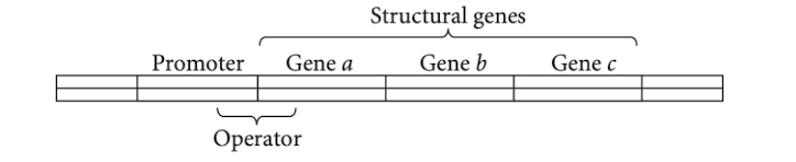
Draw a picture illustrating the general structure of an operon and identify its parts.
What is the difference between positive and negative control? What is the difference between inducible and repressible operons?
There are two basic types of transcriptional control: negative and positive. In negative control, when a regulatory protein (repressor) binds to DNA, transcription is inhibited; in positive control, when a regulatory protein (activator) binds to DNA, transcription is stimulated. Some operons are inducible; transcription is normally off and must be turned on. Other operons are repressible; transcription is normally on and must be turned off.
There are two types of transcriptional control: negative control, in which a regulatory protein is a repressor, binding to DNA and inhibiting transcription; and positive control, in which a regulatory protein is an activator, stimulating transcription. Operons can also be either inducible or repressible. Inducible operons are those in which transcription is normally off (not taking place); something must happen to induce transcription, or turn it on. Repressible operons are those in which transcription is normally on (taking place); something must happen to repress transcription, or turn it off.

Briefly describe the lac operon and how it controls the metabolism of lactose.
The lac operon consists of three structural genes—lacZ, lacY, and lacA. The lacZ gene encodes the enzyme b-galactosidase, which cleaves the disaccharide lactose into galactose and glucose and converts lactose into allolactose. The lacY gene encodes lactose permease, an enzyme necessary for the passage of lactose through the E. coli cell membrane. The lacA gene encodes the enzyme thiogalactoside transacetylase, whose function in lactose metabolism has not yet been determined. All three genes have an overlapping promoter and operator region in common. Upstream from the lactose operon, the lacI gene encodes the lac operon repressor, which binds at the operator region and inhibits the transcription of the lac operon by preventing RNA polymerase from successfully initiating transcription. When lactose is present in the cell, the enzyme b-galactosidase converts some of it into allolactose, which binds to the lac repressor, altering its shape and reducing the repressor’s affinity for the operator. Because the allolactose-bound repressor does not bind to the operator, RNA polymerase can initiate transcription of the lac structural genes from the lac promoter.
What is catabolite repression? How does it allow a bacterial cell to use glucose in preference to other sugars?
In spite of its name, catabolite repression is a type of positive control in the lac operon.The catabolite activator protein (CAP), complexed with cAMP, binds to a site near the promoter and stimulates the binding of RNA polymerase. Cellular levels of cAMP in the cell are controlled by glucose; a low glucose level increases the abundance of cAMP and enhances the transcription of the lac structural genes.
When glucose is available, genes that participate in the metabolism of other sugars are repressed, in a phenomenon known as catabolite repression. For example, the efficient transcription of the lac operon takes place only if lactose is present and glucose is absent.
In E. coli, the concentration of cAMP is regulated so that its concentration is inversely proportional to the level of available glucose. A high concentration of glucose within the cell lowers the amount of cAMP, and so little cAMP–CAP complex is available to bind to the DNA. Subsequently, RNA polymerase has poor affinity for the lac promoter, and little transcription of the lac operon takes place. Low concentrations of glucose stimulate high levels of cAMP, resulting in increased cAMP–CAP binding to DNA. This increase enhances the binding of RNA polymerase to the promoter and increases transcription of the lac genes by some 50-fold.
What changes take place in chromatin structure and what role do these changes play in eukaryotic gene regulation?
Changes in chromatin structure can result in the repression or stimulation of gene expression. The acetylation of histone proteins increases transcription. The reverse reaction, deacetylation, restores repression. Chromatin-remodeling complexes bind directly to the DNA, altering chromatin structure without acetylating histone proteins and allowing transcription to be initiated by making the promoters accessible to transcription factors. The methylation of DNA sequences represses transcription. The demethylation of DNA sequences often increases transcription.
What is the histone code?
Histones in the octamer core of the nucleosome have two domains: (1) a globular domain that associates with other histones and the DNA and (2) a positively charged tail domain that probably interacts with the negatively charged phosphate groups on the backbone of DNA. The tails of histone proteins are often modified by the addition or removal of phosphate groups, methyl groups, or acetyl groups. These modifications have sometimes been called the histone code, because they encode information that affects how genes are expressed.
Briefly explain how transcriptional activator proteins and repressors affect the level of transcription of eukaryotic genes.
Transcriptional regulatory proteins in eukaryotic cells can influence the initiation of transcription by affecting the stability or assembly of the basal transcription apparatus. Some regulatory proteins are activators and stimulate transcription; others are repressors and inhibit transcription.
What is an enhancer? How does it affect the transcription of distant genes?
An enhancer is a DNA sequence that, when bound to transcriptional activator proteins, can affect the transcription of a distant gene. Transcription at a distant gene is affected when the DNA sequence between the gene’s promoter and the enhancer loops out, bringing the promotor and the enhancer close together and allowing the transcriptional activator proteins to directly interact with the basal transcription apparatus at the promoter, which stimulates transcription.
What role does RNA stability play in gene regulation? What controls RNA stability in eukaryotic cells?
The total amount of protein synthesized depends on the amount of mRNA available for translation. The amount of available mRNA depends on the rates of mRNA synthesis and degradation. Less-stable mRNAs degrade faster than stable mRNAs, and so fewer copies of the mRNA are available as templates for translation. The 5’ cap, the 3’ poly(A) tail, the 5’ UTR, the 3’ UTR, and the coding region in an mRNA molecule affect its stability. Poly(A)-binding proteins bind at the 3’ poly(A) tail. These proteins contribute to the stability of the tail and protect the 5’ cap through direct interaction. When a critical number of adenine nucleotides have been removed from the tail, the protection is lost and the 5’ cap is removed. The removal of the 5’ cap enables 5’ -to-3’ nucleases to degrade the mRNA.
Briefly list some of the ways in which siRNAs and miRNAs regulate genes.
Through Slicer activity, which cleaves mRNA sequences; through the binding of miRNAs to complementary regions in mRNA, which prevents translation; and through transcriptional silencing, in which siRNAs play a role in altering chromatin structure.
How does bacterial gene regulation differ from eukaryotic gene regulation? How are they similar?
In eukaryotic cells, the coding regions of pre-mRNA are interrupted by introns—noncoding regions that may be much longer than the coding regions. The splicing of pre-mRNA to remove these noncoding regions is a point at which gene expression can be regulated. In most prokaryotic cells, coding regions are not interrupted by introns. In eukaryotic cells, chromatin structure plays a role in gene regulation. The process of transcription initiation is more complex in eukaryotic cells than in bacterial cells. In eukaryotes, initiation requires RNA polymerase, general transcription factors, and transcriptional activators. Bacterial RNA polymerase is either blocked or stimulated by the actions of regulatory proteins. Finally, in eukaryotes, activator proteins may bind to enhancers at a great distance from the promoter and structural gene. Distant enhancers are less common in bacterial cells. The regulation of both bacterial genes and eukaryotic genes requires the action of protein repressors and protein activators. Cascades of gene regulation in which the activation of one set of genes affects another set of genes are found in both eukaryotes and bacteria. The regulation of gene expression at the transcriptional level is common to both types of cells.
For each of the following types of transcriptional control, indicate whether the protein produced by the regulator gene will be synthesized initially as an active repressor, inactive repressor, active activator, or inactive activator.
a. Negative control in a repressible operon
b. Positive control in a repressible operon
c. Nrgative control lin an idnucible operon
d. Positive control in an inducible operon
(a) Inactive repressor;
(b) active activator;
(c) active repressor;
(d) inactive activator.
A mutation at the operator site prevents the regulator protein from binding. What effect will this mutation have in the following types of operons?
a. Regulator protein is a repressor in a repressible operon.
b. Regulator protein is a repressor in an inducible operon.
(a) The operon will never be turned off, and transcription will take place all the time.
(b) The result will be constitutive expression, and transcription will take place all the time.
A mutation prevents the catabolite activator protein (CAP) from binding to the promoter in the lac operon. What will the effect of this mutation be on the transcription of the operon?
RNA polymerase will bind the lac promoter poorly, significantly decreasing the transcription of the lac structural genes.

For E. coli strains with the lac genotypes shown below, use a plus sign (+) to indicate the synthesis of B-galactosidase and permease and a minus sign (-) to indicate no synthesis of the enzymes.


An enhancer is surrounded by four genes (A, B, C, and D), as shown in the below diagram. An insulator lies between gene C and gene D. On the basis of the positions of the genes, the enhancer, and the insulator, the transcription of which genes is most likely to be stimulated by the enhancer? Explain your reasoning.
Correct answer: Genes A, B, and C.
The enhancer can stimulate transcription of genes located both upstream and downstream of its position, even over relatively long distances.
Gene A: Can be stimulated because no insulator separates it from the enhancer.
Gene B: Can be stimulated because it is on the same side of the insulator as the enhancer.
Gene C: Can be stimulated because there is no insulator between it and the enhancer.
Gene D: Is unlikely to be stimulated because the insulator lies between it and the enhancer, blocking enhancer-promoter communication.
Key concept: Enhancers are position- and orientation-independent regulatory DNA elements, but insulators can restrict which genes they influence.
Therefore, genes A, B, and C are most likely to have their transcription stimulated, assuming their promoters are compatible with the enhancer.