protozoa: class zoomastigophora (hemoflagellates)
 Call Kai
 Learn
 Practice Test
 Spaced Repetition
 Match
 Flashcards
 Knowt Play
1/23

There's no tags or description
Looks like no tags are added yet.
24 Terms
hemoflagellate
single-celled parasite that moves by means of tail-like extensions of cytoplasm known as a flagellum that resides in blood & tissue. includes leishmania (braziliensis, donovani) & trypanosoma (brucei gambiense, brucei rhodesiense, cruzi).
blepharoplast
basal body structure located in the cytoplasm of the hemoflagellates
kinetoplast
umbrella term that refers to blepharoplast & small parabasal body located in cytoplasm of hemoflagellates
kala-azar
another name for the most severe form of visceral leishmaniasis caused by members of the leishmania donovani complex
dum dum fever
common name for the visceral leishmaniasis caused by leishmania donovani
espundia
another name for an infection resulting from leishmania braziliensis, the principle cause of mucocutaneous disease in central & south america, particularly in brazil
chaga’s disease
common name for the disease caused by trypanosoma cruzi
the 4 morphological forms from least complex to most complex
amastigote, promastigote, epimastigote, trypomastigote
amastigote
oval, non-flagellated morphologic form found in some of the hemoflagellate life cycles.
kinetoplast present, consists of dot-like blepharoplast w/ small axoneme. parabasal body located adjacent to blepharoplast. 1 nucleus, usually off center.
routinely seen in tissue, muscle, & CNS.

promastigote
long, slender hemoflagellate morphologic form containing a free flagellum.
kinetoplast, located in anterior end. single free flagellum, extending from anterior end. 1 nucleus, located in or near center.
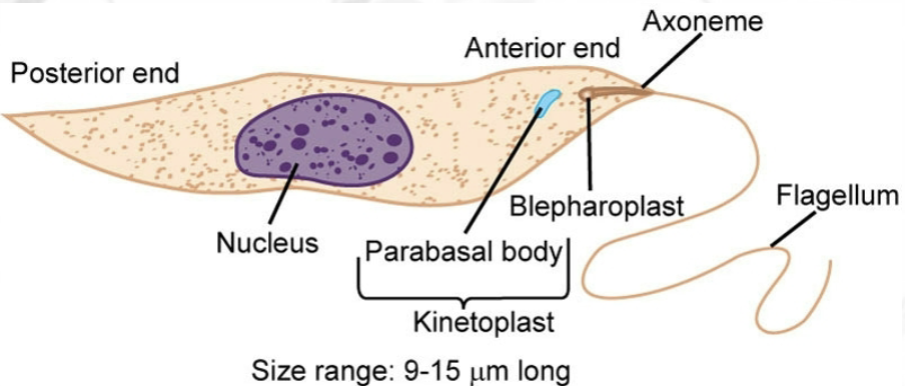
epimastigote
long, spindle-shaped, hemoflagellate morphologic form equipped with a free a flagellum and an undulating membrane that extends over ½ the body length.
kinetoplast located anterior to nucleus. free flagellum extends from anterior end. 1 nucleus located in posterior end.
seen only in arthropod vectors

trypomastigote
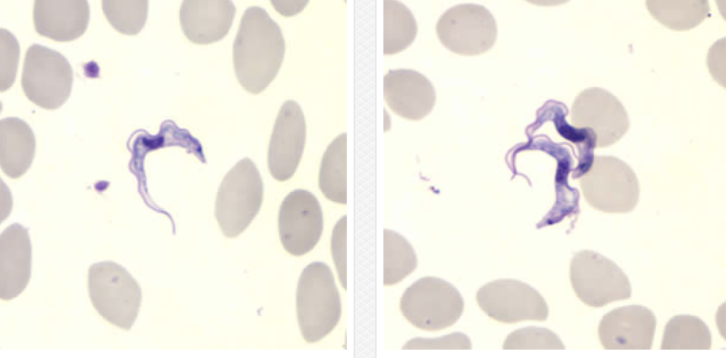
long & slender to study hemoflagellate morphologic form of trypanosoma spp that typically looks like the letter C or U on permanent stain. has full body length undulating membrane.
kinetoplast in posterior end. free flagellum extends from anterior end when present.
routinely seen in peripheral blood

symptoms of hemoflagellates
small red papule at bite site, intense itching, secondary bacterial infections, fever, diarrhea, kidney involvement, mental retardation, comatose state, death, initial skin lesions that either spontaneously heal & remain dormant for months or years
leishmania braziliensis complex
found in central & south america. transmitted by bite of sand fly. any mammal as reservoir hosts. biopsy of infected ulcer is specimen of choice. recovery of amastigotes (diagnostic stage) for microscopy.
causes mucocutaneous leishmaniasis: large ulcers in nasal mucosa, cutaneous &/or mucosal lesions. affected areas include the face & other soft parts. death from secondary bacterial infection possible

leishmania braziliensis complex life cycle
comes from arthropod vector sand fly, which injects parasite into human through blood meal. infective stage for humans: promastigotes. after entry into human, amastigotes continue to reproduce & damage tissue. promastigotes invade RTE cells & transform into amastigotes. infective stage for sand fly: picks amastigotes up during blood meal.

leishmania donovani complex
from india, pakistan, parts of africa, china, mediterranean, former soviet union, central & south america, middle east. vector: sand fly. causes visceral leishmanias: non-descript abdominal illness, hepatosplenomegaly, diarrhea, anemia, possible kidney damage, darkening of skin (kala-azar/black fever). chronic cases: possible death in 1-2 years. acute: debilitates patient & becomes lethal in only weeks
trypanosoma brucei gambiense
causes west african sleeping sickness. specimens include blood lymph node aspirations & CSF (sediment). use giemsa-stained preparations for trypomastigotes (diagnostic stage). IgM testing on CSF or serum is diagnostic. from west & central africa, esp in shaded areas along stream banks where tsetse flies breed. humans only host.
trypanosoma brucei gambiense & rhodesiense life cycle
tsetse fly (arthropod vector) infects human through blood meal. infective stage for tsetse fly: picks trypomastigotes up during blood meal. in the fly, trypomastigote convert to epimastigote during migration into salivary gland. once in salivary gland, they convert back to trypomastigotes for injection into next human during blood meal. in human, trypomastigotes migrate through bloodstream, actively multiplying. invasion of CNS may occur.

west african (gambian) sleeping sickness
painful chancre surrounded by white halo at bite site. fever, malaise, headache, weakness, anorexia, lymphadenopathy. winterbottom’s sign: enlargement of cervical lymph nodes. kerandel sign: delayed sensation to pain. CNS involvement possible, coma, death.
trypanosoma brucei rhodesiense
causes east african sleeping sickness. found in east & central africa. reservoir hosts: cattle, sheep, wild game animals. lab diagnosis by trypomastigotes in giemsa stained blood & microscopic exam of CSF. IgM studies on CSF = diagnostic. tsetse fly species: glossina morsitans & pallidipes.

east african (rhodesian) sleeping sickness
more virulent than gambiense. short incubation period. fever, myalgia, rigors, rapid weight loss, CNS involvement early in dz, mental disturbance, lethargy, anorexia, rapid & fulminating dz results. kidney damage & myocarditis, which contributes to death within 9-12 months of untreated patients
trypansoma cruzi
causes chaga’s disease/american trypanosomiasis. spread by kissing bug. lab diagnosis: giemsa blood stain trypomastigotes, giemsa lymph node biopsy & blood culture amastigotes. use PCR/ELISA for diagnosis & blood donor screening. in central & south america, highest prevalence in brazil. most commonly seen in small children

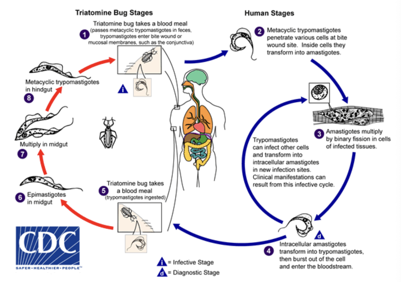
trypanosoma cruzi life cycle
kissing bug defecates near blood meal site & rubs it into skin via scratch in preparation for blood meal. other transmission modes: blood transfusion, sexual intercourse, transplacental. human infective stage: trypomastigotes. they convert into amastigotes, which multiply & destroy host cells before converting back to trypomastigotes. human diagnostic stage: trypomastigotes & amastigotes.
chaga’s disease
acute & chronic. erythematous nodule (chagoma) at infection site usually on face. edema & rash around eyes, conjunctivitis, eyelid edema (romana’s sign).
acute: fever, chills, fatigue, myalgia, malaise. can recover, transition to chronic stage, or death a few weeks later.
chronic: myocarditis, megacolon, megaesophagus, cardiomegaly, brain damage, sudden death