BSCI202 Exam 2

0.0(0)
Studied by 0 people
call kaiCall Kai
learnLearn
examPractice Test
spaced repetitionSpaced Repetition
heart puzzleMatch
flashcardsFlashcards
GameKnowt Play
Card Sorting

1/46

encourage image

There's no tags or description

Looks like no tags are added yet.

47 Terms

1
New cards

Cardiovascular System - Intro to the Heart

Pyramid Comparison

  • apex and base

    • top is apex (most narrow), base is widest portion

  • heart has base at top (widest portion)

  • heart has apex at bottom (points down and towards the left)

  • centered within the body, but skewed pointing towards the left (on patient)


Arteries

  • carries blood away from the heart

Veins

  • carries blood towards the heart


Location

  • located within the mediastinum

  • middle of body, sandwiched between left and right lung

  • on superior surface of diaphragm

  • anterior to vertebral column

  • posterior to sternum

  • medial to left and right lung


<p>Pyramid Comparison</p><ul><li><p>apex and base</p><ul><li><p>top is apex (most narrow), base is widest portion</p></li></ul></li><li><p>heart has base at top (widest portion)</p></li><li><p>heart has apex at bottom (points down and towards the left)</p></li><li><p>centered within the body, but skewed pointing towards the left (on patient)</p></li></ul><p></p><p>Arteries</p><ul><li><p>carries blood away from the heart</p></li></ul><p>Veins </p><ul><li><p>carries blood towards the heart</p></li></ul><p></p><p>Location</p><ul><li><p>located within the mediastinum</p></li><li><p>middle of body, sandwiched between left and right lung</p></li><li><p>on superior surface of diaphragm</p></li><li><p>anterior to vertebral column</p></li><li><p>posterior to sternum</p></li><li><p>medial to left and right lung</p></li></ul><p></p>
2
New cards
<p>Heart Structure</p>

Heart Structure

  • 2 types of chambers, 4 total

    • atria (plural) → upper chambers

      • right and left atrium

    • ventricles (plural) → lower chambers

      • right and left ventricle

    • chambers that blood flows through

      • surrounded by living tissues

      • blood flowing through chambers is not what nourishes the heart tissues

      • coronary circulation → blood vessels that nourish the heart

    • 2 pumps

      • act at same time, but supply blood to different portions

      • atria go to ventricles

      • leaves from ventricles to other locations

      • blood comes in the left atrium and go to left ventricle

      • contraction happening for both sides and blood coming into atria at same time (one drop of blood flows at different times though)
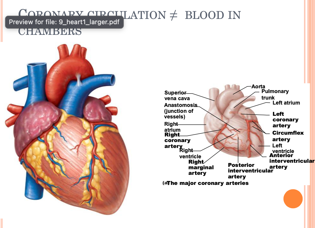

<ul><li><p>2 types of chambers, 4 total</p><ul><li><p>atria (plural) → upper chambers</p><ul><li><p>right and left atrium</p></li></ul></li><li><p>ventricles (plural) → lower chambers</p><ul><li><p>right and left ventricle</p></li></ul></li><li><p>chambers that blood flows through</p><ul><li><p>surrounded by living tissues</p></li><li><p>blood flowing through chambers is not what nourishes the heart tissues</p></li><li><p>coronary circulation → blood vessels that nourish the heart </p></li></ul></li><li><p>2 pumps</p><ul><li><p>act at same time, but supply blood to different portions</p></li><li><p>atria go to ventricles</p></li><li><p>leaves from ventricles to other locations</p></li><li><p>blood comes in the left atrium and go to left ventricle </p></li><li><p>contraction happening for both sides and blood coming into atria at same time (one drop of blood flows at different times though)</p></li></ul></li></ul></li></ul><p></p>
3
New cards

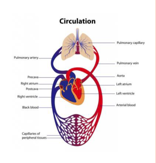
Pulmonary and Systemic Circuits

  • heart transport system; two side-by-side pumps

    • right side 1. receives oxygen-poor blood from tissues (into pulmonary circuit)

      • right side (ventricle contracts to pump blood to lungs)

        • sent through artery to lungs to get oxygenated

        • brings O2 rich blood to left side of heart

      • pumps to lungs to get rid of CO2, pick up O2

      • pulmonary circuit

    • left side 2. receives oxygenated blood from lungs (into systemic circuit)

      • left side (ventricle contracts to pump blood to body tissues)

        • travels through artery to tissues

        • delivers O2 and picks up CO2 and returns to other side of heart

      • pumps to body tissues

      • systemic circuit


<ul><li><p>heart transport system; two side-by-side pumps</p><ul><li><p>right side 1. receives oxygen-poor blood from tissues (into pulmonary circuit)</p><ul><li><p>right side (ventricle contracts to pump blood to lungs)</p><ul><li><p>sent through artery to lungs to get oxygenated</p></li><li><p>brings O2 rich blood to left side of heart</p></li></ul></li><li><p>pumps to lungs to get rid of CO2, pick up O2 </p></li><li><p>pulmonary circuit</p></li></ul></li><li><p>left side 2. receives oxygenated blood from lungs (into systemic circuit)</p><ul><li><p>left side (ventricle contracts to pump blood to body tissues)</p><ul><li><p>travels through artery to tissues</p></li><li><p>delivers O2 and picks up CO2 and returns to other side of heart</p></li></ul></li><li><p>pumps to body tissues</p></li><li><p>systemic circuit</p></li></ul></li></ul></li></ul><p></p>
4
New cards

Asymmetry of Ventricles

  • most of the time, the body is symmetrical

    • ventricles differ in symmetry

      • myocardium differs between both sides of the heart

      • left ventricle myocardium is much thicker than right ventricle

      • explained by the fact that the left ventricle must contract with a greater force to push blood to body tissues, while right ventricle must only contract enough to push blood to the lungs (shorter distance away = less force required to move)

      • contraction increases pressure (heart becomes smaller = higher pressure)

      • pumping blood to tissues requires more pressure and force (lower extremities less so, because gravity will pull blood down, however, upper extremities means blood must push against gravity)


Heart Form and Function

  • myocardium (muscles of the heart)

  • left ventricle - 3x thickness of myocardium compared to right side


<ul><li><p>most of the time, the body is symmetrical</p><ul><li><p>ventricles differ in symmetry</p><ul><li><p>myocardium differs between both sides of the heart</p></li><li><p>left ventricle myocardium is much thicker than right ventricle</p></li><li><p>explained by the fact that the left ventricle must contract with a greater force to push blood to body tissues, while right ventricle must only contract enough to push blood to the lungs (shorter distance away = less force required to move)</p></li><li><p>contraction increases pressure (heart becomes smaller = higher pressure)</p></li><li><p>pumping blood to tissues requires more pressure and force (lower extremities less so, because gravity will pull blood down, however, upper extremities means blood must push against gravity)</p></li></ul></li></ul></li></ul><p></p><p>Heart Form and Function</p><ul><li><p>myocardium (muscles of the heart)</p></li><li><p>left ventricle - 3x thickness of myocardium compared to right side</p></li></ul><p></p>
5
New cards
<p>Layers of the Heart Wall</p>

Layers of the Heart Wall

Three layers of heart wall

  1. epicardium → visceral layer of serous pericardium

    • innermost layer (visceral) of pericardium and outermost layer of heart

      • count as one of the same, not two separate layers

  2. myocardium

    • contractile cardiac muscle cells

    • cardiac skeleton: crisscrossing, interlacing layer of connective tissue

      • anchors cardiac muscle fibers

      • supports greater vessels and valves

      • limits spread of action potentials to specific paths

        • only goes through certain pathways at specific times

        • coordinates contractions

  3. endocardium → lines heart chambers and valves

    • continuous with blood vessels

    • helps reduce friction of blood against vessel walls

    • ensures continuous flow


Pericardium

  • deep two-layered serous pericardium

    • parietal layer

    • visceral layer (epicardium)

      • separated by fluid-filled pericardial cavity


<p>Three layers of heart wall</p><ol><li><p>epicardium → visceral layer of serous pericardium</p><ul><li><p>innermost layer (visceral) of pericardium and outermost layer of heart</p><ul><li><p>count as one of the same, not two separate layers</p></li></ul></li></ul></li><li><p>myocardium</p><ul><li><p>contractile cardiac muscle cells</p></li><li><p>cardiac skeleton: crisscrossing, interlacing layer of connective tissue</p><ul><li><p>anchors cardiac muscle fibers</p></li><li><p>supports greater vessels and valves</p></li><li><p>limits spread of action potentials to specific paths</p><ul><li><p>only goes through certain pathways at specific times</p></li><li><p>coordinates contractions</p></li></ul></li></ul></li></ul></li><li><p>endocardium → lines heart chambers and valves</p><ul><li><p>continuous with blood vessels </p></li><li><p>helps reduce friction of blood against vessel walls</p></li><li><p>ensures continuous flow</p></li></ul></li></ol><p></p><p>Pericardium</p><ul><li><p>deep two-layered serous pericardium</p><ul><li><p>parietal layer</p></li><li><p>visceral layer (epicardium)</p><ul><li><p>separated by fluid-filled pericardial cavity </p></li></ul></li></ul></li></ul><p></p>
6
New cards

Septum

  • interatrial septum → separates atria (in-between atria)

  • inter ventricular septum → separates ventricles (in-between ventricles)

  • divides atria and ventricles between themselves


7
New cards

Valves

  • unidirectional

  • pressure → open and close

    • areas of high pressure

    • opens one side and closes other side

      • pressure in atria → opens

      • pressure in ventricle → slams valve closed

  • open with high pressure on one side and closes with high pressure on other side

  • two atrioventricular (AV) valves

    • tricuspid valve (right)

    • mitral/bicuspid valve (left)

    • papillary muscles; chordae tendinae

      • chordae tendinae → strings that anchor into muscles within wall of heart (anchors into papillary muscles) and hold AV valve in place, preventing it from flipping and inverting

    • left atrium (bicuspid valve), right atrium (tricuspid valve)

  • two semilunar (SL) valves (blood leaving the heart)

    • aortic semilunar valve (left ventricle → aorta → systemic circuit)

    • pulmonary semilunar valve (right ventricle → pulmonary circuit)


8
New cards

AV Valves

  1. blood returns to heart, filling right atria, pressing against AV valves = increased pressure forces AV valves open

  2. as ventricle fills, AV valve flaps hang limply into ventricle

  3. atria contracts, forcing additional blood into ventricles

  4. ventricles contract, forcing blood against AV valve cusps

  5. AV valve closes

  6. papillary muscle contracts and chordae tendinae tighten, preventing valve flaps from everting into atria


9
New cards

Semilunar Valves

closes because blood falls back down on it, causing it to close


REVIEW AND ADD

10
New cards

Daily Question - Heart Sounds

  • normal heart sounds → valves closing (two sounds, because two different periods that valves close)

    • AV closing, semilunar closing (repeats after pause), producing beating heart sound

  • two sounds (Lub-dup) associated with closing of heart valves

    • first as AV valves close (lup)

    • second as SL valves close (dup)

  • heart murmurs → abnormal heart sounds; usually indicate incompetent or stenotic valves

    • mitral (bicuspid) valve regurgitation


11
New cards

Pathway of Blood

Pulmonary Circuit

  • right atrium → tricuspid valve → right ventricle

  • right ventricle → pulmonary semilunar valve → pulmonary trunk → pulmonary arteries → lungs

  • lungs → pulmonary veins → left atrium


Systemic Circuit

  • left atrium → mitral valve → left ventricle

  • left ventricle → aortic semilunar valve → aorta

  • aorta → systemic circulation (body tissues)


superior vena cava → inferior vena cava → right atrium → right ventricle → pulmonary SL valve → pulmonary artery (right and left pulmonary arteries deliver to right and left lungs) → lungs → pulmonary vein (high oxygen) → left atrium → mitral valve → left ventricle → aortic semilunar valve → aorta → body tissues

  • large quantity of blood just drops down from gravity, but other portion comes from heart muscles contracting (makes high pressure environment)

  • as ventricle fills with blood, tricuspid valve then closes (pressure builds)

  • high pressure environment within right ventricle triggers pulmonary SL valve to open

    • shoots up with a lot of force and slams back down, closing the pulmonary SL valve

    • right ventricle no longer high pressure = tricuspid valve reopens

  • oxygen picked up within lungs and brought back to heart through pulmonary vein

  • two vessels (pulmonary right and left veins) connect to left atrium

  • mitral AV valve open from higher pressure of blood filling atrium, blood enters left ventricle → pressure increases in ventricle, closing mitral valve and opening aortic semilunar valve → aorta


12
New cards

Microscopic Anatomy of Cardiac Muscle

  • Intercalated discs

    • desmosomes → lock cells in place; when mechanical force present, keeps cells together

    • gap junctions → electrically couple cells (ions flow between cells, electrically linking cells together)

      • allows heart to be functional syncytium

        • all cardiomyocytes contract as a unit, or none do

        • ensures larger force of contraction (required for ventricle to get high pressure environment for blood to eject out)

        • enables coordinated contractions

      • 99% contractile. 1% auto-rhythmic

        • contractile → contract because neighbor contracts (neighbors pass messages to contract between each other → electrically coupled cells)

        • auto-rhythmic → start message for contraction that radiates out to other cells
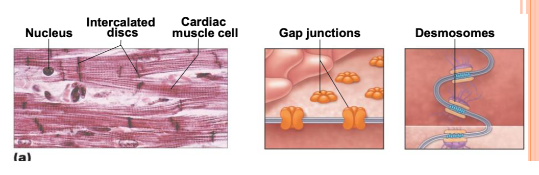

<ul><li><p>Intercalated discs</p><ul><li><p>desmosomes → lock cells in place; when mechanical force present, keeps cells together</p></li><li><p>gap junctions → electrically couple cells (ions flow between cells, electrically linking cells together) </p><ul><li><p>allows heart to be functional syncytium</p><ul><li><p>all cardiomyocytes contract as a unit, or none do</p></li><li><p>ensures larger force of contraction (required for ventricle to get high pressure environment for blood to eject out) </p></li><li><p>enables coordinated contractions</p></li></ul></li><li><p>99% contractile. 1% auto-rhythmic </p><ul><li><p>contractile → contract because neighbor contracts (neighbors pass messages to contract between each other → electrically coupled cells)</p></li><li><p>auto-rhythmic → start message for contraction that radiates out to other cells</p></li></ul></li></ul></li></ul></li></ul><p></p>
13
New cards
<p>Action Potentials - Muscle Contraction</p>

Action Potentials - Muscle Contraction

  • graph = time vs membrane potential

  • rest = -70 mV (inside cell is more negative than environment)

    • higher concentration sodium ions outside cell compared to inside and lots of potassium inside compared to outside

      • depolarization = sodium ions come in to cell (cell becomes more positive than environment) = membrane potential increases = action potential → potassium ions begin to leave (depolarization) = becomes more negative than environment and even more negative than rest → hyper-polarization → resting membrane potential


Heart

  • many electrical messages being sent

  • pathway for signaling contraction:

    • sinoatrial node → located in right atrium and contain auto-rhythmic cells

      • all around auto-rhythmic cells = contractile cells = messages spread out across cells

      • interatrial bundle (Bachman’s bundle or interatrial tract)

        • connecting sionatrial node to right atrium, triggering depolarization to spread in the right atrium and left atrium

      • internodal tract → connects atrioventricular node to right atrium

        • auto-rhymthic cells

      • 0.1 seconds later (pause → diameter changes in node = slower travel through tracts)

        • benefit: whole heart contracts at once without this delay

          • pause ensures that atria contract slightly before ventricles do = ensures atria receive message first = more atria in blood = more blood enters ventricles

      • continues down atrioventricular bundle (bundle of His) → splits into left and right bundle branches → splits into purkinje fibers (subendocardial conducting network) → spread throughout myocardium of ventricles

        • auto-rhythmic cells around purkinje fibers

  • auto rhythmic cells in both AV node and SA node → ensures that if a message isn’t sent out from SA node or AV node doesn’t receive message = ensures another place where this contraction messaging could occur

    • backup mechanism to ensure ventricles contract

  • summary: Sinoatrial node → Atrioventricular node → Atrioventricular bundle → Right and left bundle branches → Subendocardial conducting network (Purkinje fibers)

<ul><li><p>graph = time vs membrane potential </p></li><li><p>rest = -70 mV (inside cell is more negative than environment) </p><ul><li><p>higher concentration sodium ions outside cell compared to inside and lots of potassium inside compared to outside</p><ul><li><p>depolarization = sodium ions come in to cell (cell becomes more positive than environment) = membrane potential increases = action potential → potassium ions begin to leave (depolarization) = becomes more negative than environment and even more negative than rest → hyper-polarization → resting membrane potential</p></li></ul></li></ul></li></ul><p></p><p>Heart</p><ul><li><p>many electrical messages being sent</p></li><li><p>pathway for signaling contraction:</p><ul><li><p>sinoatrial node → located in right atrium and contain auto-rhythmic cells</p><ul><li><p>all around auto-rhythmic cells = contractile cells = messages spread out across cells</p></li><li><p>interatrial bundle (Bachman’s bundle or interatrial tract) </p><ul><li><p>connecting sionatrial node to right atrium, triggering depolarization to spread in the right atrium and left atrium</p></li></ul></li><li><p>internodal tract → connects atrioventricular node to right atrium</p><ul><li><p>auto-rhymthic cells </p></li></ul></li><li><p>0.1 seconds later (pause → diameter changes in node = slower travel through tracts)</p><ul><li><p>benefit: whole heart contracts at once without this delay</p><ul><li><p>pause ensures that atria contract slightly before ventricles do = ensures atria receive message first = more atria in blood = more blood enters ventricles</p></li></ul></li></ul></li><li><p>continues down atrioventricular bundle (bundle of His) → splits into left and right bundle branches → splits into purkinje fibers (subendocardial conducting network) → spread throughout myocardium of ventricles</p><ul><li><p>auto-rhythmic cells around purkinje fibers</p></li></ul></li></ul></li></ul></li><li><p>auto rhythmic cells in both AV node and SA node → ensures that if a message isn’t sent out from SA node or AV node doesn’t receive message = ensures another place where this contraction messaging could occur</p><ul><li><p>backup mechanism to ensure ventricles contract</p></li></ul></li><li><p>summary: Sinoatrial node → Atrioventricular node → Atrioventricular bundle → Right and left bundle branches → Subendocardial conducting network (Purkinje fibers)</p></li></ul><p></p>
14
New cards
<p>Cardiac Action Potentials</p>

Cardiac Action Potentials

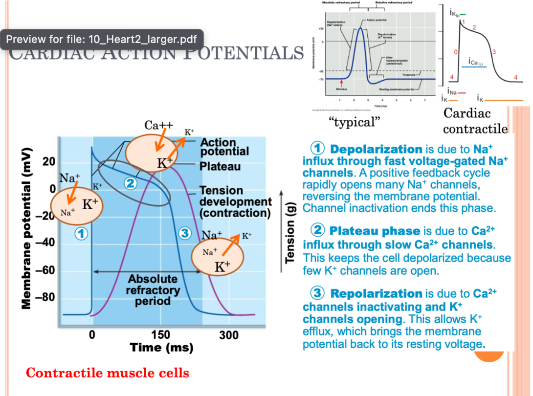
  • cardiac action potentials slightly different shape and Ca2+ plays a role

    • electrical message occurs → activates Ca2+ release → actin and myosin cross-bridge formation → contraction occurs in muscle

  • Contractile Muscle Cells

    1. resting state = negative membrane potential

    2. stimulus occurs = depolarization → Na+ influx through fast-voltage gated Na+ channels

      • positive feedback cycle rapidly open many Na+ channels, reversing membrane potential. channel inactivation ends phase

    3. plateau phase → Ca2+ influx through slow Ca2+ channels. this keeps the cell depolarized, because few K+ channels are open

      • calcium slows return to more negative state

    4. repolarization

      • due to Ca2+ channels inactivating and K+ channels opening. this allows K+ efflux, which brings membrane potential back to resting voltage.

  • Pacemaker Cells (auto-rhythmic)

    1. pacemaker potential → slow depolarization due to both opening of Na+ channels and closing of K+ channels. membrane potential is never flat line

    2. depolarization → action potential begins when pacemaker potential reaches threshold. depolarization due to Ca2+ influx through Ca2+ channels

    3. repolarization → due to Ca2+ channels inactivating and K+ channels opening. this allows K+ efflux, which brings membrane potential back to most negative charge


<ul><li><p>cardiac action potentials slightly different shape and Ca2+ plays a role</p><ul><li><p>electrical message occurs → activates Ca2+ release → actin and myosin cross-bridge formation → contraction occurs in muscle </p></li></ul></li><li><p>Contractile Muscle Cells</p><ol><li><p>resting state = negative membrane potential</p></li><li><p>stimulus occurs = depolarization → Na+ influx through fast-voltage gated Na+ channels</p><ul><li><p>positive feedback cycle rapidly open many Na+ channels, reversing membrane potential. channel inactivation ends phase</p></li></ul></li><li><p>plateau phase → Ca2+ influx through slow Ca2+ channels. this keeps the cell depolarized, because few K+ channels are open</p><ul><li><p>calcium slows return to more negative state</p></li></ul></li><li><p>repolarization</p><ul><li><p>due to Ca2+ channels inactivating and K+ channels opening. this allows K+ efflux, which brings membrane potential back to resting voltage. </p></li></ul></li></ol></li><li><p>Pacemaker Cells (auto-rhythmic)</p><ol><li><p>pacemaker potential → slow depolarization due to both opening of Na+ channels and closing of K+ channels. membrane potential is never flat line</p></li><li><p>depolarization → action potential begins when pacemaker potential reaches threshold. depolarization due to Ca2+ influx through Ca2+ channels</p></li><li><p>repolarization → due to Ca2+ channels inactivating and K+ channels opening. this allows K+ efflux, which brings membrane potential back to most negative charge</p></li></ol></li></ul><p></p>
15
New cards
<p>Electrocardiography</p>

Electrocardiography

  • electrocardiogram (ECG or EKG)

    • all action potentials at given time

  • three waves:

    • P wave → depolarization of SA node → atria

      • atrial cells become very positive

    • QRS complex → ventricular depolarization and atrial depolarization

      • atrial cells become very negative

      • ventricular cells become very positive

    • T wave → ventricular repolarization

      • ventricular cells become very negative


Missing Image:

  • Q is missing

  • labels don’t change when things are missing


<ul><li><p>electrocardiogram (ECG or EKG)</p><ul><li><p>all action potentials at given time</p></li></ul></li><li><p>three waves: </p><ul><li><p>P wave → depolarization of SA node → atria</p><ul><li><p>atrial cells become very positive</p></li></ul></li><li><p>QRS complex → ventricular depolarization and atrial depolarization</p><ul><li><p>atrial cells become very negative</p></li><li><p>ventricular cells become very positive</p></li></ul></li><li><p>T wave → ventricular repolarization</p><ul><li><p>ventricular cells become very negative</p></li></ul></li></ul></li></ul><p></p><p>Missing Image: </p><ul><li><p>Q is missing </p></li><li><p>labels don’t change when things are missing</p></li></ul><p></p>
16
New cards
<p>ECG vs Heart</p>

ECG vs Heart

  • no P wave = SA node blocked

  • AV node still depolarizes = signals atria to still contract, ventricles are still contracting, but less blood present


  • erratic signals = electrical signals still receiving messages, but muscles are contracting uncoordinated = blood not ejected from heart correctly = fibrillation (heart attack or electrical signal)


<ul><li><p>no P wave = SA node blocked</p></li><li><p>AV node still depolarizes = signals atria to still contract, ventricles are still contracting, but less blood present</p></li></ul><p></p><ul><li><p>erratic signals = electrical signals still receiving messages, but muscles are contracting uncoordinated = blood not ejected from heart correctly = fibrillation (heart attack or electrical signal)</p></li></ul><p></p>
17
New cards

Mechanical Events: Cardiac Cycle

  • systole = contraction

  • diastole = relaxation

  • cardiac cycle

    • Blood flow through heart during one complete heartbeat

    • Atrial systole and diastole → ventricular systole and diastole


<ul><li><p>systole = contraction</p></li><li><p>diastole = relaxation</p></li><li><p>cardiac cycle</p><ul><li><p>Blood flow through heart during one complete heartbeat</p></li><li><p>Atrial systole and diastole → ventricular systole and diastole</p></li></ul></li></ul><p></p>
18
New cards

Phases of Cardiac Cycle

  1. Ventricular Filling

    • AV valves are open; pressure low

    • 80% of blood passively flows into ventricles

    • Atrial systole occurs, delivering remaining 20%

    • End diastolic volume (EDV) → volume of blood in each ventricle at end of ventricular diastole

      • once ventricle is filled, before contraction occurs

      • at very end of relaxation phase of heart

      • volume in a single ventricle (about the same between left and right)

  2. Ventricular Systole

    • atrial diastole; ventricles begin to contract

    • isovolumetric contraction phase (closed valves)

    • ventricular pressure > artery pressure

      • blood moves from high pressure to low pressure

      • big difference = more blood moved faster

    • End systolic volume (ESV) → volume of blood remaining in each ventricle after systole

      • no matter how strong contraction is, some blood stays within the ventricle

      • how much blood is in a single ventricle after ventricular systole (contraction)

  3. Isovolumetric Relaxation

    • ventricles relax; atria relaxed and filling

    • blood closes SL valves

    • AV valves open; cycle begins again at step 1


Juice Box Analogy

  1. Jug = atria

  2. 1st straw = AV valve/pathway to ventricle

  3. Juice Box = ventricle

  4. 2nd straw = artery


Total Blood Volume Output = End diastolic volume - end systolic volume


Impacts on Blood Out:

  1. Force of contraction

  2. How much filled

  3. Cardiac Output


<ol><li><p>Ventricular Filling</p><ul><li><p>AV valves are open; pressure low</p></li><li><p>80% of blood passively flows into ventricles</p></li><li><p>Atrial systole occurs, delivering remaining 20%</p></li><li><p>End diastolic volume (EDV) → volume of blood in each ventricle at end of ventricular diastole</p><ul><li><p>once ventricle is filled, before contraction occurs</p></li><li><p>at very end of relaxation phase of heart</p></li><li><p>volume in a single ventricle (about the same between left and right)</p></li></ul></li></ul></li><li><p>Ventricular Systole</p><ul><li><p>atrial diastole; ventricles begin to contract</p></li><li><p>isovolumetric contraction phase (closed valves) </p></li><li><p>ventricular pressure &gt; artery pressure</p><ul><li><p>blood moves from high pressure to low pressure</p></li><li><p>big difference = more blood moved faster</p></li></ul></li><li><p>End systolic volume (ESV) → volume of blood remaining in each ventricle after systole</p><ul><li><p>no matter how strong contraction is, some blood stays within the ventricle</p></li><li><p>how much blood is in a single ventricle after ventricular systole (contraction)</p></li></ul></li></ul></li><li><p>Isovolumetric Relaxation</p><ul><li><p>ventricles relax; atria relaxed and filling</p></li><li><p>blood closes SL valves</p></li><li><p>AV valves open; cycle begins again at step 1</p></li></ul></li></ol><p></p><p>Juice Box Analogy</p><ol><li><p>Jug = atria</p></li><li><p>1st straw = AV valve/pathway to ventricle</p></li><li><p>Juice Box = ventricle</p></li><li><p>2nd straw = artery</p></li></ol><p></p><p>Total Blood Volume Output = End diastolic volume - end systolic volume</p><p></p><p>Impacts on Blood Out:</p><ol><li><p>Force of contraction</p></li><li><p>How much filled</p></li><li><p>Cardiac Output</p></li></ol><p></p>
19
New cards

Cardiac Output (CO)

  • volume of blood pumped by each ventricle in one minute

  • CO = heart rate (HR) * stroke volume (SV)

    • HR = number of beats per minute

    • SV = volume of blood pumped out by one ventricle with each beat

  • Normal - 5.25 L/min

    • CO (ml/min) = HR (75 beats/min) * SV (70 ml/beat)

    • = 5.25 L/min


SV DOESN’T equal end diastolic volume


TPS: If we have 100 ml of blood in a ventricle prior to contraction and 10 ml remained in the ventricle after the contraction, then what is the stroke volume, end diastolic volume, and end systolic volume?

  • EDV = 100 ml

  • ESV = 10 ml

  • SV = 100 ml - 10 ml = 90 ml

  • SV = EDV - ESV


20
New cards

Cardiac Output Variation

  • CO increases if either/both SV or HR increase

    • maximal CO is 4-5 times resting CO (~25 L/min)

      • up to 35 L/min in trained athletes

    • Cardiac reserve → difference between resting and maximal CO


21
New cards

Regulation of Stroke Volume

  • SV = EDV - ESV

    • EDV → length of ventricular diastole & venous pressure

    • ESV → arterial BP & ventricular contraction strength

  • Three main SV impactors:

    • preload

    • contractility

    • afterload


Preload

  • degree of stretch of cardiac muscle cells before they contract (Frank-Starling law of heart)

    • venous return → amount of blood returning to heart

      • increased venous return distends (stretches) ventricles


Contractility

  • contractile strength

  • Increased Contractility

    • sympathetic stimulation → increased Ca2+ influx → more cross bridges

  • Decreased Contractility

    • calcium channel blockers = fewer cross bridges formed = weaker contraction


Afterload

  • pressure ventricles must overcome to eject blood

  • hypertension = increased after load = increased ESV = decreased SV

    • high pressure = larger hurdle to get over = less blood moves = higher ESV

  • high pressure → low pressure


<ul><li><p>SV = EDV - ESV</p><ul><li><p>EDV → length of ventricular diastole &amp; venous pressure</p></li><li><p>ESV → arterial BP &amp; ventricular contraction strength</p></li></ul></li><li><p>Three main SV impactors: </p><ul><li><p>preload</p></li><li><p>contractility</p></li><li><p>afterload</p></li></ul></li></ul><p></p><p>Preload</p><ul><li><p>degree of stretch of cardiac muscle cells before they contract (Frank-Starling law of heart) </p><ul><li><p>venous return → amount of blood returning to heart</p><ul><li><p>increased venous return distends (stretches) ventricles</p></li></ul></li></ul></li></ul><p></p><p>Contractility</p><ul><li><p>contractile strength</p></li><li><p>Increased Contractility</p><ul><li><p>sympathetic stimulation → increased Ca2+ influx → more cross bridges</p></li></ul></li><li><p>Decreased Contractility </p><ul><li><p>calcium channel blockers = fewer cross bridges formed = weaker contraction</p></li></ul></li></ul><p></p><p>Afterload</p><ul><li><p>pressure ventricles must overcome to eject blood</p></li><li><p>hypertension = increased after load = increased ESV = decreased SV</p><ul><li><p>high pressure = larger hurdle to get over = less blood moves = higher ESV</p></li></ul></li><li><p>high pressure → low pressure </p></li></ul><p></p>
22
New cards
<p>Cardiac Output &amp; Heart Rate</p>

Cardiac Output & Heart Rate

  • CO = heart rate (HR) * stroke volume (SV)

  • CO increases if either/both SV or HR increases

  • positive chronotropic factors = increase heart rate

  • negative chronotropic factors = decrease heart rate


Regulation of Heart Rate

  • Fear → sympathetic nervous system → epinephrine → B1 receptors of SA node (receptors on auto-rhythmic cells) → Na+ & Ca2+ (channels opened through receptor binding) → causes depolarization = message for contraction to be sent, if muscle works, it will contract (more action potentials) → increased heart rate & stroke volume

    • epinephrine → positive chronotropic factor

      • if administered clinically → chronotropic drug

  • Parasympathetic system returns heart rate to normal

    • Vagus nerve → ACh (interacts with auto-rhythmic cell in AV and SA nodes) → binds to M2 (muscarinic acetylcholine/cholinergic) receptors on SA node → opens K+ channels and closes Ca2+ channels (harder to reach peak = reaches peak less often = less action potentials) → decreased heart rate & stroke volume


Broad Factors Influencing Heart Rate

  • Age (size)

    • fetus has fastest HR

  • Sex (size)

    • Females faster than males

      • on average, females have lower body mass = higher HR

  • Exercise

    • increases HR

  • Body Temperature

    • HR increases with body temperature

  • Chemical Factors

    • Intra- and extracellular ion concentrations (ca2+ and K+)

      • Hypocalcemia (ca2+) → depresses HR

      • Hyper-calcemia (ca2+) → increases HR and contractility

      • Hyper-kalemia (potassium) → alters electrical activity, cardiac arrest

      • Hypokalemia (potassium) → feeble heartbeat, arrhythmia

    • Hormones

      • epinephrine from adrenal medulla increases HR and contractility

      • thyroxine enhances effects of norepinephrine and epinephrine (stronger impact of epinephrine → synergistic)


<ul><li><p>CO = heart rate (HR) * stroke volume (SV)</p></li><li><p>CO increases if either/both SV or HR increases</p></li><li><p>positive chronotropic factors = increase heart rate</p></li><li><p>negative chronotropic factors = decrease heart rate</p></li></ul><p></p><p>Regulation of Heart Rate</p><ul><li><p>Fear → sympathetic nervous system → epinephrine → B1 receptors of SA node (receptors on auto-rhythmic cells) → Na+ &amp; Ca2+ (channels opened through receptor binding) → causes depolarization = message for contraction to be sent, if muscle works, it will contract (more action potentials) → increased heart rate &amp; stroke volume</p><ul><li><p>epinephrine → positive chronotropic factor </p><ul><li><p>if administered clinically → chronotropic drug</p></li></ul></li></ul></li><li><p>Parasympathetic system returns heart rate to normal</p><ul><li><p>Vagus nerve → ACh (interacts with auto-rhythmic cell in AV and SA nodes) → binds to M<sub>2</sub> (muscarinic acetylcholine/cholinergic) receptors on SA node → opens K+ channels and closes Ca2+ channels (harder to reach peak = reaches peak less often = less action potentials) → decreased heart rate &amp; stroke volume</p></li></ul></li></ul><p></p><p>Broad Factors Influencing Heart Rate</p><ul><li><p>Age (size) </p><ul><li><p>fetus has fastest HR</p></li></ul></li><li><p>Sex (size)</p><ul><li><p>Females faster than males</p><ul><li><p>on average, females have lower body mass = higher HR</p></li></ul></li></ul></li><li><p>Exercise </p><ul><li><p>increases HR</p></li></ul></li><li><p>Body Temperature </p><ul><li><p>HR increases with body temperature</p></li></ul></li><li><p>Chemical Factors</p><ul><li><p>Intra- and extracellular ion concentrations (ca2+ and K+)</p><ul><li><p>Hypocalcemia (ca2+) → depresses HR</p></li><li><p>Hyper-calcemia (ca2+) → increases HR and contractility</p></li><li><p>Hyper-kalemia (potassium) → alters electrical activity, cardiac arrest</p></li><li><p>Hypokalemia (potassium) → feeble heartbeat, arrhythmia </p></li></ul></li><li><p>Hormones</p><ul><li><p>epinephrine from adrenal medulla increases HR and contractility</p></li><li><p>thyroxine enhances effects of norepinephrine and epinephrine (stronger impact of epinephrine → synergistic)</p></li></ul></li></ul></li></ul><p></p>
23
New cards

Heart Rate Regulation Summary

  • cardioacceleratory center → sympathetic nerve fibers (SS → epinephrine → B receptors → Na+ and Ca2+ channels → increased HR and stroke volume)

  • PS → vagus nerve → ACh → M2 receptors → opens K+ channels and closes Ca2+ channels → hyper polarized → decreased heart rate and decreased stroke volume

    • redundancy in innervating SA and AV nodes

    • other backups on purkinje fibers (can be chemically stimulated)


TPS:

  • SA node intrinsically fires 100/min

  • Typical resting HR: ~ 75 beats/min

  • Why difference?

    • Vagal tone → at rest, parasympathetic nervous system is more active than sympathetic nervous system = suppresses firing rate of SA node


<ul><li><p>cardioacceleratory center → sympathetic nerve fibers (SS → epinephrine → B receptors → Na+ and Ca2+ channels → increased HR and stroke volume) </p></li><li><p>PS → vagus nerve → ACh → M2 receptors → opens K+ channels and closes Ca2+ channels → hyper polarized → decreased heart rate and decreased stroke volume</p><ul><li><p>redundancy in innervating SA and AV nodes</p></li><li><p>other backups on purkinje fibers (can be chemically stimulated)</p></li></ul></li></ul><p></p><p>TPS:</p><ul><li><p>SA node intrinsically fires 100/min</p></li><li><p>Typical resting HR: ~ 75 beats/min</p></li><li><p>Why difference?</p><ul><li><p>Vagal tone → at rest, parasympathetic nervous system is more active than sympathetic nervous system = suppresses firing rate of SA node</p></li></ul></li></ul><p></p>
24
New cards

Heart Rate Homeostatic Imbalances

  • tachycardia → abnormally fast HR (>100 beats/min)

    • if persistent, may lead to fibrillation

  • bradycardia → heart rate slower than 60 beats/min

    • may result in inadequate blood circulation

    • other factors might makeup for slower heart rate (venous return, lower blood pressure, etc)


25
New cards
<p>Blood Flow Through Body</p>

Blood Flow Through Body

  • highway system with many exits (big highways lead to smaller exits = leads to smaller vessels, etc)

  • gets to capillaries = site of exchange

  • blood returns to heart going from smaller vessels → larger vessels

  • gets close to most cells of the body to deliver oxygen, nutrients, etc and then pickup waste, chemical messengers, etc


Impacts on Blood Flow

  1. Vasodilators

    • low oxygen and increased carbon dioxide

  2. Vasoconstrictors

  3. Temperature

    • as temperature rises:

      • hypothalamus signals → warm blood flushes into capillary beds → heat radiates from skin (heat lost to environment = blood turns colder)

    • sweat also causes vasodilation via bradykinin in perspiration

      • more blood sent to site where blood is being cooled

    • as temperature decreases:

      • blood is shunted to deeper, more vital organs (avoids heat loss to environment)

      • blood goes to more vital areas of the body and less blood to areas with more contact with environment


<ul><li><p>highway system with many exits (big highways lead to smaller exits = leads to smaller vessels, etc)</p></li><li><p>gets to capillaries = site of exchange</p></li><li><p>blood returns to heart going from smaller vessels → larger vessels</p></li><li><p>gets close to most cells of the body to deliver oxygen, nutrients, etc and then pickup waste, chemical messengers, etc</p></li></ul><p></p><p>Impacts on Blood Flow</p><ol><li><p>Vasodilators</p><ul><li><p>low oxygen and increased carbon dioxide</p></li></ul></li><li><p>Vasoconstrictors</p></li><li><p>Temperature</p><ul><li><p>as temperature rises:</p><ul><li><p>hypothalamus signals → warm blood flushes into capillary beds → heat radiates from skin (heat lost to environment = blood turns colder) </p></li></ul></li><li><p>sweat also causes vasodilation via bradykinin in perspiration</p><ul><li><p>more blood sent to site where blood is being cooled</p></li></ul></li><li><p>as temperature decreases:</p><ul><li><p>blood is shunted to deeper, more vital organs (avoids heat loss to environment) </p></li><li><p>blood goes to more vital areas of the body and less blood to areas with more contact with environment</p></li></ul></li></ul></li></ol><p></p>
26
New cards

ADD MISSING LECTURE

27
New cards
<p>The Respiratory System</p>

The Respiratory System

  • Major Function: Respiration

    • supply O2/remove CO2 associated with cellular respiration;

    • four processes associated respiratory and circulatory systems

    • Olfaction & Speech

      • odor molecules bind

      • speech → production of sound through force of air moving out of body

    • Purify, Humidify, and Warm Incoming Air

      • warm, clean, and make wetter


<ul><li><p>Major Function: Respiration</p><ul><li><p>supply O2/remove CO2 associated with cellular respiration;</p></li><li><p>four processes associated respiratory and circulatory systems</p></li><li><p>Olfaction &amp; Speech</p><ul><li><p>odor molecules bind</p></li><li><p>speech → production of sound through force of air moving out of body</p></li></ul></li><li><p>Purify, Humidify, and Warm Incoming Air</p><ul><li><p>warm, clean, and make wetter</p></li></ul></li></ul></li></ul><p></p>
28
New cards

Processes of Respiration

Respiratory System

  • Pulmonary Ventilation

    • breathing → air in and out of lungs

    • external and internal environment interacting

  • External Respiration

    • O2 and CO2 between lungs and blood

    • movement within the lungs to the blood and blood to within the lungs


Circulatory System

  • Transport

    • O2 and CO2 in blood

  • Internal Respiration

    • O2 and CO2 between systemic blood vessels and tissues


Pulmonary Ventilation → Transport → External Respiration → Transport → Internal Respiration


<p>Respiratory System</p><ul><li><p>Pulmonary Ventilation</p><ul><li><p>breathing → air in and out of lungs</p></li><li><p>external and internal environment interacting</p></li></ul></li><li><p>External Respiration</p><ul><li><p>O2 and CO2 between lungs and blood</p></li><li><p>movement within the lungs to the blood and blood to within the lungs</p></li></ul></li></ul><p></p><p>Circulatory System</p><ul><li><p>Transport</p><ul><li><p>O2 and CO2 in blood</p></li></ul></li><li><p>Internal Respiration</p><ul><li><p>O2 and CO2 between systemic blood vessels and tissues</p></li></ul></li></ul><p></p><p>Pulmonary Ventilation → Transport → External Respiration → Transport → Internal Respiration </p><p></p>
29
New cards
<p>Functional Zones</p>

Functional Zones

Respiratory Zone → site of gas exchange

  • microscopic structures

    • respiratory bronchioles, alveolar ducts, and alveoli

      • external respiration


Conducting Zone → conduits to gas exchange sites

  • all other respiratory structures;

  • cleanses, warms, and humidifies incoming air

    • dehumidifies air leaving (and reclaims heat)


Diaphragm and other respiratory muscles promote ventilation


30
New cards

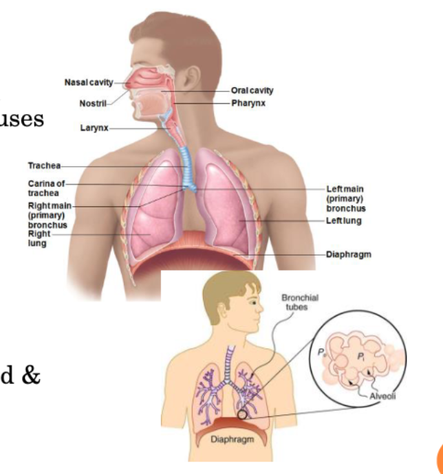
Respiratory System - Functional Anatomy

  • Major Organs

    • nose, nasal cavity, and paranasal sinuses

    • pharynx

    • larynx

    • trachea

    • bronchi and their branches

    • lungs and alveoli

  • Upper Airways → Head & Neck (up to pharynx)

  • Respiratory Tract → Larynx to Lungs


<ul><li><p>Major Organs</p><ul><li><p>nose, nasal cavity, and paranasal sinuses</p></li><li><p>pharynx</p></li><li><p>larynx </p></li><li><p>trachea </p></li><li><p>bronchi and their branches</p></li><li><p>lungs and alveoli</p></li></ul></li><li><p>Upper Airways → Head &amp; Neck (up to pharynx) </p></li><li><p>Respiratory Tract → Larynx to Lungs</p></li></ul><p></p>
31
New cards
<p>The Nose</p>

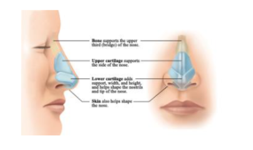
The Nose

  • Two regions-external nose and nasal cavity

  • Functions:

    • Airway

    • Moisten and Warms entering air

      • respiratory mucosa

    • Filters and Cleans inspired air

      • respiratory mucosa & vibrissae (hairs)

    • Chamber to resonate speech

      • only certain sounds

      • sound still being produced, but not bouncing off chamber

    • Olfactory receptors

      • olfactory mucosa on superficial surface
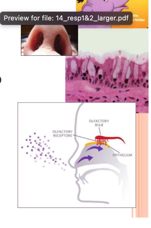

<ul><li><p>Two regions-external nose and nasal cavity</p></li><li><p>Functions:</p><ul><li><p>Airway </p></li><li><p>Moisten and Warms entering air</p><ul><li><p>respiratory mucosa</p></li></ul></li><li><p>Filters and Cleans inspired air</p><ul><li><p>respiratory mucosa &amp; vibrissae (hairs) </p></li></ul></li><li><p>Chamber to resonate speech</p><ul><li><p>only certain sounds</p></li><li><p>sound still being produced, but not bouncing off chamber</p></li></ul></li><li><p>Olfactory receptors</p><ul><li><p>olfactory mucosa on superficial surface </p></li></ul></li></ul></li></ul><p></p>
32
New cards

Mucosa

  • Olfactory Mucosa

    • olfactory epithelium

  • Respiratory Mucosa

    • pseudo stratified ciliated columnar epithelium

    • mucous and serous secretions

    • inspired air warmed by capillaries and veins


<ul><li><p>Olfactory Mucosa</p><ul><li><p>olfactory epithelium</p></li></ul></li><li><p>Respiratory Mucosa</p><ul><li><p>pseudo stratified ciliated columnar epithelium</p></li><li><p>mucous and serous secretions</p></li><li><p>inspired air warmed by capillaries and veins </p></li></ul></li></ul><p></p>
33
New cards
<p>Conchae</p>

Conchae

  • Nasal Conchae → superior, middle, and inferior

    • protrude medially from lateral walls

    • increase mucosal area

    • enhance air turbulence

  • Conchae and Nasal Mucosa

    • During Inhalation:

      • Filter, heat, and moisten air

    • During Exhalation:

      • Reclaim heat and moisture


Inhalation: Adding Obstacles/Protrusions to the Wall (lined with respiratory mucosa) → slow down things moving through the nose and force them to touch the walls of the structures to get cleaned, warmed, etc


Exhalation: Air takes heat and water, gets to lungs (warm, wet, clean), leave lungs (warm, wet, clean), on way back out - surfaces are colder (heat and water goes to lower concentration/colder), so reclaims heat and water

<ul><li><p>Nasal Conchae → superior, middle, and inferior</p><ul><li><p>protrude medially from lateral walls</p></li><li><p>increase mucosal area</p></li><li><p>enhance air turbulence</p></li></ul></li><li><p>Conchae and Nasal Mucosa</p><ul><li><p>During Inhalation:</p><ul><li><p>Filter, heat, and moisten air</p></li></ul></li><li><p>During Exhalation:</p><ul><li><p>Reclaim heat and moisture</p></li></ul></li></ul></li></ul><p></p><p>Inhalation: Adding Obstacles/Protrusions to the Wall (lined with respiratory mucosa) → slow down things moving through the nose and force them to touch the walls of the structures to get cleaned, warmed, etc </p><p></p><p>Exhalation: Air takes heat and water, gets to lungs (warm, wet, clean), leave lungs (warm, wet, clean), on way back out - surfaces are colder (heat and water goes to lower concentration/colder), so reclaims heat and water </p>
34
New cards
<p>Paranasal Sinuses</p>

Paranasal Sinuses

  • cavities

  • in frontal, sphenoid, ethmoid, and maxillary bones

  • lighten skull; resonate sound; secrete mucus; help to warm and moisten air


35
New cards

Pharynx (Throat)

  • muscular passage

    • connects nasal cavity to larynx

  • three regions:

    • nasopharynx: superior region behind the nasal cavity

      • only air

      • air passes through this cavity if inhaled through nose

    • oropharynx: middle region behind mouth

      • food - digestive system

      • air - respiratory system

    • laryngopharynx: inferior region attached to larynx

      • food - digestive system

      • air - respiratory system


<ul><li><p>muscular passage</p><ul><li><p>connects nasal cavity to larynx</p></li></ul></li><li><p>three regions:</p><ul><li><p>nasopharynx: superior region behind the nasal cavity</p><ul><li><p>only air</p></li><li><p>air passes through this cavity if inhaled through nose</p></li></ul></li><li><p>oropharynx: middle region behind mouth</p><ul><li><p>food - digestive system </p></li><li><p>air - respiratory system</p></li></ul></li><li><p>laryngopharynx: inferior region attached to larynx</p><ul><li><p>food - digestive system</p></li><li><p>air - respiratory system</p></li></ul></li></ul></li></ul><p></p>
36
New cards

Larynx (Voice Box)

  • plays a role in speech

  • routes air and food into proper channels

  • eight rigid hyaline cartilages and spoon-shaped flap of elastic cartilage (epiglottis)


<ul><li><p>plays a role in speech</p></li><li><p>routes air and food into proper channels</p></li><li><p>eight rigid hyaline cartilages and spoon-shaped flap of elastic cartilage (epiglottis)</p></li></ul><p></p>
37
New cards
<p>Larynx Structures</p>

Larynx Structures

  • Epiglottis

    • routes food to the esophagus and air towards the trachea

  • Laryngeal Prominence

    • thyroid cartilage

    • adam’s apple

  • Vocal Folds (True Vocal Cords)

    • vibrate with expelled air to create sound (speech)

    • vestibular fold (false vocal cord)

  • Glottis

    • opening between vocal cords


<ul><li><p>Epiglottis</p><ul><li><p>routes food to the esophagus and air towards the trachea</p></li></ul></li><li><p>Laryngeal Prominence</p><ul><li><p>thyroid cartilage</p></li><li><p>adam’s apple </p></li></ul></li><li><p>Vocal Folds (True Vocal Cords) </p><ul><li><p>vibrate with expelled air to create sound (speech)</p></li><li><p>vestibular fold (false vocal cord) </p></li></ul></li><li><p>Glottis</p><ul><li><p>opening between vocal cords</p></li></ul></li></ul><p></p>
38
New cards

Trachea (Wind Pipe)

  • Four-inch-long-tube

    • connects larynx with bronchi

  • Walls reinforced with C shaped hyaline cartilage

    • gives more plasticity

  • lined with pseudo stratified ciliated columnar epithelium

    • cilia beating creates a current that moves dirt/debris back towards external environment and away from lungs
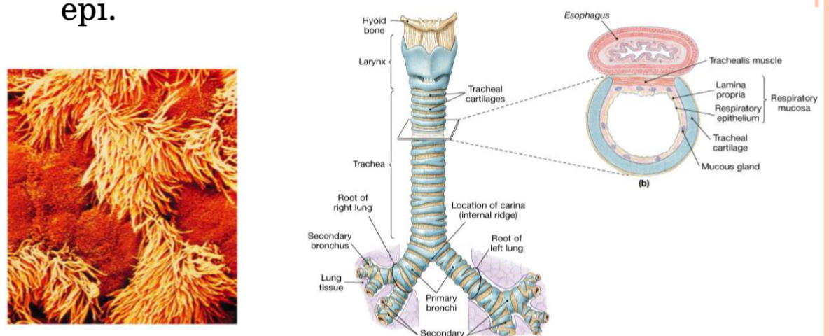

<ul><li><p>Four-inch-long-tube</p><ul><li><p>connects larynx with bronchi</p></li></ul></li><li><p>Walls reinforced with C shaped hyaline cartilage</p><ul><li><p>gives more plasticity</p></li></ul></li><li><p>lined with pseudo stratified ciliated columnar epithelium</p><ul><li><p>cilia beating creates a current that moves dirt/debris back towards external environment and away from lungs</p></li></ul></li></ul><p></p>
39
New cards

Lungs

  • thoracic cavity

  • apex is near the clavicle (superior portion)

  • base rests on the diaphragm (inferior portion)

  • each lung is divided into lobes by fissures

    • left lung, two lobes

      • cardiac notch (where heart lies near lungs) is what gives left side only two lobes

    • right lung, three lobes


<ul><li><p>thoracic cavity</p></li><li><p>apex is near the clavicle (superior portion)</p></li><li><p>base rests on the diaphragm (inferior portion)</p></li><li><p>each lung is divided into lobes by fissures</p><ul><li><p>left lung, two lobes</p><ul><li><p>cardiac notch (where heart lies near lungs) is what gives left side only two lobes</p></li></ul></li><li><p>right lung, three lobes</p></li></ul></li></ul><p></p>
40
New cards
<p>Bronchi</p>

Bronchi

  • all but the smallest of these passageways have reinforcing cartilage in their walls

    • primary bronchi

    • secondary bronchi

    • tertiary bronchi

    • bronchioles

    • terminal bronchioles


<ul><li><p>all but the smallest of these passageways have reinforcing cartilage in their walls</p><ul><li><p>primary bronchi</p></li><li><p>secondary bronchi</p></li><li><p>tertiary bronchi</p></li><li><p>bronchioles</p></li><li><p>terminal bronchioles</p></li></ul></li></ul><p></p>
41
New cards
<p>Alveoli</p>

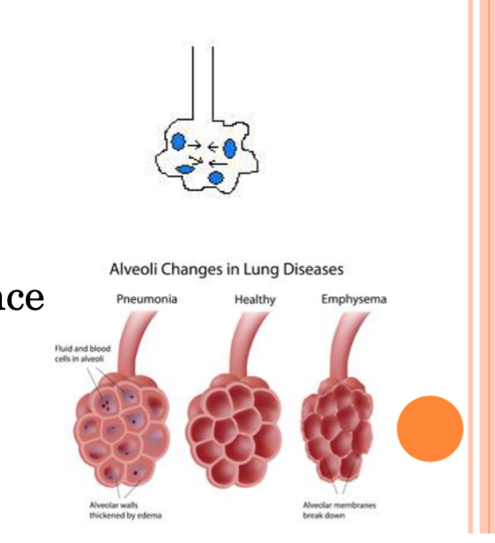
Alveoli

  • alveoli = site of gas exchange

    • 300 million alveoli/lung

  • rich blood supply-capillaries from sheet over alveoli

  • type I alveolar cells → most cells of the walls

    • make up wall of alveoli

    • single layer epithelial cells

  • type II alveolar cells

    • secrete surfactant


<ul><li><p>alveoli = site of gas exchange</p><ul><li><p>300 million alveoli/lung</p></li></ul></li><li><p>rich blood supply-capillaries from sheet over alveoli</p></li><li><p>type I alveolar cells → most cells of the walls </p><ul><li><p>make up wall of alveoli</p></li><li><p>single layer epithelial cells </p></li></ul></li><li><p>type II alveolar cells </p><ul><li><p>secrete surfactant</p></li></ul></li></ul><p></p>
42
New cards
<p>Alveoli and Surface Tension</p>

Alveoli and Surface Tension

  • surface tension: attracts liquid molecules to one another at gas-liquid interface

    • water → high surface tension (due to hydrogen bonds)

      • coats alveolar walls

  • surfactant → prevents alveoli from collapsing when we breathe out

    • lipid and protein complex produced by type II alveolar cells

    • reduces surface tension of alveolar fluid and discourages alveolar collapse

    • prevents water molecules from forming hydrogen bonds with other water molecules on opposite walls


<ul><li><p>surface tension: attracts liquid molecules to one another at gas-liquid interface</p><ul><li><p>water → high surface tension (due to hydrogen bonds) </p><ul><li><p>coats alveolar walls</p></li></ul></li></ul></li><li><p>surfactant → prevents alveoli from collapsing when we breathe out</p><ul><li><p>lipid and protein complex produced by type II alveolar cells</p></li><li><p>reduces surface tension of alveolar fluid and discourages alveolar collapse</p></li><li><p>prevents water molecules from forming hydrogen bonds with other water molecules on opposite walls </p></li></ul></li></ul><p></p>
43
New cards

Surfactant Image

  • contains hydrophilic and hydrophobic ends

  • fetal development → without surfactants, premature fetuses often experience alveolar collapse and are less likely to survive


<ul><li><p>contains hydrophilic and hydrophobic ends</p></li><li><p>fetal development → without surfactants, premature fetuses often experience alveolar collapse and are less likely to survive</p></li></ul><p></p>
44
New cards
<p>Respiratory Volumes and Capacities</p>

Respiratory Volumes and Capacities

  • used to assess respiratory statues

    • tidal volume (TV) → normal in or normal out

    • inspiratory reserve volume (IRV) → normal in (EXTRA in)

      • if measure both inspiratory capacity ( = TV + RV)

    • Expiratory reserve volume (ERV) - Normal in. Normal Out. EXTRA OUT

    • Residual volume (RV) → amount you can’t get out

  • Vital capacity

    • total amount of exchangeable air

    • vital capacity = TV + IRV + ERV

  • Functional Residual Capacity

    • amount of air in lungs after normal breath out

    • ERV + RV

  • Total Lung Capacity

    • max amount of air that could be in the lungs

    • TV + IRV + ERV + RV


<ul><li><p>used to assess respiratory statues</p><ul><li><p>tidal volume (TV) → normal in or normal out</p></li><li><p>inspiratory reserve volume (IRV) → normal in (EXTRA in)</p><ul><li><p>if measure both inspiratory capacity ( = TV + RV) </p></li></ul></li><li><p>Expiratory reserve volume (ERV) - Normal in. Normal Out. EXTRA OUT</p></li><li><p>Residual volume (RV) → amount you can’t get out</p></li></ul></li><li><p>Vital capacity</p><ul><li><p>total amount of exchangeable air</p></li><li><p>vital capacity = TV + IRV + ERV </p></li></ul></li><li><p>Functional Residual Capacity</p><ul><li><p>amount of air in lungs after normal breath out</p></li><li><p>ERV + RV</p></li></ul></li><li><p>Total Lung Capacity</p><ul><li><p>max amount of air that could be in the lungs</p></li><li><p>TV + IRV + ERV + RV</p></li></ul></li></ul><p></p>
45
New cards

Dead Space

  • anatomical dead space

    • no contribution to gas exchange

    • air remaining in passageways (~150 ml)

  • alveolar dead space

    • nonfunctional alveoli due to collapse/obstruction

  • total dead space

    • sum of anatomical dead space + alveolar dead space


<ul><li><p>anatomical dead space</p><ul><li><p>no contribution to gas exchange</p></li><li><p>air remaining in passageways (~150 ml) </p></li></ul></li><li><p>alveolar dead space</p><ul><li><p>nonfunctional alveoli due to collapse/obstruction</p></li></ul></li><li><p>total dead space </p><ul><li><p>sum of anatomical dead space + alveolar dead space</p></li></ul></li></ul><p></p>
46
New cards

Factors Impacting Pulmonary Ventilation

  • Lung Compliance → stretchability of the lung and openness of alveoli

    • Low Compliance Causes:

      • scar tissue

      • high surface tension

    • High Compliance Causes

      • emphysema


<ul><li><p>Lung Compliance → stretchability of the lung and openness of alveoli</p><ul><li><p>Low Compliance Causes:</p><ul><li><p>scar tissue </p></li><li><p>high surface tension </p></li></ul></li><li><p>High Compliance Causes</p><ul><li><p>emphysema</p></li></ul></li></ul></li></ul><p></p>
47
New cards

Daily Questions - Respiratory System

  • Why does your nose run when its cold outside?

    • humid environment with really cold water bottle → water on outside of water bottle (warm air with lots of water gets closer to cold water bottle, air around water bottle becomes colder and can’t hold as much water, so it sticks to the water bottle)

    • nose → very warm in body and in really cold environment (short distance and large temperature difference)

      • nasal cavity moves from warmer environment to colder environment (large shift)

      • breathe in → put water into air and make warmer

      • exhale → warm air holds more water, but as it moves out the cavity, temperature declines, so some water vapor gets deposited onto walls of nose (lots of water = some runs out and doesn’t get absorbed quick enough)


  • Elephant Shrew Nose (Lung Nose) Selective Advantage?

    • longer nose = looses less water with each breath out

    • in desert = important to keep as much water as possible