lecture 11 - brain: overview and protective layers
 Call Kai
 Learn
Learn Practice Test
 Spaced Repetition
 Match
Match Flashcards
 Knowt Play
1/195

There's no tags or description
Looks like no tags are added yet.
Name | Mastery | Learn | Test | Matching | Spaced | Call with Kai | Chat |
|---|
No analytics yet
Send a link to your students to track their progress
196 Terms
What is the anterior portion of the brain?
The top portion of the brain when viewing an inferior view

What is the posterior portion of the brain?
The bottom portion of the brain when viewing an inferior view

What is an inferior view of the brain?
Looking at the brain from the bottom up

How can cranial nerves help identify an inferior view of the brain?
Cranial nerves are on the inferior side of the brain

How can a cross-section of the spinal cord help identify an inferior view of the brain?
The spinal cord is beneath the brain; seeing a cross-section of the spinal cord indicates that you are looking up toward the brain
What features can indicate the posterior side of the brain?
Features on the posterior portion of the brain that are on the inferior side, such as the cerebellum and brain stem
What is the brain?
Part of the central nervous system; the portion contained in the cranial cavity, inside the skull
What are the four main regions of the brain?
brain stem
cerebellum
diencephalon
cerebrum

What is the brain stem?
The first region encountered when moving upward from the spinal cord; connects the spinal cord to the other portions of the brain

What is one key function of the brainstem?
Integration of reflexes necessary for survival
What regulatory centers are contained in the brain stem?
Centers for:
respiration
cardiovascular control
digestion
How does the brain stem relate to homeostasis?
Sensory information comes in, is integrated by the control center, and motor responses come out; the brain stem contains many control centers, especially those key for survival

What backup systems can exist for functions necessary for survival?
Systems necessary for survival often have backup systems; heart rate is controlled by nerves from the brainstem and can also be regulated by hormones released in the body

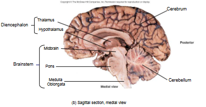
Where is the cerebellum located?
Posterior to the brainstem and inferior to the cerebrum; branches off the brainstem

What does the cerebellum resemble?
A mini cerebrum because it has right and left hemispheres, or right and left halves

How folded is the cerebellum?
It is extensively folded

What percentage of the brain's mass does the cerebellum contain?
About 10% of the brain's mass

What percentage of the brain's neurons does the cerebellum contain?
Almost 50% of the brain's neurons

What are the main jobs of the cerebellum?
Control of locomotion, balance, and posture

How does the cerebellum work with the cerebrum?
Works with the cerebrum to coordinate skeletal muscle movements

Does the cerebellum initiate movements or create motor programs?
No; the cerebrum initiates the movements and creates the motor programs
What role does the cerebellum play during movement?
Acts as a referee or judge of movements
What information does the cerebellum gather during movements?
Information from the internal environment, including muscle movement, muscle force, and joint position

What does the cerebellum do with information gathered during movement?
Gathers and integrates the information, then gives advice to the cerebrum on how to carry out movements more smoothly

Why is the cerebellum important for locomotion and walking?
Locomotion requires coordination of many muscle groups, and the cerebellum controls and coordinates these movements to produce fluid movements

How does the cerebellum develop coordination through movement?
It constantly gathers sensory information as movements occur; subsequent movements become more coordinated as it learns from the movements and environment

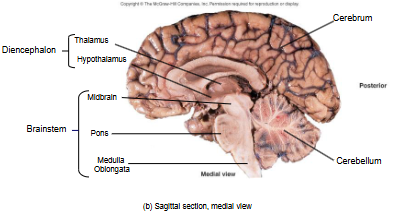
What is the diencephalon?
A region deep within the brain that sits on top of the brainstem

Why is the diencephalon difficult to picture?
It is deep within the brain and cannot actually be seen unless the brain is cut through

What functions is the diencephalon associated with?
output of the pituitary gland

How does the diencephalon connect the nervous and endocrine systems?
Acts as the nervous system connection to some endocrine functions in the body

What are the four main regions of the diencephalon?
Thalamus, epithalamus, hypothalamus, and subthalamus

What differences may exist between references regarding the diencephalon?
Some references do not discuss the diencephalon as a region on its own, and some do not mention the subthalamus; this course follows the textbook's presentation

What functions does the diencephalon have?
regulates day and night cycles
controls moods and emotions
stimulates thirst and hunger
controls body temperature
contains a relay center for sensory information from the internal and external environment
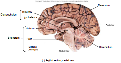
What is the cerebrum?
The largest part of the brain

What does the cerebrum contain?
Billions of neurons and two large hemispheres, or halves

What higher-order or executive functions does the cerebrum perform?
planning
reasoning
analyzing
storing and accessing memories
perceiving and interpreting sensory information
coordinating motor function
conscious thought and control
How does the cerebrum contribute to movement?
Creates the motor program in the first place and works with the cerebellum to coordinate movements so they are fluid

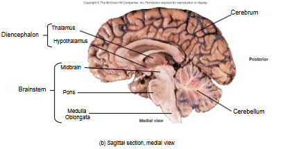
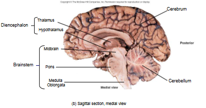
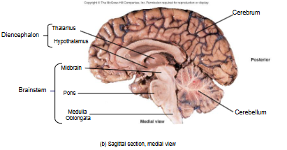
What type of brain section is shown when viewing the brain from the side along the midline?
Sagittal section; medial view

What are the three components of the brainstem?
Medulla oblongata, pons, and midbrain

What is the medulla oblongata?
The most inferior portion of the brainstem

What is the pons?
An enlarged mass sitting on top of the medulla oblongata that points anteriorly
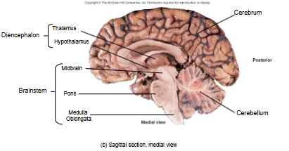
What is the midbrain?
The most superior portion of the brainstem

Where is the diencephalon located relative to the brainstem?
Sitting like a little cap on top of the brainstem
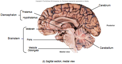
Which diencephalon regions are highlighted in the lecture's image?
Hypothalamus and thalamus
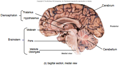
Where is the epithalamus located?
A little more posterior than the highlighted hypothalamus and thalamus

Where is the subthalamus located?
It is also part of the diencephalon

Where is the cerebellum located relative to the brainstem?
Posterior to the brainstem

How is the cerebellum connected to the brainstem and cerebrum?
Attached to the brainstem and has many connections to the brainstem; it does not directly connect to the cerebrum, so information has to go through the brainstem to reach the cerebrum

What is the largest portion of the brain?
Cerebrum

What are the main protective layers/features of the brain?
Cranial bones, cranial meninges, and cerebrospinal fluid

What are the cranial bones?
The bones of the skull that make up the cranial cavity

How many main cranial bones are there?
Eight

What are the cranial meninges?
Connective tissue layers that surround the brain and extend down to surround the spinal cord

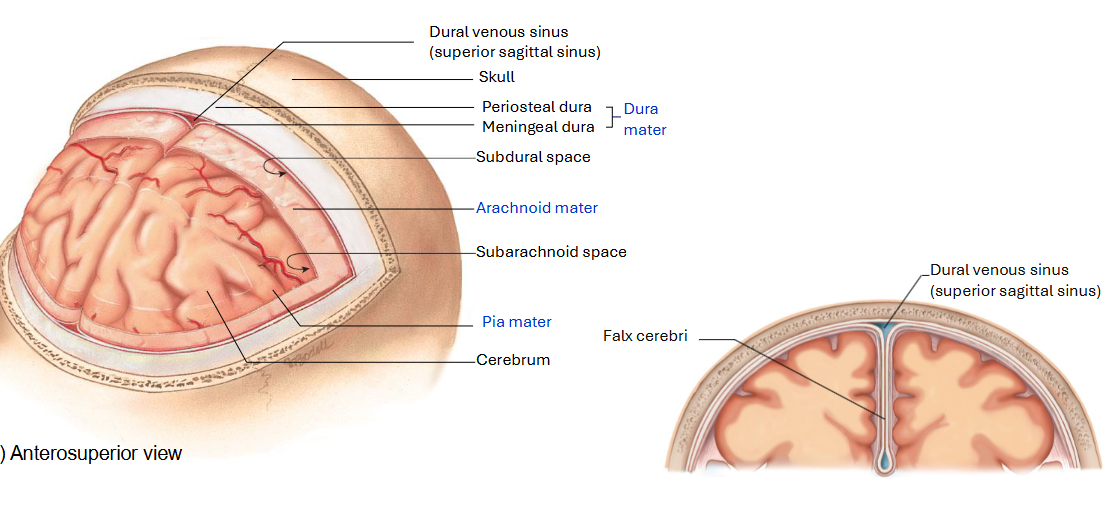
What are the three main layers of meninges?
Pia mater, arachnoid mater, and dura mater

How many layers does the dura mater have?
Two layers that are connected to each other
What is the final level of protection for the brain?
Cerebrospinal fluid
What are the meninges?
Connective tissue layers that surround the brain and extend to surround the spinal cord

What is meningitis?
Inflammation of the connective tissue layers of the meninges

Why can meningitis be dangerous?
The connective tissue layers closely adhere to the nervous tissue that makes up the central nervous system

What is the periosteal dura?
The first portion of the dura mater

What is the periosteum?
A connective tissue layer covering the outside surface of bones

What makes the cranial periosteum different?
It is highly associated with or connected to the other layer of the dura mater, the meningeal dura

What is the meningeal dura?
The other layer of the dura mater that connects with the periosteal dura

What do the periosteal dura and meningeal dura form?
One functional layer called the dura mater

What does "dura mater" mean?
"Tough mother"
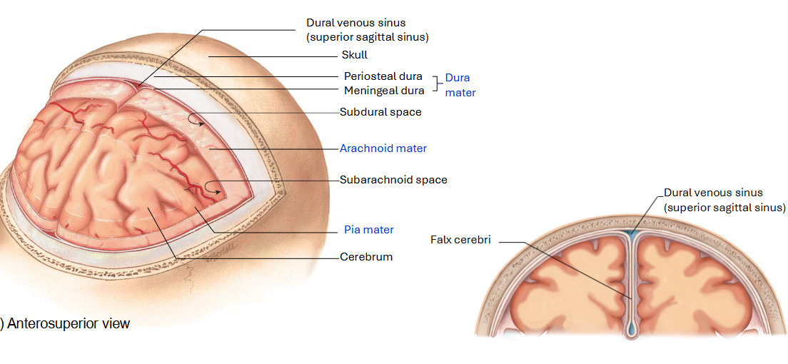
Why is the dura mater called "tough mother"?
The connective tissue layers are made of dense, irregular connective tissue, making them quite strong

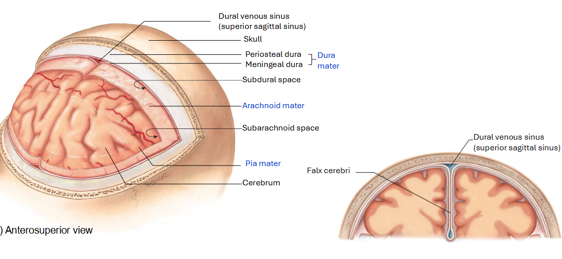
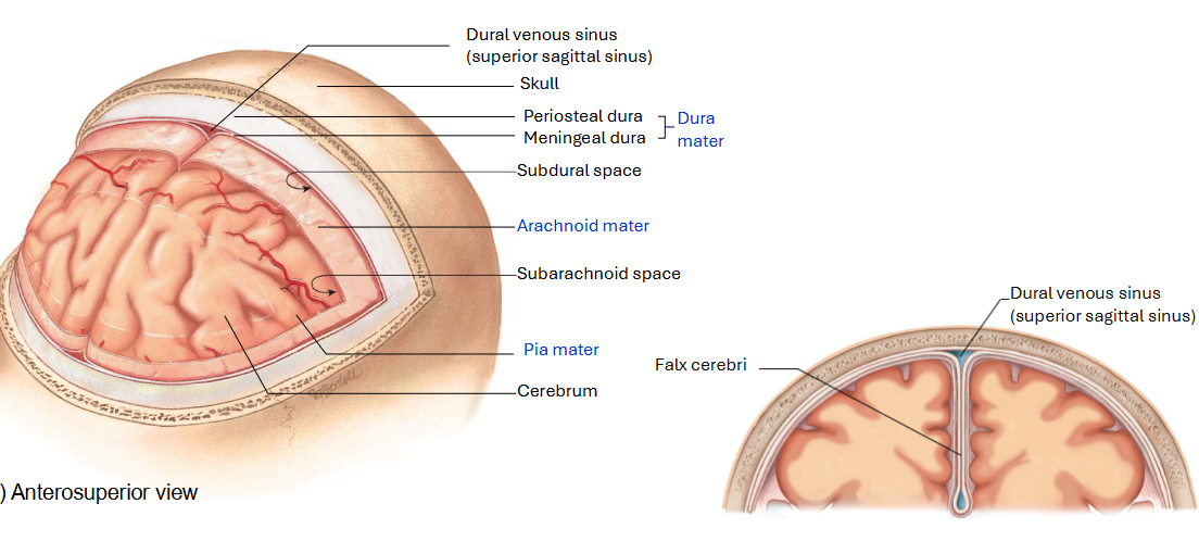
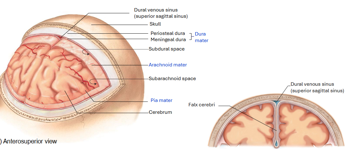
What happens to the meningeal dura at deep grooves in the brain?
The meningeal dura pulls away from the periosteal layer, which remains stuck to the bone

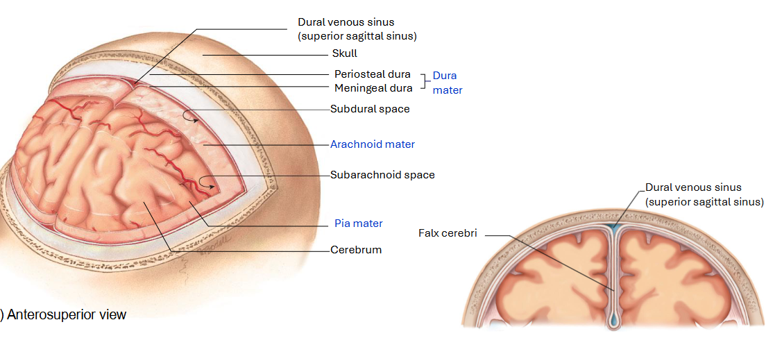
What is the longitudinal fissure?
A very deep groove between the two hemispheres of the brain

What happens to the meningeal dura at the longitudinal fissure?
It goes down into the groove, folds upon itself, and comes back up again

What do the folds of the dura mater create?
dural venous sinuses

What are dural venous sinuses?
Spaces created by the dural layer that carry venous blood

How is blood drained from the cranial cavity?
Through spaces in the dura mater known as venous sinuses

What is the superior sagittal sinus?
A dural venous sinus at the top between the two hemispheres of the brain

What is the inferior sagittal sinus?
A dural venous sinus at the bottom

What is the falx cerebri?
The folded region of the meningeal layer of the dura mater that extends into the longitudinal fissure between the two cerebral hemispheres

What is the purpose of the dural folds?
They create anchor points so the cranial meninges do not move around inside the cranial cavity relative to the brain
What is the falx cerebelli?
A fold that goes between the hemispheres of the cerebellum
What is the tentorium cerebelli?
A fold that goes between the cerebellum and cerebrum

What is the arachnoid mater?
A thin layer of fibrous membrane with web-like structures moving from it; closely adheres to the dura mater

Why is the arachnoid mater called "arachnoid"?
Its web-like structures look like spiderwebs

What is the pia mater?
A very thin membrane that closely adheres to every bump and groove of the cerebrum and spinal cord

What is the subarachnoid space?
The space between the arachnoid mater and pia mater; contains web-like projections and is filled with cerebrospinal fluid

What is the subdural space?
The space between the arachnoid mater and dura mater; a relatively small space containing a small amount of serous fluid
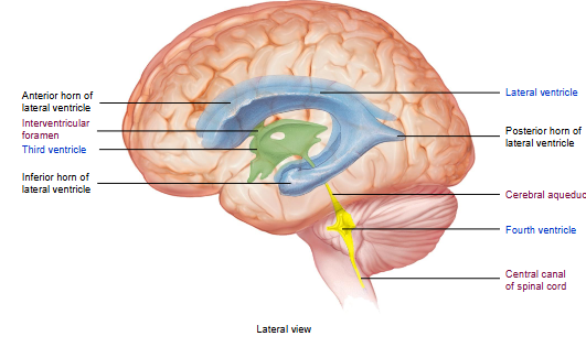
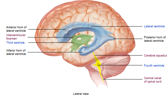
What are the ventricles?
Spaces or cavities inside different regions of the brain involved in producing cerebrospinal fluid
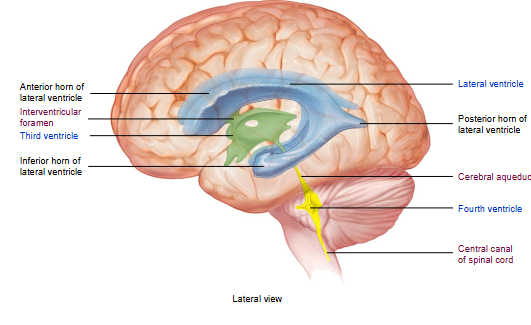
What do the ventricles look like in images?
They may look like actual structures, but they are actually spaces or cavities inside the brain

What are the lateral ventricles?
Two large, C-shaped spaces or cavities in the cerebrum, one in each hemisphere, located on either side of the corpus callosum

What is the corpus callosum?
The area of the brain that attaches the two hemispheres of the cerebrum to each other

How many lateral ventricles are there?
Two; they are bilateral

What are the regions of the lateral ventricles?
Anterior horn, posterior horn, and inferior horn

Where is cerebrospinal fluid first produced?
In the lateral ventricles

What is the third ventricle?
A ventricle that surrounds the region of the diencephalon

What is the interventricular foramen?
A space running from each lateral ventricle into the third ventricle; there are two, one from each lateral ventricle

Where does the interventricular foramen run?
From the floor of each lateral ventricle into the third ventricle

What is the cerebral aqueduct?
A small, narrow duct through which cerebrospinal fluid moves from the third ventricle toward the fourth ventricle

Where does the cerebral aqueduct run?
Through the midbrain of the brainstem

What is the fourth ventricle?
A space where the pons and medulla oblongata come into contact with the cerebellum

What is the embryological origin of the ventricles?
The neural tube was hollow inside; as it folded to create brain structures, some of the original space remained and became the brain ventricles and central canal of the spinal cord

Why are the ventricles continuous with each other?
They were all connected as the original inside of the neural tube
What cells line the ventricles?
Specialized ependymal cells

What do ependymal cells produce?
Cerebrospinal fluid