Red meat inspection practical 2
 Call Kai
 Learn
 Practice Test
 Spaced Repetition
 Match
 Flashcards
 Knowt Play
1/36

There's no tags or description
Looks like no tags are added yet.
37 Terms
How do you inspect sheep lungs & what is mandatory for incision?
Visually inspected
Palpation & incision of bronchial & mediastinal LNs only necessary in case of indications of poss risk to human / animal health or animal welfare
based on FCI analysis & additional epi data / other data from farms & or ante-mortem inspection findings, and / or animal welfare rule compliance checks and/or post-mortem inspection findings by mandatory procedures
Describe Muellerius capilaris in sheep lungs
Invades alveoli → inflam, characterised by incr no. of eosinophils → lesions
On palpation —> feels like lead shot (inflam + parasite → fibrosis)

Describe Dictyocaulus filaria in sheep lungs
Invades lumen of small bronchi, interferes w/ air being expelled → gives rise to localised emphysema —> appears raised & paler areas in lungs

What is the ABP category of Muellerius capillaris vs Dictyocaulus filaria?
Muellerius capillaris —> local rejection, ABP category 3
Dictyocaulus filaria —> local rejection, ABP category 2
What does blood splash look like and how does it occur?
Ruptured alveolar-capillary network during clonic contractions ass. w/ stunning
Or after electrical stunning due to sudden incr in BP
predis factors = stress & dehyd @ time of stunning
Or aspiration/blood entering trachea

What is the ABP category for blood splash?
Local rejection —> ABP category 2 or 3 depending on extension of lesions
Describe mycoplasma-like pneumonia distribution
Chronic, non-progressive bronchopneumonia w/ changes more freq in cr-ventral portions of lung → become hepatic-like & red-brown to grey in colour, collapsed & firmed than adjacent unaffected.
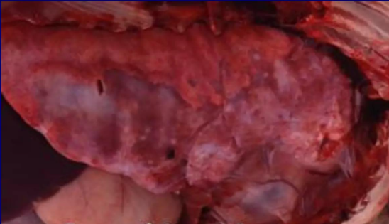
Which sheep is mycoplasma-like pneumonia seen in?
Lambs in high density stocking, ass. w/ reduced growth rates
What are the different poss causes of mycoplasma-like pneumonia?
Mycoplasma ovipneumoniae (most often isolated)
Also Mannheimia haemolytica, parainfluenza-3 virus & resp syncytial virus
Describe pasteurella-like lesions on the lungs
Variable & dependent on stage
Acute —> lobular to lobar pattern, can be haemorrhagic w/ blood-tinged foam seen in airways, & / or fibrino-necrotising
Lesions often extend into pleura
Subacute to chronic —> fibrinopurulent bronchopneumonia, absessation & fibrous pleurisy → adhesion formation

What causes pasteurella-like pneumonia?
Pasteurellacae (Mannheimia haemolytica, Histophilus somni, Pasteurella multocida) ass. pneumonia —> develops following stress factors:
changes in weather
poor handling
de-worming/dipping
What is the ABP category for Pasteurella-like pneumonia?
Local rejection (organ) —> ABP category 2
How do you inspect bovine lungs and what is mandatory for incision?
<8mo:
mandatory to do visual inspection followed by palpation of lungs & visual insection of trachea.
Palpation of mediastinal & bronchial LNs
If indication of poss risk to human/animal health or animal welfare, then lungs, trachea, main branches of brochial & mediastinal LNs should be incised
>8mo:
Mandatory to do visual inspection followed by palpation of lungs, visual inspection of trachea & mediastinal LNs, bronchial LNs incised
If indication of poss risk to human/animal health or animal welfare, then lungs, trachea, main branches of brochial & mediastinal LNs should be incised
Describe what pneumonia lesions look like on the bovine lungs
What is the ABP category?
Affected lungs firmer & darker than adjacent unaffected tissue & cut surface diffusely denser cf to adjacent tissue → liver-like appearance
Occ. agonal atelectasis can have similar appearance but if affected lung cut, changes can be seen to be restricted to subpleural parenchyma
Local rejection of organ, ABP category 2
Describe pleurisy of lungs
Inflam of pleura involving either or both parietal (serosa lining thoracic cavity) or visceral (serosa lining lungs) portion.
What is the ABP category of melanosis?
Category 3
What can pleurisy be ass. w/ (depending on stage)?
Fibrinous tags, loose and/or adherent to plurae
Variable amounts of fluid than can be purulent
Fibrous, which can create attachments between plurae & lead to atelectasis of lung in affected areas / attachement of lung to thoracic cavity & interfere w/ animals breathing
What is the ABP category of pleurisy?
Rejection of affected parts ABP category 2 —> if not septic
What other signs will be present in other organs if septicaemia is present as well as pleurisy? What is the ABP category?
Multiple abscesses in diff organs
Total rejection, ABP category 2
How do you inspect pig lungs & what is mandatory for incision?
Mandatory to do visual inspection of lungs & trachea
If indications of a possible risk to human/animal health or animal welfare, then lungs, trachea, main branches of bronchi should be incised; lungs, mediastinal LNs & bronchial LNs palpated
Describe the presentation of pneumonia in pigs
Depends on causes, duration & severity:
Darker tissue
Rubbery (e.g. mycoplasma spp w/ affected tissue resembling thymus) to firm consistency (e.g. Pasteurella spp)
Fibrin deposits often resulting adhesions (e.g. actinobacillus spp)
Haemorrhage
Necrosis
Purulent material that can be contained within an abscess, often when bacterial cause (e.g. Pasteurella spp).
If multifocal abscesses present then haematogenous spread should be suspected & investigation should focus in ascertaining if septicaemia present
Tubercles —> in TB
Fibrosis —> seen in long standing processes
Development of pleurisy in adjacent pleura
What is the ABP category for pneumonia in pigs?
Rejection of affected parts
ABP category 2 —> if not septic
What are the causes of pneumonia in pigs?
Multifactorial including:
Viruses —> e.g. porcine repro & resp syndrome virus, influenza A virus & porcine circovirus 2
bacteria —> e.g. mycoplasma spp. actinobacillus spp, salmonella spp.
parasites —> e.g. pneumocystis carinii
adverse weather conditions
aspiration of foreign material / penetration of FB, often ass w/ necrotising pneumonia
How do you inspect the heart of sheep & what is mandatory?
Visual inspection
Incision only necessary in case of indications of poss risk to human/animal health or animal welfare
Vertical incision through intraventricular septum should be carried out if suspicions of cysticercus ovis
Describe cysticercus ovis in the sheep heart
Larval form of dog tapeworm taenia ovis found in muscle of sheep & goats
Describe the appearance of cysticercus ovis in sheep heart
Depends on state of visibility within diff stages being poss in one animal
Cysts oval & 3-8mm in size
Visible cysts exhibit clear fluid & gradually degenerate becoming caseous & greenish yellow in colour then paler as they calcify
Prediliction sites heart, diaphragm, masseter muscles, tongue
What is the ABP category of cysticercus ovis?
Rejection of affected parts, ABP category 2
Describe cysticercus tenuicollis
Cystic stage of dog tapeworm taenia hydatigena found in liver, not heart
sheep ingest eggs, developed larvae migrate through intestinal wall to liver → migrate briefly through parenchyma until reach abdo cavity → remain there & form cysts
What is the ABP category of cysticercus tenuicollis?
Rejection of affected parts, ABP category 2
What would be done if cysticercus bovis is found in the bovine heart?
(Cystic stage of taenia saginata → found in m. of cattle & zoonotic)
If cyst found only in one location (namely the heart), would be rejected ABP category 2 & rest of carcass would be cold treated
minimum 3wks @ temp <7C or
minimum of 2wks @ temp < -10C
What is the ABP category if multiple cysticercus bovis cysts are found in more than one bovine area?
Carcass & offal should be rejected @ category 2 (total rejection)
What does pericarditis look like in the sheep heart?
Presence of fluid can be:
clear —> infection / hypoproteinaemia
fibrinous & / or suppurative —> due to bacterial infection e.g. mycoplasma spp
composed of blood —> following rupture within heart or vascular supply
fibrosis —> in long standing conditions
pericardium becomes thicker, opaque & firmer & often will be adherent to heart
What is the ABP category of pericarditis in bovine heart?
Rejection of affected parts, ABP category 2 —> if no signs of infection spreading (not septicaemic)
How do you inspect bovine heart and what is mandatory for incision?
<8mo —> visually inspect heart, incision only necessary in poss risk of human/animal health or animal welfare
>8mo —> visually inspect along with mandatory lengthways incision of heart to open ventricles & cut through interventricular septum
How does traumatic pericarditis occur in bovines & what changes can be seen?
Can occur when sharp FB reaches heart
Result of perforation of reticulum, by object such as nail or wire that cow has ingested
Once in abdo cavity, object can migrate through neighbouring diaphragm & into thoracic cavity where can reach heart
Changes seen dependent on when animal was able to circumscribe FB & prevent its further migration
changes seen in peritoneum, lungs, heart etc.
What is the ABP category of traumatic pericarditis in bovines?
Rejection of affected parts, ABP category 2 if no signs of infection spreading (not septicaemic)
How do you inspect the pig heart & what is mandatory for incisi