Chest & Lower Respiratory Tract Disorders Part 1
 Call Kai
 Learn
Learn Practice Test
 Spaced Repetition
 Match
Match Flashcards
 Knowt Play
1/74

There's no tags or description
Looks like no tags are added yet.
Name | Mastery | Learn | Test | Matching | Spaced | Call with Kai | Chat |
|---|
No analytics yet
Send a link to your students to track their progress
75 Terms
Lower Respiratory Tract
Trachea (windpipe)
Bronchi
Bronchioles
Alveoli
Lungs
Diaphragm

Lower Respiratory A&P
Alveoli is where the gas exchange takes place- these are the grapelike clusters .
Trachea → carries air to the lungs
Bronchi → carry air into each lung
Bronchioles → smaller airways
Alveoli → gas exchange (O₂ in, CO₂ out)
Lungs → organs for breathing
Diaphragm → main muscle used for breathing
Pleura → membranes surrounding the lungs
Air pathway: Trachea → Bronchi → Bronchioles → Alveoli
Inhale: diaphragm contracts ↓ → air enters
Exhale: diaphragm relaxes ↑ → air leaves
Lung Dynamics - Compliance
Ability to accept gases
Lung Dynamics - Resistance
Resistance to inflow of gases
Lung Dynamics - Elastic Recoil
Ability to accept gas and return to original state
Mechanics of Breathing
Ventilation
Diffusion
Gas Transport
Ventilation
Mechanical movement of gas/air into and out of the lungs via movement of chest wall and diaphragm
Air pressure changes, compliance & resistance to air flow
Requires neurologic control, intact muscle and skeletal support
movement of air in and out of lungs
Inhale O₂ → exhale CO₂
Diffusion
O₂ moves from high concentration in alveoli → low concentration in capillaries
CO₂ moves from high concentration in capillaries → low concentration in alveoli
Requires an intact alveolar-capillary membrane
movement of gases between alveoli and blood
O₂ → blood
CO₂ → alveoli
Gas Transport
O2 supplied & CO2 removed via circulation
O2 supplied from blood to cell (arterial)
CO2 removed from cell to blood (venous)
Requires intact circulatory system and sufficient
movement of gases through the blood
O₂ mainly carried by hemoglobin
CO₂ mainly carried as bicarbonate
Respiration
gas exchange between the lungs and blood and between the blood and tissues
Perfusion
the filling of the pulmonary capillaries with blood
Problems of Ventilation
Pleuritis (pleurisy)
Pleural effusion
Pneumothorax
Chest Trauma
Problems of Ventilation - Related Nursing Diagnoses
Ineffective airway clearance
Ineffective breathing pattern
Pleuritis/Pleurisy - Pathophysiology
Inflammation of pleural layers
Caused by:
Upper & lower respiratory infections*
e.g. pneumonia, TB
Collagen disease
Chest trauma
Pulmonary infarction or Pulmonary embolus (PE)
Primary & metastatic cancers
s/p Thoracotomy
Pleuritis/Pleurisy

Inflammation of the pleura
Inflammation → swelling → more inflammation (cycle)
Pleura has intercostal nerve endings, making it painful
With breathing, the pleural layers rub against each other → pain
Deep breath and holding it can temporarily lessen the pain
Pleuritis/Pleurisy - Clinical Manifestations
Pleuritic pain – worse with breathing/movement
Sharp, knifelike pain
Worse with inspiration
Improves when breath is held (less pleural movement)
Usually unilateral and localized to affected side
May radiate to shoulder or abdomen
Deep breathing, coughing, or sneezing makes pain worse
Pain may decrease as pleural fluid collects
Pleural friction rub may be heard early
Parietal pleura has nerve endings → causes pain
Visceral pleura has no pain-sensitive nerve endings
Assess pain using PQRST (what makes it better/worse, location, quality, severity, timing)
Pleuritis/Pleurisy - Assessment
Pleural friction rub –before fluid collects
Listen with your stethc
Pleuritis/Pleurisy - Diagnostics
CXR (chest X-ray) – most common
CT scan – identifies cause/complications
EKG – rule out cardiac cause of chest pain
CBC – check for infection/inflammation
Cardiac enzymes/troponin – evaluate for cardiac injury
Pleuritis/Pleurisy - Medical Mgmt
Treat the underlying cause
Analgesics – relieve pain
Anti-inflammatory medications – e.g., indomethacin
Intercostal nerve block – for severe pain; uncommon
Avoid opioids
Pleuritis/Pleurisy - Nursing Management
Provide comfort and pain relief
Splint the chest
Turn onto the affected side → decreases depth of inspiration and pain
Hold a pillow against the chest when coughing/deep breathing
Give analgesics and anti-inflammatory medications
Encourage TCDB (turn, cough, deep breathe), despite discomfort
Monitor for pleural effusion
Look for guarding of the affected side
Assess lung sounds
q2–4 hr depending on acuity
q1 hr if acutely ill
Pleural Effusion - Pathophysiology
Excess fluid in the pleural space
Normally, there is a small amount of fluid (<15 mL) to lubricate the pleural surfaces
Often occurs secondary to another problem, such as pleuritis
Common causes:
Heart failure
TB
Respiratory infections - espically viral
Pneumonia
SLE/connective tissue disorders
Nephrotic syndrome
Pulmonary embolism
Neoplasms
Pleural Effusion

Pleural Effusion - Fluid characteristics: Clear/Serous
Hydrothorax
Clear or straw-colored fluid
Pleural Effusion - Fluid characteristics: Sanguinous
Hemothorax
Blood in the pleural space
Pleural Effusion - Fluid characteristics: Purulent
Empyema
Thick, greenish/pus-like fluid
Often caused by pneumonia or lung abscess
Patient may be acutely ill
May require surgical drainage/removal
Pseudomonas can be a cause
Pleural Effusion - Clinical Manifestations
Symptoms depend on the underlying cause
Severity depends on:
Amount of fluid
Speed of fluid collection
Underlying disease
Common symptoms:
Chest pain
Cough
Fever
Dyspnea
Tachypnea
Pleural Effusion - Assessment
Decreased/absent breath sounds
Decreased tactile fremitus
less vibration felt through the chest when the patient speaks.
when the patient says “99,” you feel less vibration on the affected side.
Dull/flat percussion
Dyspnea/respiratory distress if large effusion
Tracheal deviation away from affected side if large effusion
Pleural Effusion - Diagnostics
CXR – detects fluid in the pleural space
Chest CT – gives a more detailed view
Thoracentesis – removes pleural fluid for testing
Pleural fluid analysis:
C&S – culture & sensitivity
Gram stain – checks for bacteria
AFB – checks for TB
Cell count
Glucose
Protein
Amylase
LDH
Cytology – checks for abnormal/cancer cells
pH
Pleural biopsy – removes a small tissue sample to identify the cause
Pleural Effusion - Medical Management
Treat the underlying cause
Infection → antibiotics
Prevents further fluid buildup
To relieve discomfort
Thoracentesis
Removes pleural fluid
May be guided by ultrasound or at the BS
Chest tube
Used for large or complicated effusions
Pleurodesis
Chemical irritant (e.g., talc) closes the pleural space
Helps prevent fluid from reaccumulating
Often used for malignant/recurrent effusions & require repeated thoracentesis (taps)
Surgical pleurectomy
Removes part of the pleura
Drainage or shunt
Usually considered with malignant effusions
Pleural Effusion - Nursing Management
Care based on underlying condition and symptoms
Manage pain
Prepare for procedures
Document drainage amount
Collect and properly label specimens
Monitor chest tubes
Suction
Drainage
Water seal
Provide comfort measures and position of comfort
Administer analgesics as ordered
Educate patient about procedures and pleural catheter/drain care
Support the HCP and monitor the patient’s response to treatment
Provider obtains informed consent
RN may witness consent and reinforce teaching
RN does not obtain the informed consent for the procedure
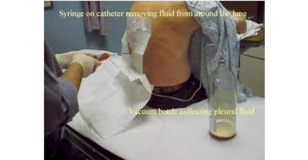
Thoracentesis
Invasive procedure to remove fluid from the pleural space
Performed by the HCP, not the RN
RN provides supportive care and may assist/observe
Fluid can be sent to the lab for analysis
Documentation: Describe the drainage by amount, color, and consistency.
Pneumothorax - Pathophysiology
Collapsed/deflated lung- partial or complete and air accumulates in pleural space
Loss of negative pressure leads to entry of positive atmospheric pressure allowing air in
Air enters the pleural space
Causes the lung to partially or completely collapse
Normally, pleural pressure is negative, keeping the lung inflated
A breach in the pleura allows air to enter → loss of negative pressure
Positive pressure causes the lung to collapse
Lung collapse → decreased gas exchange/oxygenation
Pneumothorax - Types
Simple
Traumatic
Tension
Simple Pneumothorax
Spontaneous
occurs without trauma
Often from rupture of a bleb (small air-filled blister) or bronchopleural fistula
Can occur in otherwise healthy people
More common in tall, thin males
Simple Pneumothorax - Clinical Presentation
Symptoms depend on size of pneumothorax
Sudden chest pain
Dyspnea
Tachypnea
Tachycardia
Decreased/absent lung sounds on affected side
Hyperresonance on percussion on affected side
Simple Pneumothorax - Risks factors:
Smoking
Emphysema
Tall, thin stature in a healthy person
Marfan syndrome - genetic d/o that affects the connective tissue
Pregnancy
Familial pneumothorax
Traumatic Pneumothorax
Pleural disruption from a chest wall wound
Air enters through chest wall & trachea
Medical Emergency!
Traumatic Pneumothorax - Causes
Blunt trauma
Penetrating trauma (e.g., knife wound)
Medical procedures that puncture the pleura
Traumatic Pneumothorax - Open Pneumothorax
Air moves in and out through the chest wall opening
May cause a “sucking chest wound”
Can cause mediastinal shift/swing:
Inspiration → mediastinum shifts toward the unaffected side
Expiration → shifts back toward the affected side
Traumatic Pneumothorax - Clinical presentation:
Same signs and symptoms as simple
Presence of OPEN SUCKING CHEST WOUND on inspiration
Hemothorax
Pleural disruption → bleeding
Blood collects in the pleural space
Usually caused by trauma
Can cause rapid, massive blood loss
May lead to:
Lung collapse
Hypovolemic shock
Treatment focuses on removing the blood, usually with a chest tube
Hemopneumothorax = air + blood in the pleural space
Severe cases may require immediate surgical intervention
Hemothorax - Clinical Presentation
Same as simple pneumo
Signs of shock!
Fast HR
Low BP
Pale, cool, clammy skin
Weak pulse
Fast breathing
Confusion/restlessness
↓ urine output
Diminished breath sounds on affected side
Dullness on percussion affected side
Tension Pneumothorax
Life-threatening emergency!
Air enters pleural space but cannot escape on expiration
Air becomes trapped
With each breath → pressure increases
Increased intrathoracic pressure causes:
Lung collapse on affected side
Mediastinal shift toward the unaffected side
Compression of the heart and great vessels
↓ venous return
↓ cardiac output (CO)
Can lead to PEA (pulseless electrical activity)
Tension Pneumothorax - Clinical Presentation
Severe difficulty breathing (dyspnea)
Chest pain (CP)
Markedly diminished/absent breath sounds on affected side
Hypotension → ↓ cardiac output
Distended neck veins (JVD) → compression of great vessels
Late signs:
Cyanosis
Tracheal deviation AWAY from the affected side (toward opposite side)
Pneumothorax - Assessment
depends on severity. Some simple pneumothoraces may resolve on their own, but patients may be observed/admitted for monitoring.
Oxygenation
Pallor → early sign
Cyanosis/duskiness → late sign
Skin temperature
Cool if patient is in shock
Vital signs
↑ HR (tachycardia)
↑ RR (tachypnea), often rapid/shallow
↓ BP if severe/shock
Tracheal alignment
Deviates away from affected side → tension pneumothorax
Chest expansion
Decreased and asymmetrical
Breath sounds
Diminished or absent on affected side
Percussion
Hyperresonance → pneumothorax (air)
Dullness → hemothorax (blood)
Subcutaneous emphysema
Air escapes into tissue under the skin
Feels/sounds like crackling or “crunching” (crepitus)
“Air Hunger”
Anxious
Agitated/restless
Diaphoretic
Short of breath
Subcutaneous Emphysema
Air escapes the pleural space and enters the subcutaneous tissue under the skin.
Usually occurs after chest trauma or injury to the pleura/lung.
Can affect the thorax, neck, and/or head.
Area may appear swollen/edematous due to trapped air.
Tactile crepitus when palpated → feels like Rice Krispies/crackling under the skin.
If air spreads into the neck, it may compromise the airway.
Can be distressing for the patient.
Usually resolves over time as the air is absorbed or expelled.
Pneumothorax - Diagnostics
SpO2 and/or ABGs
Chest X-ray - everyday
Chest CT*
US
Pneumothorax - Medical Management
Based on severity, type, and cause
Goal: remove blood/air from the pleural space and to allow reinflation of the lung
Supplemental O₂
Chest tube insertion
Removes air or fluid/blood
Smaller tube → usually for air
Larger tube → usually for blood/fluid
Connected to suction as ordered
blood more dense so at bottom, air on top
Antibiotics → may be given to prevent/treat infection
Needle decompression → emergency treatment for tension pneumothorax
Used to rapidly release trapped air and decrease pressure
Followed by chest tube placement
Autotransfusion → may be used with significant hemothorax to return the patient's own blood
Emergency Thoracotomy
Thoracotomy = surgical opening of the chest
thoracotomy if >1500 ml of blood is aspirated or chest tube has > 200 ml/hr
Pneumothorax - Nursing Management
Monitor Vitals/respiratory status
Reassess for signs of distress or hypoxia
Monitor SpO2
Watch for Shock
Assess for pain/anxiety
provide comfort measures
administer/analgesia
Manage and Monitor chest chest tubes- amount/type of drainage
Chest Tubes & Drainage - 3 chamber system
Collection chamber
Seal chamber
Suction chamber
Chest Tubes & Drainage - 3 chamber system: Collection chamber
Collects pleural drainage
Monitor the amount, color, and consistency of drainage
Chest Tubes & Drainage - 3 chamber system: Seal chamber
Allows air to leave the patient
Prevents air from entering the chest through the chest tube
Acts as a one-way valve
Traditional water seal: fill to 2 cm sterile water
Dry seal: uses a mechanical one-way valve
Chest Tubes & Drainage - 3 chamber system: Suction chamber
Controls/limits the amount of suction
Wet suction: uses sterile fluid
Dry suction: uses a mechanical device
Usually set to 20 cm H₂O
With low-vacuum suction, you may see bubbling in the suction chamber
Chest Tubes & Drainage - One way valve
7-12 Fr allows air out but no air in
Smaller pneumothorax
Chest Drainage Systems - Water seal and wet suction

Chest Drainage Systems - Water seal and dry suction

Chest Tube Nursing Management
Action | Reason |
|---|---|
1. Fill water-seal chamber with sterile water to the level specified by manufacturer (usually 2 cm). | Allows pleural air to escape while preventing air from going back into the chest. |
2. Fill water-suction chamber to the ordered level, usually 20 cm H₂O. | Controls the amount of suction. If using dry suction, set the dial to the prescribed level. |
3. Attach chest tube to collection chamber and tape all connections. | Keeps the system closed and prevents air leaks/disconnection. |
4. Connect suction chamber to wall suction until gentle bubbling is present. | With wet suction, the amount of suction is determined by the water level (e.g., 20 cm H₂O), not by how much it bubbles. |
5. Measure, mark, and record drainage. | Helps monitor blood/fluid loss. Frequency depends on how quickly the patient is draining (e.g., q15 min vs. q1 hr). Drainage should generally decrease over time. |
Water seal = keeps air from going back in
Suction = controls negative pressure
Collection = measures drainage
Chest Tubes and Drainage - Nursing Management
Insertion Site
Cover with occlusive, petroleum-based dressing
Secure with tape
Document tube placement based on post-procedure chest X-ray
Keep tubing coiled on the bed
Keep tubing free of kinks and dependent loops
Water-Seal Chamber
Maintain fluid level at 2 cm
Keep drainage unit upright
Monitor for:
Fluid fluctuation (tidaling)
Air leaks
Collection Chamber
Keep drainage unit below heart level
Notify provider if:
Initial drainage >1,000 mL
Hourly drainage >200 mL/hr
Document amount and characteristics/color of drainage
Suction Chamber
Check fluid level
Check suction gauge
Check for bubbling
FOCA
Assess and document fluctuation, output, color of drainage, and air leak
Emergency dislodgement-immerse tubing in sterile water
Chest Tubes and Drainage - Special Considerations
Chest Tube Clamping
Avoid clamping unless specifically ordered or required by hospital policy.
Clamping can cause tension pneumothorax by trapping air.
Chest Tube Milking/Stripping
Avoid unless specifically ordered/per hospital policy.
Can create excessive negative pressure in the pleural space.
Changing the Drainage System
Change when the collection chamber is full or according to facility policy.
Chest Tubes and Drainage - If the tube comes OUT of the patient:
Cover the insertion site with an occlusive dressing.
use a 4 × 4 dressing and tape 3 sides, leaving one side untaped to allow air to escape.
Notify the provider/emergency team.
Chest Tubes and Drainage - If the tubing becomes disconnected from the drainage system:
Place the end of the chest tube into sterile water about 2 cm deep to create a temporary water seal.
Reconnect to a new drainage system as soon as possible.
Flail Chest - Pathophysiology
Usually results from blunt chest trauma
Examples: MVC, steering wheel, airbag injury
3 or more adjacent ribs are fractured in 2 or more places
Creates a free-floating/flail segment of the chest wall
Chest wall becomes unstable
Causes paradoxical chest movement: opposite of rest of chest
Inspiration → flail segment moves IN
Expiration → flail segment moves OUT
Leads to:
Severe difficulty breathing (DIB)
Inability to take a deep breath
↓ Lung compliance
↓ Gas exchange/oxygenation
Mediastinum shifts toward the affected side → ↓ cardiac output
Flail Chest

Flail Chest - Clinical Presentation
Dyspnea
Chest wall pain
Hypoxemia/hypercarbia
Hypotension
1st signs = paradoxical chest movement & Mediastinum
Flail Chest - Assessment (depends on severity)
Paradoxical chest movement –moves in during inspiration and out during expiration
Tenderness on palpation with bony crepitus
Use of accessory muscles and increased work of breathing---- resp. distress
Tachypnea, Hypotension
Cyanosis
Flail Chest - Diagnostics
Chest x-ray
ABG
usually acidosis
Flail Chest - Medical Management
Based on severity
Pain Control-intercostal nerve blocks, high thoracic epidural blocks, IV opioids
Controlling secretions
Mild- moderate
-clear airway by positioning, pain management, Chest PT
Severe-
- intubation, mechanical ventilation with positive pressure -stabilizes chest wall, allows healing of fractures and pulmonary contusions, and improves gas exchange
rare surgical repair
Flail Chest - Nursing Management
Assess respiratory status
Monitor oxygenation- SpO2
Reassess pain and administer analgesia
Monitor VS, I & O
Manage secretions-positioning and suctioning
Manage care needs re: intubation/ventilator
Pulmonary Contusion - Pathophysiology
Lung injury from direct trauma
Usually blunt trauma
Can also occur from deceleration or high-velocity bullet wounds
Damage causes blood to leak into the lung tissue (parenchyma)
Leads to:
Hemorrhage
Edema
Protein/plasma leaking into the interstitial and alveolar spaces
Frequently associated with flail chest
Can affect one or both lungs
Severity depends on:
Size of the contusion
Damage to the alveolar-capillary membrane
Development of atelectasis/infiltrates
Respiratory status can worsen gradually, so patients need close monitoring.
Pulmonary Contusion
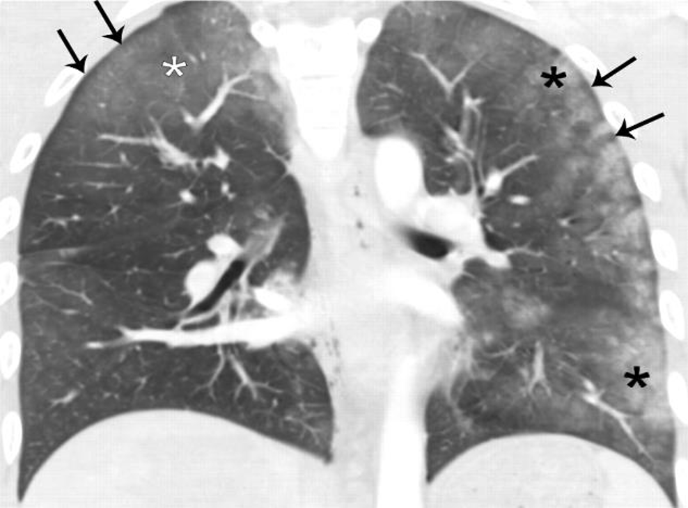
bruises
Pulmonary Contusion - Clinical Presentation
May begin with subtle S&S
Ranges from mild to severe
Dyspnea
Ineffective cough at clearing secretions
Hemoptysis/frank bloody secretions - coughing up blood
Chest pain
Pulmonary Contusion - Assessment
AIRWAY
Chest wall contusions or abrasions
Decreased breath sounds, crackles
Tachypnea and tachycardia
Hypoxemia
Cyanosis
Mental status changes-combativeness
Pulmonary Contusion - Diagnostics
Chest x-ray-initial may be normal until infiltrates appear 1-2 days later
ABG
SpO2
Hypercapnia (high carbon dioxide in the blood)
Pulmonary Contusion - Medical Management
Goals: Maintain airway, provide adequate oxygenation, and control pain.
Mild
Supplemental O₂
IV fluids and PO fluids to loosen secretions → avoid fluid overload
Chest physiotherapy
Coughing/deep breathing (TCDB)
Incentive spirometer
Tracheal suctioning as needed
Pain control: intercostal nerve blocks or PCA opioids
Antibiotics
Moderate
Bronchoscopy → removes secretions
Intubation + mechanical ventilation with PEEP
Diuretics + fluid restriction → decrease pulmonary edema
NGT → decompress stomach
Severe
Respiratory failure may be present
Intubation/mechanical ventilation
PEEP
Fluid restriction + diuretics → reduce pulmonary edema
IV fluids as needed to treat hypovolemia
Antibiotics → prevent/treat infection and pneumonia
Pulmonary Contusion - General Nursing Management
Frequent reassessment and vital signs for signs of respiratory compromise
Manage secretions-positioning and suctioning
Strict I & O
subtract to see if negative or positive
Reassess pain and administer analgesics
Manage ventilator support
Administer antibiotics