Micro Lab 2 PowerPoint Questions Fall 2026
 Call Kai
 Learn
Learn Practice Test
 Spaced Repetition
 Match
Match Flashcards
 Knowt Play
1/93

There's no tags or description
Looks like no tags are added yet.
Name | Mastery | Learn | Test | Matching | Spaced | Call with Kai | Chat |
|---|
No analytics yet
Send a link to your students to track their progress
94 Terms
True or False? The transfer of genetic material from parents to their offspring is called horizontal gene transfer.
True
False
False
Two organelles that are theorized to have evolved from prokaryotic cells are ______________ and _________________.
mitochondria; chloroplasts
mitochondria; ribosomes
ribosomes and nucleoli
chloroplasts, nucleoli
mitochondria; chloroplasts
True or False? Organisms in domain archaea are more closely genetically related to organisms in domain bacteria than domain eukarya.
True
False
False
Which of the following support the endosymbiotic theory of mitochondria and chloroplast evolution? Select all that apply.
mitochondria and chloroplasts are found in both prokaryotes and eukaryotes
mitochondria and chloroplasts possess pseudopeptidoglycan
mitochondria and chloroplasts have 70S ribosomes
mitochondria and chloroplasts have a single double-stranded circular chromosome
mitochondria and chloroplasts have a single double-stranded circular chromosome
mitochondria and chloroplasts have 70S ribosomes
What molecule is a component of both Gram-positive and Gram-negative bacterial cell walls?
pseudomurin
LPS
pseudopeptidoglycan
peptidoglycan
peptidoglycan
Organisms in domain archaea have cell walls that contain _______________.
pseudopeptidoglycan
LPS
peptidoglycan
mycolic acid
pseudopeptidoglycan
Many extremophiles such as halophiles, thermophiles, and acidophiles belong to domain _______________.
archaea
bacteria
animalia
eukarya
archaea
Prokaryotes reproduce asexually mainly by a process called ____________.
partitioning
binary fission
mitosis
fractionation
binary fission

septum formation
3
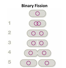
daughter cells separate
5

cell partitions
4

DNA replication
1

cell elongation
2

nucleoid
G

plasma membrane
B

cell wall
C

fimbriae
D

capsule
I

flagellum
E

ribosome
A

streptococci
B

vibrio
G

pallisade
F

diplococci
A

filamentous
K

streptobacilli
E

spiral
H

diplobacilli
D

spirochete
I

stella
J

pleomorphic
L

staphylococci
C

What is the shape and arrangement of the circled bacterial cells?
diplococci
pleomorphic cocci
staphylococci
streptococci
diplococci

What is the shape and arrangement of the circled bacterial cells?
pleomorphic bacilli
streptococci
staphylococci
filamentous bacilli
staphylococci

What is the shape and arrangement of the circled bacterial cells?
diplococci
staphylococci
cocci in tetrads
a palisade arrangement
cocci in tetrads

What is the shape and arrangement of these bacterial cells?
diplococci
staphylococci
streptococci
helical cocci
streptococci
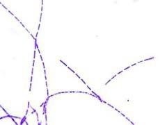
What is the shape and arrangement of these bacterial cells?
streptobacilli
staphylobacilli
coccobacilli
a palisade arrangement
streptobacilli
A patient presents with nausea and diarrhea, and informs you that he was recently in Africa and drank water from a stream. Tests for various parasitic infections were negative, so the illness is likely caused by a bacterial infection. After further testing the patient is diagnosed with chlorea caused by the bacterium, Vibrio cholerae. Using the information ascertained from the bacteria's name, what shape do you think this organism is?
filamentous strand
comma shaped
short rod shape
circle shaped
comma shaped

rough ER
C

smooth ER
E

centrioles
I

golgi appartatus
K

mitochondrion
F

nucleus
D

ribosome
G
When preparing a slide for bacterial staining, the _________________ step attaches the bacteria to the slide and kills the microorganisms.
heat fixing
sample drying
mordant
sample deposition
heat fixing
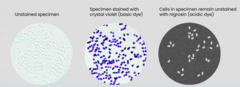
Unstained bacteria are hard to see in a bright-field light microscope. Based on these images, what do you think the two stained specimens have in common? In both samples....
the cells are dyed
the contrast between the cells and background is decreased
the contrast between the cells and background is increased
the background is dyed
the contrast between the cells and background is increased
Using your knowledge about the charge of various bacterial cellular components, predict what charge an acidic dye has.
the ion is negatively charged
the ion is positively charged
the ion has no charge
the ion is negatively charged
examples are crystal violet and methylene ble
Basic dye
stains microbial cells
Basic dye
colorful portion of the dye is negatively charged
Acidic dye
colorful portion of the dye is positively charged
Basic dye
examples include nigrosin and India ink
Acidic dye
repelled by microbial cells
Acidic dye
heat-fix the smear
Step 3
rinse smear with deionized water
Step 5
air-dry the smear
Step 2
spread the cells on a clean slide
Step 1
blot the slide dry
Step 6
flood smear with methylene blue
Step 4
Which of the following actions should be taken when preparing a bacterial smear for staining with a basic dye? Select all that apply.
make the smear as thick as possible to ensure you will see cell during microscope observation
cool the wire loop sufficiently after flame sterilization prior to taking your bacterial sample
blow on the smear to ensure faster drying times
adequately heat-fix the specimen by holding the slide in the Bunsen burner flame for at least 5 minutes
air-dry the smear completely prior to heat-fixing
cool the wire loop sufficiently after flame sterilization prior to taking your bacterial sample
air-dry the smear completely prior to heat-fixing
Your medical laboratory receives a clinical specimen from a patient believed to have inhalation anthrax. Your lab assistant has stained the specimen with methylene blue and observes rods arranged in chains. Which of the following arrangements best describes the observed cellular shape and arrangement?
palisade
diplobacilli
streptobacilli
streptococci
streptobacilli
You are preparing a bacterial smear from a broth culture for simple staining. You just spread a loopful of cells on a glass slide. What is the next step in the process?
flood the smear with dye
heat-fix the slide
air-dry the slide
blot the slide dry
air-dry the slide
Your clinical lab technician misplaced the stock of methylene blue. The technician want to stain the cells directly and asks if it would be acceptable to substitute acid fuchsin, an acidic dye, for methylene blue. Would this be an acceptable substitution?
Yes, both methylene blue and acid fuchsin are acidic dyes and will stain the cells.
No, methylene blue is a basic dye which stains cells and acid fuchsin is an acidic dye that stains the background.
No, methylene blue is a negatively charged dye which stains cells and acid fuchsin is a positively charged dye that stains the background.
Yes, both methylene blue and acid fuchsin are both positively charged and will stain the cells.
No, methylene blue is a basic dye which stains cells and acid fuchsin is an acidic dye that stains the background.
Your eager lab partner decides to skip the step of resuspending the cells in a broth culture tube, saying, "I see a sediment at the bottom of the tube; I will just pick up cells from there." How should you respond?
Report your partner to the lab instructor.
The sediment at the bottom of the tube is composed of dead cells; you should sample from the live cells at the top of the tube.
Resuspending the culture ensures even sampling and appropriate smear thickness.
Actually, your lab partner is correct; this step isn't necessary.
Resuspending the culture ensures even sampling and appropriate smear thickness.
Correctly order the procedural steps in the left-hand column to indicate how the inoculum to prepare a smear is aseptically obtained.
replace tube
Step 5
uncap culture tube
Step 1
flame-sterilize tube lip
Step 2
flame-sterilize tube lip and recap tube
Step 4
obtain one loopful of culture
Step 3
You just transferred one loopful of inoculum to a slide. What would be the likely result if you added several more loopfuls of culture. Select all that apply.
The increased cell density would require a longer period of time for heat-fixing.
Cell layering would make cellular morphology more difficult to determine.
Heat-fixation may be inadequate for some cells, resulting in staining inconsistencies.
The increased number of cells present would make it easier to determine cell morphology.
Cell layering would make cellular morphology more difficult to determine.
Heat-fixation may be inadequate for some cells, resulting in staining inconsistencies.
How is a solid culture smear preparation different from a broth culture smear preparation?
the slide should not be air-dried prior to heat-fixation
water must first be added to the slide
the amount of time for heat-fixation is significantly longer
the cells are not spread in a circular motion on the slide
water must first be added to the slide
You are preparing a bacterial smear from a solid culture for simple staining. You just transferred deionized water onto the glass slide. What should you do next?
flame sterilize and cool the loop
touch the loop to a single bacterial colony on the culture plate
heat-fix your sample
stain your sample with methylene blue
flame sterilize and cool the loop
Your impatient lab partner doesn't want to wait for the slide to fully dry before heat-fixing the specimen. What do you advise?
Skipping the air-drying step may cause the cells to boil, distorting their morphology.
Skipping the air-drying step will prevent cells from adhering to the slide during heat-fixation.
The slide must be dry, but to save time you can blow on it or wave it in the air.
Let's skip the air-drying step. It is better to heat fix the slide while still wet to avoid cellular distortion.
Skipping the air-drying step may cause the cells to boil, distorting their morphology.
Where in the Bunsen burner flame should you heat-fix your specimen?
in the inner cone of the flame
above the flame
in the outer cone of the flame
in the outer cone of the flame
What is the proper way to heat-fix a slide?
hold it in the flame for one minute
sweep it through the flame three times
sweep it through the flame for three minutes
hold it in the flame for five minutes
sweep it through the flame three times

Liliana has a suspected case of meningitis. The doctor performs a lumbar puncture to collect cerebrospinal fluid (CSF) for simple staining to determine if the meningitis is bacterial in nature. The image shows the results of the CSF simple stain. Does Liliana's specimen contain bacterial cells?
No; the specimen does not contain any cells.
Yes; the bacterial cells have been directly stained by the methylene blue.
Yes; the colorless bacterial cells are visible against a blue background.
No; the specimen only shows the presence of stained white blood cells.
Yes; the bacterial cells have been directly stained by the methylene blue.

Based on the CSF micrograph shown and the informational table provided, which pathoge(s) are a possible cause of Liliana's illness? Select all that apply.
Escherichia coli
Streptococcus agalactiae
Listeria monocytogenes
Neisseria meningitidis
Haemophilus influenzae
Streptococcus pneumoniae
Neisseria meningitidis
Streptococcus pneumoniae

Bacterial cells were detected in a patient's CSF sample, so additional tests were ordered including culturing the CSF on blood agar. After examining the cultural characteristics of the colonies growing on the plate, you are concerned that more than one species may be present. Would a simple stain of each colony (A and B) be able to determine if there are two bacterial species growing on the blood agar plate?
No; simple stains do not stain cells and hence not able to differentiate between cell types.
Possibly; simple stains can provide information about certain bacterial characteristics and hence be able to distinguish species that differ in morphology.
No; simple stains stain all cells the same color and hence are not able to distinguish between cell types.
Yes; simple stains can stain cells different colors depending on their cell wall properties.
Possibly; simple stains can provide information about certain bacterial characteristics and hence be able to distinguish species that differ in morphology

Smear preparations and simple stains are performed on both the Colony A and Colony B isolates from the blood agar plate. The cell morphology of Colony B is similar to the organism observed in the CSF sample. However, the cell morphology of Colony A differs, indicating that it is not the same species as Colony B. Based on the cell morphology listed in the table, Colony A is most likely which organism?
Bacillus subtilis
Micrococcus luteus
Staphylococcus epidermidis
Streptococcus salivarius
Staphylococcus epidermidis
Lilliana's CFS sample was stained with methylene blue. What type of information can this simple stain provide about the bacterial pathogen? Select all that apply.
the pathogen's definitive identity
cellular shape
cellular size
cellular arrangement
cellular shape
cellular size
cellular arrangement
A simple stain was performed on bacterial colony using methylene blue, a dye which stains cells blue. Which of the following statements are true? Select all that apply.
methylene blue is an acidic dye
methylene blue is a positively charged dye
methylene blue is a negatively charged dye
methylene blue is a basic dye
methylene blue is a positively charged dye
methylene blue is a basic dye
A simple stain is performed on a bacterial colony using India ink, a brown-colored acidic dye. How do you expect the specimen to appear after staining?
both the cells and background will appear brown
both the cells and background will appear colorless
the cells will be brown and the background will be colorless
the cells will appear colorless and the background will be brown
the cells will appear colorless and the background will be brown

Troubleshoot the following:
Your laboratory technician is having difficulty identifying the morphology of this stained specimen and asks for your advice. How should you respond?
the cells were overly heat-fixed
the smear was made overly thick
the specimen was rinsed too long with deionized water
too much methylene blue was added during the staining process
the smear was made overly thick
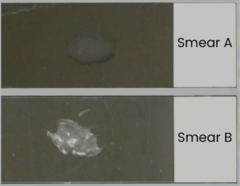
Before air-drying and heat-fixing this specimen, your lab technician asks you which smear to use. Which smear (A or B) has an appropriate thickness for staining?
Smear A is better; its liquid appearance ensures that the cells will settle in several layers.
Smear A is better; it contains fewer cells, resulting in more accurate staining and more clearly visible cell morphology.
Smear B is better; it contains more cells so there is less likelihood the cells would wash away during staining.
Smear B is better; it contains more cells which will be easier to find when using the microscope.
Smear A is better; it contains fewer cells, resulting in more accurate staining and more clearly visible cell morphology.
Troubleshoot the following:
Your lab partner is preparing a smear for simple staining from a solid culture. Oddly, the stained slide doesn't appear to have any cells on it. What went wrong?
too much stain was applied to the specimen
the sample was heat-fixed for too long
the cells were rinsed with deionized water for too long
the cells were inadequately heat-fixed
the cells were inadequately heat-fixed
Various errors can occur when simple staining. Match the scenarios in the left-hand column to the error that likely occured during the procedure.
slide shatters
over-heating during heat-fixation
large groups of stained cells prevent accurate determination of cellular morphology and arrangement
smear made overly thick
no cells present
inadequate heat-fixation
only unstained cells are observed
smear made overly thick
stained cells are visible, but their cellular morphology is distorted
over-heating during heat-fixation
Which of the following procedural errors is most likely to result in distorted cellular morphology?
the broth culture is not properly suspended prior to obtaining a sample
the smear is not sufficiently air-dried prior to heat-fixing
the smear is inadequately heat-fixed
the loop fully swipes the surface of a colony when obtaining a sample
the smear is not sufficiently air-dried prior to heat-fixing
Which of the following problems may occur when a smear is made overly thick? Select all that apply.
the cells are harded to find
it's difficult to determine cellular morphology
the cells are inconsistently stained
the slide is more likely to shatter
not all cells will properly adhere to the slide
it's difficult to determine cellular morphology
the cells are inconsistently stained
not all cells will properly adhere to the slide
When heat-fixing a smear, you quickly pass the slide once through the Bunsen burner flame. Which of the following will most like occur because of this action?
the slide will shatter
an abundance of stained cells will be visible with minimal cellular distortion
a greater amount of cellular distortion will occur
the cells will not be visible under the microscope
the cells will not be visible under the microscope