Exam 3: Toes, Foot, Ankle, Calcaneus, Tib/Fib, Knee, & Patella
 Call Kai
 Learn
 Practice Test
 Spaced Repetition
 Match
 Flashcards
 Knowt Play
1/268

There's no tags or description
Looks like no tags are added yet.
269 Terms

Label 6
third metatarsal, right foot
AP Oblique foot medial rotation criteria
Tuberosity of the fifth metatarsal
= amount of space between shafts of 2nd-5th metatarsals
Closed joint space b/w medial and intermediate cuneiforms
Open joint spaces around cuboid and the sinus tars
Degree of rotation of the toes for an AP oblique medial rotation projection?
30-45 degrees
Degree of tube angulation for an AP axial projection of the foot
10 degrees posterior

label 18
lateral cuneiform
Centering point for plantodorsal projection of the calcaneus
entering plantar surface near the base of the third metatarsal
Which tarsal bone articulates with the distal tibia and fibula to form the mortise joint?
talus
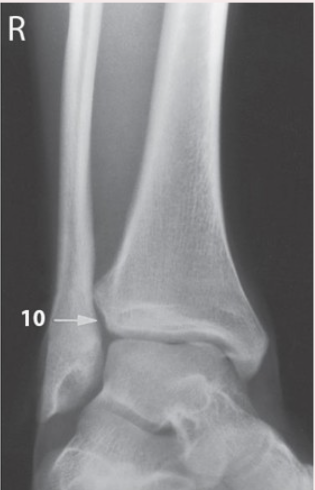
Ankle positioning in this image
right leg/foot obliqued medially 45 degrees
AP oblique ankle rotation
15-20 degree medial rotation
An AP oblique ankle rotation with 15-20 degree medial rotation will place the intermalleolar plane of the ankle ______ with the plane of the IR.
parallel
The AP ankle stress view is useful in verifying:
ligamentous tears
Degree of tube angulation for an AP axial projection of the toes to best demonstrate the joint spaces?
15 degrees CR angulation
How much foot rotation is needed for an AP oblique projection of the foot with medial rotation?
30 degrees medial rotation

Label 1
Talus
CR for an AP oblique projection, medial rotation of the toes (to include all toes)?
perpendicular to IR, entering the 3rd MTP joint
What tube angle will reduce foreshortening in the phalanges and metatarsals of the foot?
angling tube 10 degrees towards the heel

How was the central ray set up to produce this image?
40 degree angle towards the heel
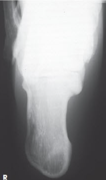
What projection and anatomy is this?
Calcaneus axial (plantodorsal), Harris view

What projection and anatomy is this?
AP oblique ankle

What positioning is used to achieve this image?
the lower extremity rotated medially 45 degrees

the lower extremity rotated medially 45 degrees best demonstrates the _____
Distal tibiofibular joint open

What projection and anatomy is this?
AP oblique projection

This AP oblique projection of the right ankle was performed with what rotation of the lower leg?
15-20 degrees medial
The standard projection for a lateral ankle is the:
mediolateral
Where should the CR be placed for the routine lateral calcaneus?
1" inferior to medial malleolus
Each foot consists of ________ bones.
26
What portion of the 5th metatarsal is frequently fractured? (Jones Fracture)
proximal/base
Gout
condition that appears radiographically as uric acid and deposits in joint space (leading to destruction of joint space)
Centering point for an AP axial foot projection?
base of 3rd MT (metatarsal)
degree and direction of CR angle for the AP axial foot
10 degrees posterior ( towards the heel)
What is the direction of rotation for the AP Oblique foot projection?
medial/internal rotation
Which is the preferred lateral projection of the foot?
mediolateral
SID for a lateral weight-bearing foot projections?
40'“
benefit of doing an AP axial toe projection instead of an AP toe projection?
opens up IP joint spaces and reduces foreshortening
What is the (1) degree of tube angulation, (2) the direction of the central ray, and (3) centering point for an AP axial toe projection?
15 degrees posteriorly, centered to the MTP joint
Name tarsal bones in the midfoot that are less superimposed when doing an AP oblique medial rotation projection of the foot versus an AP foot
navicular, lateral cuneiform, cuboid
With which projection are the cuboid and its articulations best demonstrated
AP oblique foot in medial rotation position

What special projection of the lower extremity is demonstrated?
Tangential projection (Holly method), sesamoid bones
What part of the foot should be perpendicular to the IR on a lateral projection of the foot?
plantar surface
Centering point for each toe 2-5 in a lateral projection?
perpendicular to PIP of interest
Which joint spaces are demonstrated open on the AP axial projection of the toes?
IP joints and MTP joints
To evaluate the sesamoid bones in the toes, the tangential sesamoids projection requires the CR to be:
perpendicular to IR, directed to posterior aspect of 1st MTP joint
What type of leg/foot rotation is done routinely with AP oblique ankles? (medial rotation or lateral rotation)
medial rotation
How much rotation is required on the AP oblique Mortise projection of the ankle?
15-20 degrees medial rotation
How should the patient's foot & ankle be positioned for an axial projection of the calcaneus?
dorsiflexed
CR angle for an axial projection of the calcaneus?
40 degrees, towards the heel
Under what circumstances might you perform a (less common) lateromedial ankle projection?
trauma, patent can't move from supine, image will be a crosstable projection
centering point for an axial projection of the calcaneus?
base of third metatarsal
Which projection of the ankle will place the intermalleolar plane of the ankle parallel with the plane of the IR?
AP oblique mortise (15-20 degrees medial rotation)
When looking at images, what is one way you can tell the mediolateral lateral projections of the calcaneus vs the ankle?
Centering: The calcaneus will be 1" distal to the medial malleolus whereas the ankle centering will be on the medial malleolus.
Collimation: The calcaneus should be more closely collimated.
How do you get the mortise joint "open" on an image?
medially rotate the foot/leg 15-20 degrees
What type of fracture is commonly seen at the base of the 5th metatarsal?
Jones fracture
What 2 tarsal bones make up the hindfoot?
talus and calcaneus
What joint should be well visualized on a properly positioned axial calcaneus projection?
talocalcaneal (subtalar) joint
CR for a lateral projection of the calcaneus?
1" distal to the medial malleolus
For the mortise joint to be demonstrated, the lower limb must be positioned in
15-20 degree medial oblique
Which clothing items might cause an artifact during imaging of the lower extremity?
socks, shoes, jeans, jewlery
Which name is given to a fracture at the fifth metatarsal base?
Jones Fracture
Which tarsal bone articulates with the distal tibia and fibula to form the mortise joint?
Talus

Which positioning maneuver was required to produce this image?
Dorsiflexion to place plantar surface perpendicular to IR

What projection and anatomy is this
plantodorsal axial calcaneus projection

What was the CR orientation for this projection?
40 degrees toward the heel
Which malleolus is more posterior?
lateral
Which projection will open the tibiofibular joint space?
AP oblique 45 degree internal rotation
SID for the AP axial calcaneus?
40"
A grid should always be used for AP and Lateral projections of the ankle
false
The CR is directed ____ to the IR for the AP oblique Mortise ankle projection
perpendicular

The joint space highlighted in the image below is the:
talofibular joint
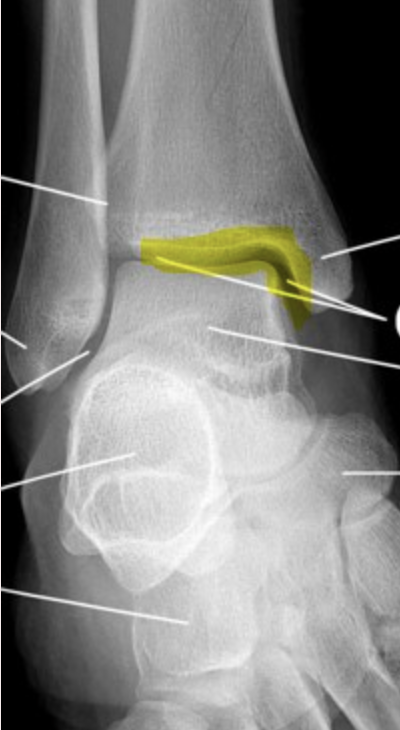
The joint space highlighted in the image below is the:
tibiotalar joint

The joint space highlighted in the image below is the:
mortise joint

The joint space highlighted in the image below is the:
distal tibiofibular joint
CR angle when radiographing the knee joint space in the AP projection on patient whose ASIS – tabletop measurement distance is 23.5 cm ?
perpendicular

This is a ____________ projection of the _______________
AP; left knee
Which of the following structures serves as a shock absorber within the knee joint?
Meniscus

3
fibular head

1
medial epicondyle

2
patella

4
intercondylar tubercles

7
right lateral tibial plateau

8
right femoral intercondylar fossa
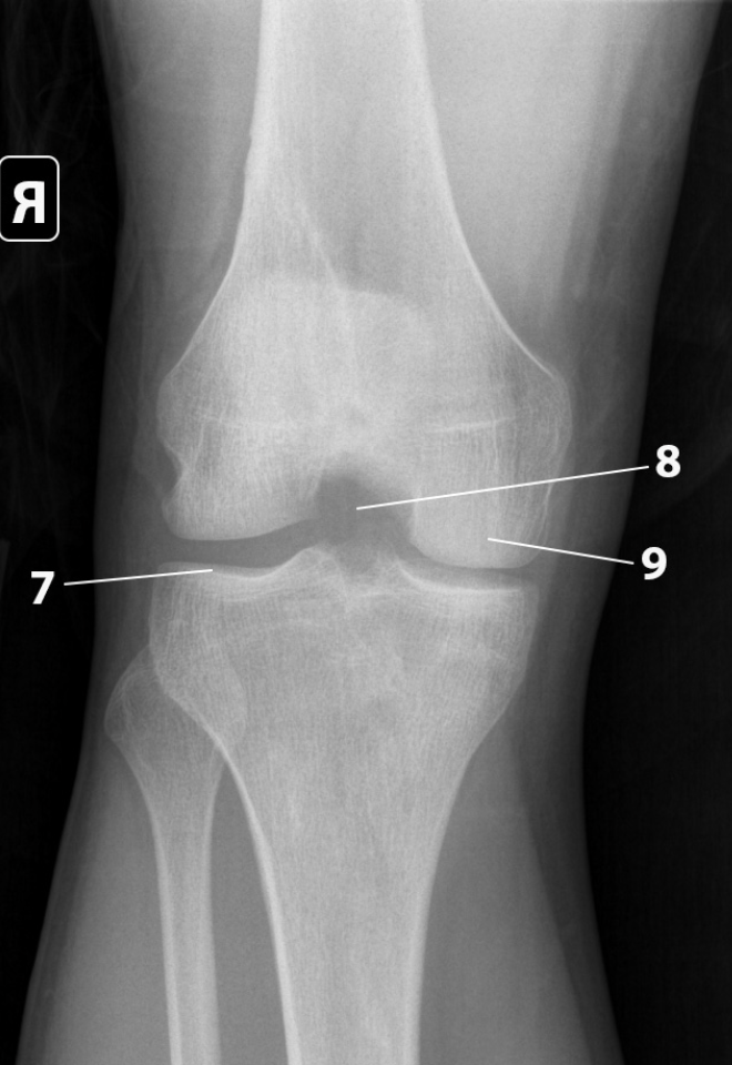
9
right medial femoral condyle
Why is a long SID recommended for the Merchant’s Method?
to decrease magnification

1
Patella

3
lateral femoral condyle

4
medial femoral condyle

5
Fibula
How much will you oblique the patient knee for an AP oblique with medial or lateral rotation?
45 degrees

What anatomy is indicated by the white line?
Medial tibial condyle
For a lateral projection of the knee, the plane through the femoral epicondyles is positioned in what orientation to the plane of the IR?
perpendicular
What of the following options will best place the knee in true AP position?
rotate lower limb internally 3-5 degrees
What projection best reveals narrowing of the joint space at the knee (tibiofemoral) joint?
AP weight-bearing method of the knees
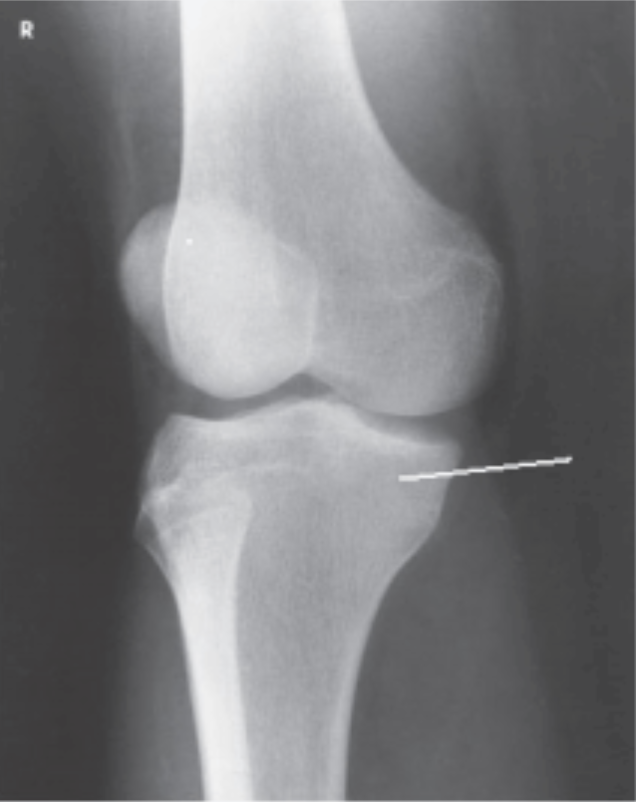
What projection and position are demonstrated?
AP oblique projection of the knee, lateral rotation position
anatomy & positioning for a Camp-Coventry method image?
PA axial projection of the intercondylar fossa

what method is this
camp-coventry

1
left medial femoral epicondyle
The PA projection of the patella provides improved spatial resolution over the AP projection because:
it uses a smaller OID on the PA projection

Which statement best describes the correct central ray for this projection?
angle of 5-7 degrees cepahlic
Describe the location of the apex and base on the patella.
the apex is pointed inferior and base is the superior border of the patella