General Anatomy 2 Midterms
 Call Kai
 Learn
 Practice Test
 Spaced Repetition
 Match
 Flashcards
 Knowt Play
1/382

There's no tags or description
Looks like no tags are added yet.
383 Terms
Fascial layers
They create anatomical spaces that influence the spread of infections
Superficial cervical fascia
A layer of fatty connective tissue located between the skin and the first deep fascial layer
Contains:
Cutaneous nerves
Blood and lymphatic vessels
Superficial lymph nodes
Variable amounts of fat
Superficial cervical fascia
In which layer is the platysma muscle embedded in?
Facial expression (depresses lower jaw, draws down lip & mouth corner)
Tenses the skin (shaving and easing a tight collar)
Functions of the superficial cervical fascia
Cervical branch of the facial nerve (CN VII)
Innervation of the superficial cervical fascia
Main cutaneous nerves of neck
External jugular vein
Deep to the platysma, we find _____ (innervation)
Superficial cervical fascia
Attachments:
Superior: Deep fascia covering pectoralis major & deltoid
Inferior:
Mandible
Skin
Subcutaneous tissues of lower face
Deep cervical fascia
Supports and compartmentalizes cervical structures
Three main layers:
Investing fascia
Pretracheal fascia
Prevertebral fsascia
*Carotid sheath is also part
Investing layer
Layer of Deep Fascia
Encircles the entire neck, deep to skin & superficial fascia
Encloses trapezius and SCM muscle
Contains:
Inferior ends of anterior jugular veins
Jugular venous arch
Fat and deep lymph nodes
Investing layer
Layer of Deep Fascia
Forms a fibrous capsule around:
Submandibular gland (splitting below mandible)
Parotid gland (splitting behind mandible)
Investing layer
Which layer of the deep cervical fascia forms the suprasternal space?
Investing layer
Attachments:
Superior:
Superior nuchal line
Mastoid process
Zygomatic arch
Mandible, hyoid bone, cervical vertebrae
Inferior:
Manubrium of sternum
Clavicles
Scapulae
Suprasternal space
Space located between the sternal heads of the SCM
Pretracheal layer (Anterior deep fascia)
Layer of Deep Fascia
Located only in the anterior neck
Extends from hyoid bone to thorax (merging w/ fibrous pericardium)
2 Parts:
Muscular part — encloses infrahyoid muscles
Visceral part — encloses thyroid gland, trachea, esophagus
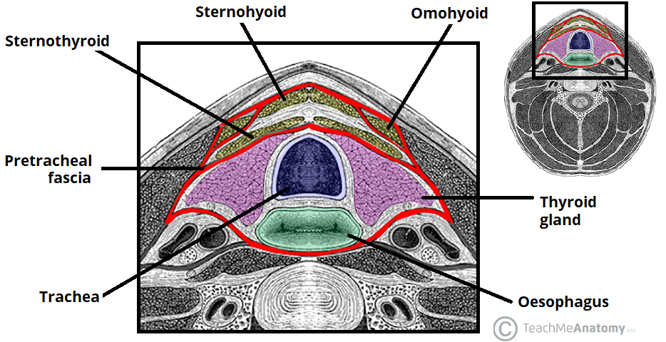
Muscular part
Which part of the pretracheal layer encloses infrahyoid muscles?
Visceral part
Which part of the pretracheal layer encloses thyroid gland, trachea, and esophagus?
Buccopharyngeal fascia
Superiorly, the pretracheal layer blends with the ______
Carotid sheath
Laterally, the pretracheal layer blends with the _____
Mediastinum
Inferiorly, the pretracheal layer communicates with the _____
Pretracheal layer
Specialization: Forms pully (trochlea) for the intermediate tendon of the digastric muscle
Prevertebral layer (Deepest deep fascia)
Layer of Deep Fascia
Surrounds vertebral column and associated muscles:
Longus colli
Longus capitis
Scalene muscles
Deep cervical muscles
Encloses the cervical part of the sympathetic trunk
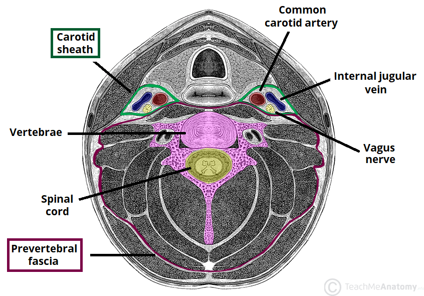
Prevertebral layer
Attachments:
Superiorly: Cranial base
Inferiorly: Blends with endothoracic fascia at T3 vertebra
Laterally: Extends as axillary sheath (encloses axillary vessels & brachial plexus)
Carotid sheath
Tubular fascial structure that extends from the cranial base to the root of the neck
Carotid sheath
Contents:
Common and internal carotid arteries
Internal jugular vein
Vagus nerve (CN X)
Deep cervical lymph nodes
Carotid sinus nerve & sympathetic fibers
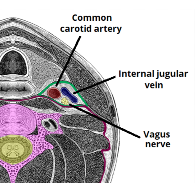
Carotid sheath
Fascial connections:
Anterior: Investing & pretracheal fascia
Posterior: Prevertebral fascia
Inferior: Mediastinum
Retropharyngeal space
Largest potential space in the neck
Between prevertebral fascia (posteriorly) and buccopharyngeal fascia (anteriorly)
Retropharyngeal space
Allows pharynx, esophagus, larynx, and trachea to move freely during swallowing
Retropharyngeal space
Boundaries:
Superior: Cranial base
Lateral: Carotid sheath
Inferior: Communicates w/ mediastinum
True retropharyngeal space, Danger space
What are the 2 divisions of the retropharyngeal space?
True retropharyngeal space
Division of RP space
Anterior to alar fascia
Extends from skull base to C7
Danger space
Division of RP space
Between alar fascia and prevertebral fascia
Extends from skull base to diaphragm
Infections here can spread to the thoracic cavity
Superficial infections
Spread of infections is limited to the superficial fascia
Pretracheal space infections
Infections here may spread to the anterior mediastinum
Retropharyngeal space infections
Infections here may extend to the superior mediastinum
Danger space infections
Infections here can reach the posterior mediastinum and diaphragm, leading to serious complications
Base of the cranium and clavicles
The neck is the anatomical region between the _____ and the _____
Anterior triangle
Boundaries:
Anterior: Midline of the neck.
Posterior: Anterior border of the sternocleidomastoid (SCM).
Superior: Inferior border of the mandible.
Roof: Subcutaneous tissue containing the platysma.
Floor: Pharynx, larynx, thyroid gland.
Apex: Located at the jugular notch of the manubrium
Submental
Submandibular
Carotid
Muscular
What are the subdivisions of the anterior triangle of the neck?
Submental triangle
Anterior Triangle Subdivision
Located below the chin
Boundaries:
Inferior: Hyoid bone.
Lateral: Right and left anterior bellies of the digastric muscles.
Apex: Mandibular symphysis.
Floor: Mylohyoid muscles meeting in a median fibrous raphe.
Contents:
Submental lymph nodes
Small veins that unite to form anterior jugular vein
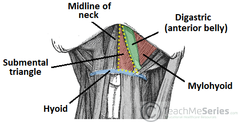
Submandibular triangle
Anterior Triangle Subdivision
Boundaries:
Superior: Inferior border of the mandible.
Anteroinferior: Anterior belly of the digastric muscle.
Posteroinferior: Posterior belly of the digastric muscle.
Roof: Investing layer of deep cervical fascia.
Floor: Mylohyoid and hyoglossus muscles.
Contents:
Submandibular gland & lymph nodes
Hypoglossal nerve (CN XII)
Mylohyoid nerve
Facial artery & vein
Marginal mandibular branch of facial nerve (in 20% of individualss)
Hypoglossal nerve (CN XII)
C1 fibers
Ansa cervicalis
Innervation of the submandibular triangle
Ansa cervicalis (C1-C3)
Supplies omohyoid, sternothyroid, and sternohyoid muscles
Hypoglossal nerve
Passes deep to the posterior belly of the digastric muscle
Provides motor innervation to intrinsic and extrinsic tongue muscles
C1 fibers
They travel with the hypoglossal nerve (CN XII) and give off the thyrohyoid branch to the thyrohyoid muscle
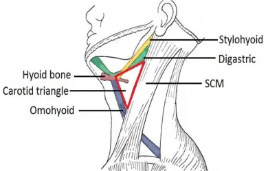
Carotid triangle
Anterior Triangle Subdivision
Boundaries:
Inferomedial: Superior belly of the omohyoid muscle.
Superior: Posterior belly of the digastric muscle.
Inferolateral: Anterior border of the SCM.
Common carotid artery
External carotid artery
Internal jugular vein
Blood vessels of the carotid triangle
Vagus nerve (CN X)
Hypoglossal nerve (CN XII)
Superior root of ansa cervicalis
Spinal accessory nerve (CN XI)
Branches of the cervical plexus
Innervation of the carotid triangle
Thyroid gland
Larynx
Pharynx
Deep cervical lymph nodes
Contents of the viscera of the carotid triangle
Carotid sheath
Surrounds the carotid arteries, internal jugular vein, and vagus nerve
Muscular triangle
Anterior Triangle Subdivision
Boundaries;
Superolaterally: Superior belly of the omohyoid muscle
Inferolaterally: Anterior border of the SCM
Medially: Median plane of the neck
Muscular triangle
Infrahyoid muscles (sternohyoid, omohyoid, sternothyroid, thyrohyoid), thyroid and parathyroid glands are part of which triangle?
Common carotid artery (bifurcates into internal & external)
Blood supply of the anterior triangle
Brachiocephalic trunk
The right common carotid artery originates from the _____
Aortic arch
The left common carotid artery originates from the _____
Upper border of thyroid cartilage
Where does the common carotid artery bifurcate?
Internal carotid artery
No branches in the neck.
Enters the carotid canal and supplies the brain
Carotid sinus (baroreceptor) and carotid body (chemoreceptor) are located at the bifurcation
External carotid artery
Supplies most structures external to the cranium.
Terminal branches: Maxillary artery and superficial temporal artery.
Branches: “Some Anatomists Like Freaking Out Poor Medical Students”
Superior thyroid artery
Ascending pharyngeal artery
Lingual artery
Facial artery
Occipital artery
Posterior auricular artery
Maxillary artery (terminal)
Superficial temporal artery (terminal)
Internal jugular vein
Venous drainage of the anterior triangle
Maxillary artery and superficial temporal artery
What are the terminal branches of the external carotid artery?
Internal jugular vein
Largest vein in the neck
Begins at the jugular foramen as a continuation of the sigmoid sinus
Descends within the carotid sheath
Merges with the subclavian vein to form the brachiocephalic vein
Tributaries:
Inferior petrosal sinus
Facial vein
Lingual vein
Pharyngeal veins
Superior and middle thyroid veins
Bicuspid valve
The inferior bulb of the IJV contains a _____ to prevent backflow of blood
Suprahyoid and infrahyoid muscles
What are the muscles of the anterior triangle?
Mylohyoid
Geniohyoid
Stylohyoid
Digastric
What are the suprahyoid muscles?
Sternohyoid
Omohyoid
Sternothyroid
Thyrohyoid
What are the infrahyoid muscles?
Posterior triangle
Formed mainly by the sternocleidomastoid (SCM) and trapezius muscles
Boundaries:
Anterior: Posterior border of the SCM
Posterior: Anterior border of the trapezius.
Inferior: Middle third of the clavicle (between SCM and trapezius).
Roof: Investing layer of deep cervical fascia.
Floor: Formed by prevertebral muscles covered by the prevertebral layer of deep cervical fascia.
Apex: Where SCM and trapezius meet at the superior nuchal line of the occipital bone.
Spinal accessory nerve (CN XI) and fibers from C2, C3
Innervation of the SCM and trapezius muscle
Sternocleidomastoid
Actions:
Unilateral contraction: Lateral flexion (same side) and rotation (opposite side).
Bilateral contraction:
Extends the neck at the atlanto-occipital joint.
Flexes the neck, bringing the chin to the manubrium.
Elevates the clavicles and ribs during forced respiration.
Trapezius
Actions:
Superior fibers: Elevate scapula.
Middle fibers: Retract scapula.
Inferior fibers: Depress scapula.
Superior + inferior fibers: Rotate scapula superiorly.
Bilateral contraction: Extends the neck.
Unilateral contraction: Laterally flexes the neck.
Splenius capitis
Levator scapulae
Anterior scalene
Middle scalene
Posterior scalene
Floor muscles of the posterior triangle
Scalene muscles
These muscles elevate the ribs
Occipital triangle (Superior) and Omoclavicular triangle (Inferior)
What are the subdivisions of the posterior triangle?
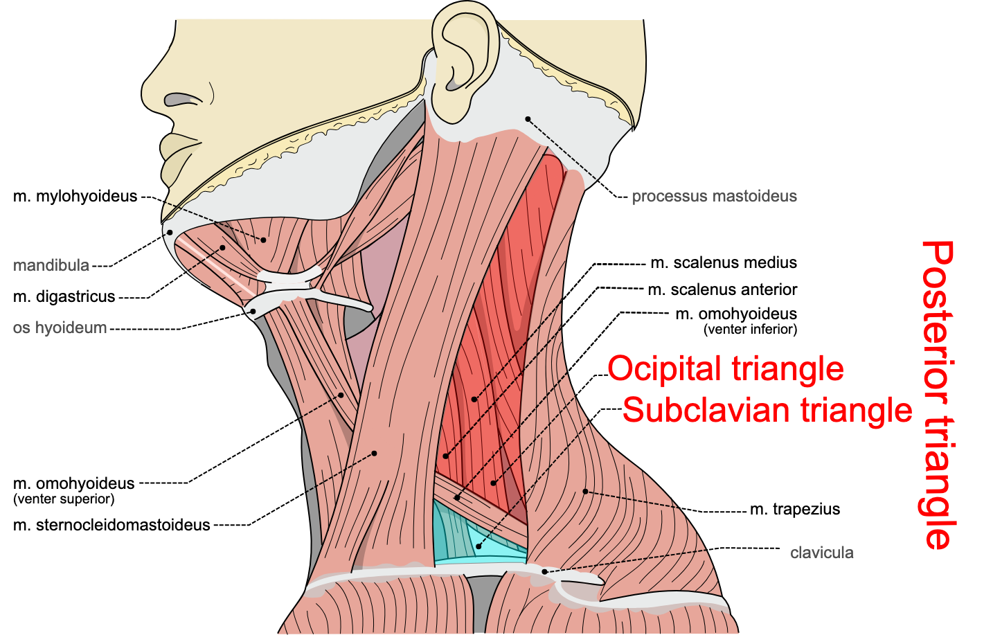
Occipital triangle
Contents:
Occipital artery at its apex
Spinal accessory nerve (CN XI)
Omoclavicular (Subclavian) triangle
Boundaries:
Inferior belly of omohyoid (posterosuperiorly)
Posterior border of SCM (anteriorly)
Middle third of clavicle (inferiorly)
Contains:
Inferior part of the external jugular vein
Third part of the subclavian artery
Suprascapular artery
Cervicodorsal trunk (Transverse cervical artery)
Third part of subclavian artery
Occipital artery
Arteries in the posterior triangle
Suprascapular artery
Artery that runs across the anterior scalene muscle
Cervicodorsal trunk (Transverse cervical artery)
Blood supply to the trapezius
Occipital artery
A branch of the external carotid artery that supplies the posterior scalp
Third part of subclavian artery
Artery that lies deep in the inferior part of the omoclavicular triangle
External jugular vein
Vein in the posterior triangle
Begins near the angle of the mandible.
Formed by the union of the retromandibular vein and posterior auricular vein
Drains into the subclavian vein
Retromandibular vein and posterior auricular vein
What union forms the external jugular vein?
Spinal accessory nerve (CN XI)
Cervical plexus
Brachial plexus
Nerves in the posterior triangle
Brachial plexus
Formed by anterior rami of C5–T1
Emerges between the anterior and middle scalene muscles
Passes inferolaterally through the posterior triangle
Suprascapular nerve arises from the superior trunk of the plexus
Cervical plexus
Formed by anterior rami of C1–C4
Provides sensory and motor innervation to the neck
Superficial branches emerge at the posterior border of SCM
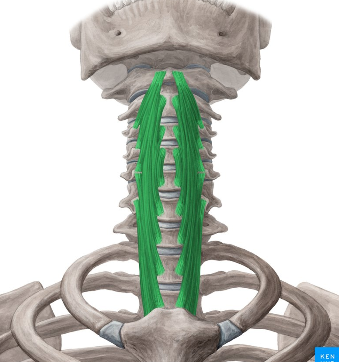
Longus colli
Longus capitis
Rectus capitis anterior
Anterior scalene
Anterior prevertebral muscles
Longus colli
Origin:
• Bodies of C5–T3 vertebrae.
• Transverse processes of C3–C5 vertebrae.
Insertion:
• Anterior tubercle of C1.
• Bodies of C2–C4 vertebrae.
• Transverse processes of C3–C6.
Innervation: Anterior rami of C2–C6 spinal nerves.
Action:
• Bilateral contraction: Flexion of the neck.
• Unilateral contraction: Rotation of the neck to the opposite side.
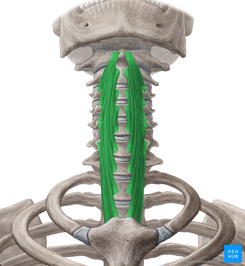
Anterior rami of C2-C6 spinal nerves
Innervation of longus colli muscle
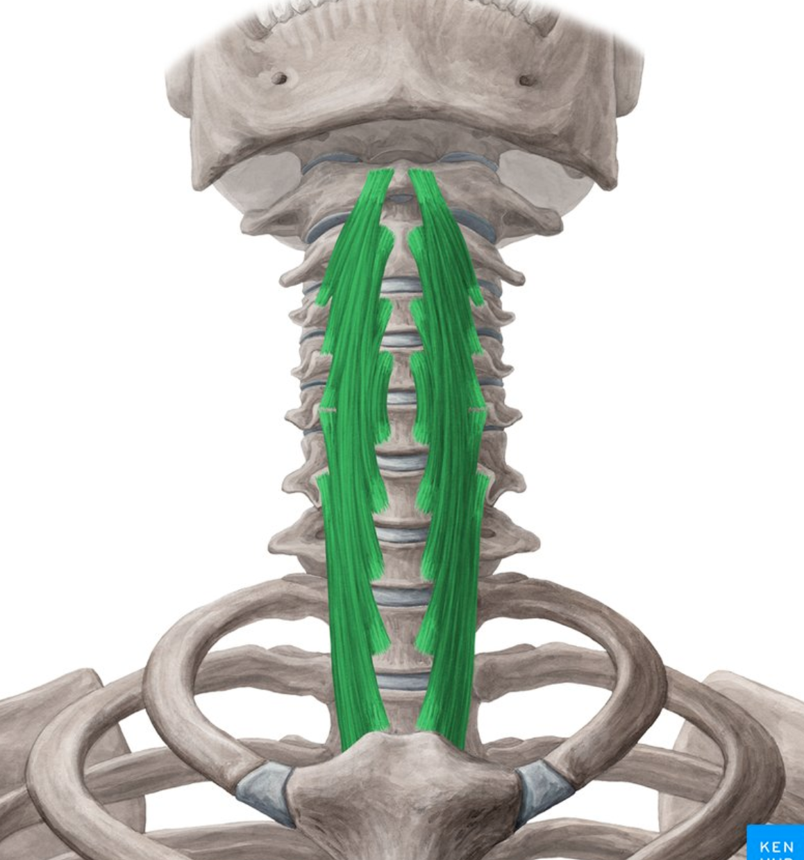
Flexion and rotation of the neck
Action of longus colli muscle
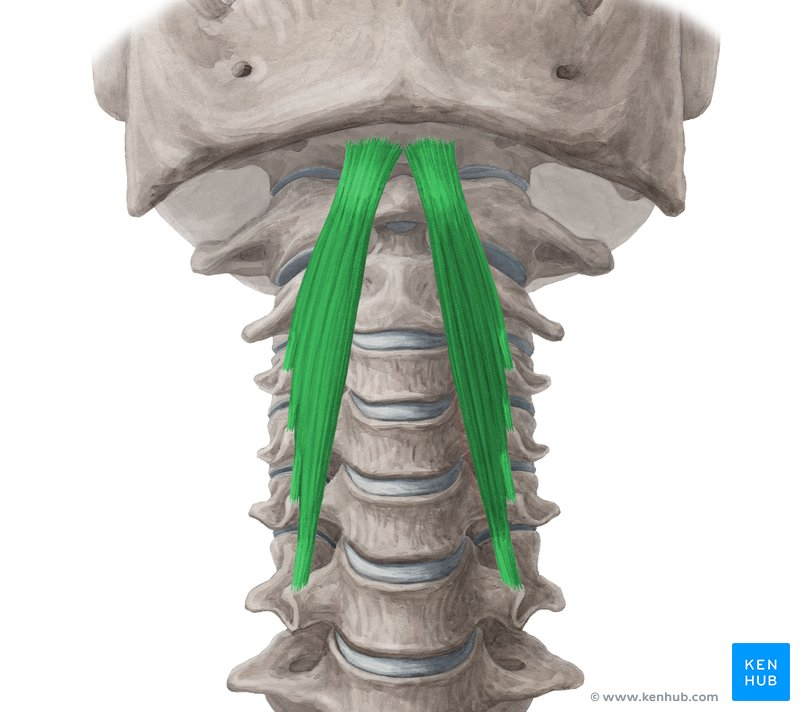
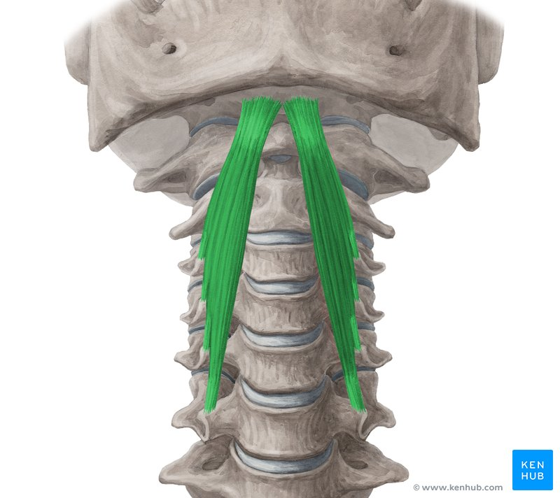
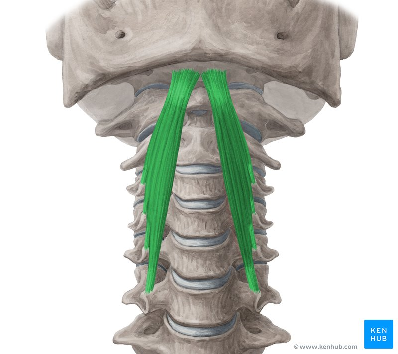
Longus capitis
• Origin: Anterior tubercles of transverse processes of C3–C6.
• Insertion: Basilar part of occipital bone.
• Innervation: Anterior rami of C1–C3 spinal nerves.
• Action: Flexion of the head at the atlanto- occipital joint.
Anterior rami of C1-C3 spinal nerves
Innervation of longus capitis muscle
Flexion of head at atlanto-occipital joint
Action of longus capitis muscle
Rectus capitis anterior
• Origin: Anterior surface of the lateral mass of C1 (Atlas).
• Insertion: Base of the cranium, anterior to the occipital condyles.
• Innervation: Branches from the loop between C1 and C2 spinal nerves.
• Action: Flexion of the head at the atlanto-occipital joint.
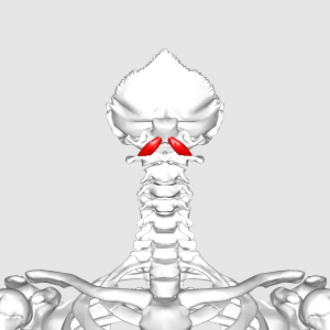
Branches from loop between C1 and C2 spinal nerves
Innervation of rectus capitis anterior muscle
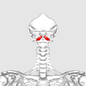
Flexion of head at atlanto-occipital joint
Action of rectus capitis anterior muscle
Anterior scalene
• Origin: Anterior tubercles of transverse processes of C3–C6.
• Insertion: First rib.
• Innervation: Anterior rami of C4–C6 spinal nerves.
• Actions:
Elevates the first rib during forced inspiration.
Bilateral contraction: Flexion of the neck.
Unilateral contraction: Lateral flexion of the neck.
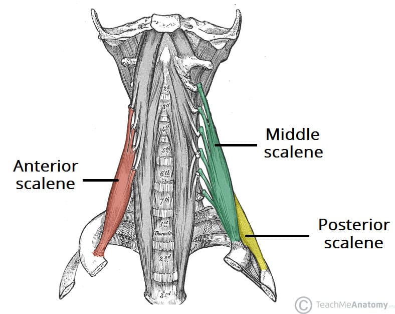
Anterior rami of C4-C6 spinal nerves
Innervation of anterior scalene muscle
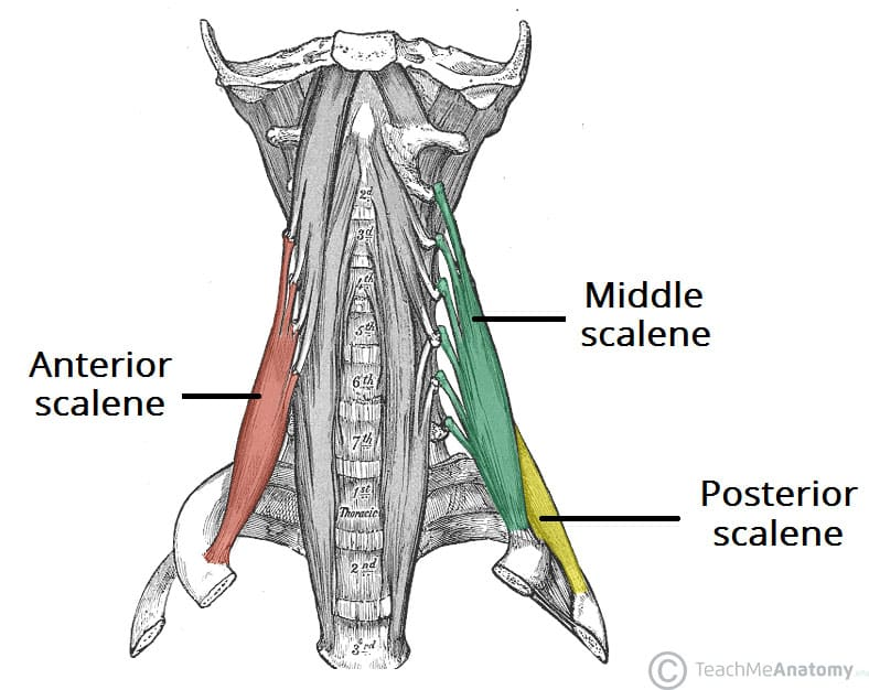
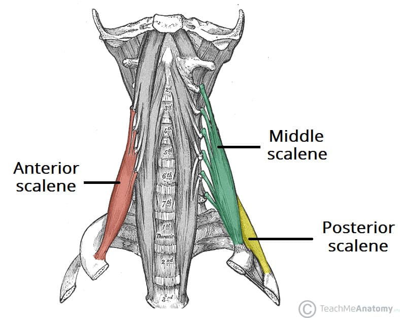
Elevates 1st rib during forced inspiration
Neck flexion and lateral flexion
Action of the anterior and middle scalene muscles
Rectus capitis lateralis
Middle scalene
Posterior scalene
Levator scapulae
Splenius capitis
Lateral prevertebral muscles
Rectus capitis lateralis
• Origin: Transverse process of C1 (Atlas).
• Insertion: Jugular process of the occipital bone.
• Innervation: Branches from the loop between C1 and C2 spinal nerves.
• Action: Stabilizes and flexes the atlanto-occipital joint
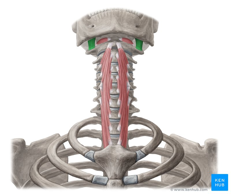
Branches from the loop between C1 and C2 spinal nerves
Innervation of rectus capitis lateralis
Stabilizes and flexes the atlanto-occipital joint
Action of the rectus capitis lateralis