Vascular Exam 2 - Peripheral Arterial
 Call Kai
 Learn
 Practice Test
 Spaced Repetition
 Match
 Flashcards
 Knowt Play
1/95

There's no tags or description
Looks like no tags are added yet.
Name | Mastery | Learn | Test | Matching | Spaced | Call with Kai | Chat |
|---|
No analytics yet
Send a link to your students to track their progress
96 Terms
Which vessel wall layer regulates arterial size?
Tunica media
Which vessel wall layer has valves?
Tunica intima
Which vessel wall layer contains the vasa vasorum?
Tunica adventitia

Upper Extremity
Three branches of the aorta:
_____ (_____) artery
_____ _____
_____ _____ artery
Brachiocephalic artery branches into the _____ _____ and _____ _____ artery
Right subclavian artery continues laterally, passing below the _____ to become the _____ artery
Axillary artery continues down the bicep to become the _____ artery
Brachial artery bifurcates at the _____ to become the _____ and _____ arteries
Brachiocephalic, Innominate, Left CCA, Left subclavian, right CCA, right subclavian, clavicle, axillary, brachial, elbow, radial, ulnar
Upper Extremity – About
_____ = border of first rib; becomes axillary
Can be found either above or below the clavicle depending on the patient
_____ = passes behind the clavicle
_____ = main blood supply to the arm, forearm, and hand
_____ = thumb side; terminal branch that forms the deep palmar arch
_____ = pinky side; terminal branch forms the superficial palmar arch
Palmar arches AKA _____ arch – not always complete!
_____ Test
Subclavian, Axillary, Brachial, Radial, Ulnar, volar, Allen’s
Allen’s Test is a physical exam that assesses the patency of the…
palmar arch
The state of being open, clear, and unobstructed
Patency
Palmar arch may be “_____”
If a palmar arch is _____, that means that it is not connecting back to the other side of the _____
incomplete, incomplete, wrist

Palmar Arches
_____ = supplied primarily by the radial artery with contribution from the ulnar artery
_____ = supplied primarily by the ulnar artery with contribution from the radial artery
If one of these arches were to be _____, the radial artery wouldn’t be connected to the ulnar artery and vice versa
_____ branch → main blood supply
Deep, Superficial, incomplete, Terminal
The difference between the right and left upper extremity is that the _____ _____ is a branch of the brachiocephalic artery and the _____ _____ is a branch directly from the aortic arch. All of the other branches after the _____ artery are the same on both sides.
right subclavian, left subclavian, subclavian

Lower Extremity – Thigh to Knee
Aorta bifurcates into the left and right _____ _____ arteries
Common iliac arteries bifurcate into _____ and _____ iliac arteries
_____ iliac artery AKA hypogastric artery
External iliac artery continues medially down the thigh passing below the _____ _____ to become the _____ _____ _____ (_____)
CFA bifurcates into the _____ (_____) and _____ _____ (_____) arteries
With ultrasound, we only really image the _____ profunda
SFA continues down the leg, then passes behind the _____ where it becomes the _____ artery (_____)
common iliac, internal, external, internal, inguinal ligament, common femoral artery, CFA, profunda, deep, superficial femoral, SFA, proximal, knee, popliteal, Pop A
Lower Extremity – Knee to Ankle
The _____ continues down the leg, then passes behind the knee where it becomes the popliteal artery (Pop A)
Popliteal artery terminates into a _____:
_____ _____ artery (_____) - Lateral
_____ _____ artery (_____) - Medial
_____ artery (_____) - Middle
^^^All part of the Tibial/Peroneal trunk
***The trifurcation is technically just _____ branches
A “trunk” which bifurcates into the _____ artery and _____
The _____
ATA crosses the ankle and becomes the _____ _____ artery (_____) on top of the _____
The DPA is the first major branch of the _____ _____ artery (arch)
The peroneal artery is _____ than the PTA
SFA, trifurcation, Anterior tibial, ATA, Posterior tibial, PTA, Peroneal, Pero A, two, peroneal, PTA, ATA, doralis pedis, DPA, foot, deep plantar, deeper
State the location of each vessel in regards to the leg and each other:
Anterior tibial artery (ATA) - _____
Posterior tibial artery (PTA) - _____
Peroneal artery (Pero A) - _____
Lateral, Medial, Middle
Lower Extremity – Foot
_____ _____ and _____ form the plantar arch
_____ plantar artery (distal branch of the DPA)
Medial and lateral plantar artery (branches of the _____)
Deep plantar and lateral plantar arteries unite to form the _____
Plantar and dorsal metatarsal arteries arise from the plantar arch and supply blood to the _____
Dorsalis pedis, PTA, Deep, PTA, arch, toes
Lower Extremity
Understand the “_____” of vessels (where does one vessel become the next):
EIA to CFA
At the _____ _____
CFA becomes the _____ artery and _____
The SFA is _____ to the initial origin of the profunda artery, but the profunda then passes behind (posterior) and deep to run more _____ or posterolaterally as it descends.
We only image the _____ profunda
We image the entire _____
SFA becomes the _____ artery
At the _____
Pop A and the trunk
_____, _____, _____
change, inguinal ligament, profunda, SFA, medial, medially, proximal, SFA, popliteal, knee, ATA, PTA, peroneal

Normal Sono Appearance for Peripheral Arteries
2D and color will be similar to your normal _____ appearance
_____ lumen free of defects
Smooth, thin, _____ walls
Complete, even _____ fill – varies with the _____ _____! _____ flow!
_____: waveform will dip below the baseline
Spectral Doppler
_____ or _____ waveform → _____ = normal, _____ = abnormal
Spectral _____ present
Normal peripheral arterial waveforms will have bigger _____ _____ and thinner _____ than carotid waveforms
carotid, Anechoic, echogenic, color, cardiac cycle, PULSATILE, Variations, Triphasic, multiphasic, Multiphasic, Monophasic, window, spectral windows, tracings
Arterial Hemodynamics
Factors that control arterial flow:
_____ _____
How much blood is being pushed out of the heart
Intraluminal wall _____
Are the vessels healthy and elastic or are this rigid and calcified
Total body _____
Arterial wall _____
The ability of an arterial wall to expand and recoil passively in response to changes in blood pressure and volume
Arteriolar _____ or _____
Remember that arterioles control _____ and _____
Cardiac output, resistance, resistance, compliance, vasoconstriction, dilation, vasoconstriction, dilation

At rest, arterioles are _____
With movement, arterioles are _____
Peripheral artery Doppler waveforms demonstrate _____
_____ is reflected by the projections above and below the _____
Blood flows towards the feet in _____ (away from the heart) (first above baseline peak), back towards the heart in _____ _____ (below baseline), then towards the feet again before the next _____ begins (above baseline)
Shown on image
A normal waveform should be _____ or _____
_____ waveforms are also considered multiphasic and normal
vasoconstricted, dilated, resistance, Resistance, baseline, systole, early diastole, cycle, triphasic, multiphasic, Biphasic
At rest…
_____ demand for blood supply
_____ occurs at the arteriole level
Little blood going to the _____ _____
Most blood is going to the _____/_____ circulation
_____ = organs
_____-resistance flow observed
During exercise…
Metabolic demand of skeletal muscles _____
Heart rate and cardiac output _____
_____ occurs at the arteriole level
Blood is shunted from the _____ to the _____ _____
Substantial _____ in blood volume in peripheral arteries and blood volumes may _____ significantly
As a result, arterial Doppler waveforms will exhibit _____ resistance flow
Low, Vasoconstriction, skeletal muscles, viscera, cerebral, Viscera, High, increases, increase, Vasodilation, viscera, skeletal muscles, increase, increase, low

During exercise…
Peripheral resistance dramatically _____ during this time, and blood flows in an _____ direction throughout the cardiac cycle. Distal intraluminal _____ is maintained during vasodilation due to the increase in _____. Remember Poiseuille’s law:
Q ↑ (_____) x R ↓ (_____) = distal P (_____ _____)
_____ in the lower vascular beds occurs quickly with the onset of even mild exercise
decreases, antegrade, pressure, volume, increases, decreases, no change, Vasodilation
Increase in blood flow to tissues or organs
Can be normal or abnormal
Normal = in response to exercise or digestion
Abnormal = in response to infection or heart failure
Two types:
Active – result of increased metabolic activity
Passive – result of obstructed blood flow
Hyperemia
Two types of hyperemia:
_____ – result of increased metabolic activity
_____ – result of obstructed blood flow
Active, Passive
When scanning the upper and lower arterial protocols, if the _____ waveform is normal, there is a pretty good chance that everything _____ is normal as well.
distal, proximal
The distal SFA will _____ similar to the distal ICA
It will look “_____” like the distal ICA when you add color
dive, junky
You will see various vessels at the proximal popliteal artery and the vessel will be going _____ the screen
The distal popliteal artery will be going _____ the screen
up, down
What is the difference between a stenosis and occlusion?
A stenosis is the _____ of a vessel that still lets some blood flow go through. An occlusion is the complete _____ of a vessel that doesn’t let any blood flow through it.
narrowing, blockage
According to Bernoulli, how are pressure and velocity related?
Inversely

What do we expect to find, in terms of waveform and velocity, when we encounter a stenosis?
At a stenosis, you should expect to see a smaller _____ and increased _____, which would cause the waveform to dip below the _____. The velocity in a stenosis _____. You generally see _____ flow (waveform) after a stenosis and a _____ velocity.
Proximal to stenosis: _____ _____ may or may not be altered depending upon the severity and where the sample is obtained
Stenosis: velocity _____, the flow becomes disorganized - spectral _____
Post-stenosis: _____ (e.g., flow reversal, eddy currents, vortices)
radius, resistance, baseline, increases, turbulent, decreased, flow pattern, increases, broading, turbulence
With proximal disease, what will happen to our waveforms downstream (distally)? Why?
The waveform will have a _____ _____ shape due to decreased blood flow through a stenosis.
An arterial occlusion or stenosis may result in decreased _____ distally (depends on the severity)
Vessels will _____ to maintain flow volume
If tissue demand exceeds available blood volume, _____ will result
Dilation of the vessels _____ pressure and resistance, but _____ flow volume
Peripheral resistance will _____ as a result (depending on severity and demand)
Delayed _____ _____ (_____) – time interval between onset of systole and PSV; how long does it take from the start of systole to reach max velocity
tardus parvus, pressure, vasodilate, vasodilation, decreases, increases, decrease, acceleration time, AT
What should a normal waveform look like in a peripheral artery?
_____phasic with an open spectral _____ and _____ tracing
Peripheral artery Doppler waveforms demonstrate _____
_____ is reflected by the projections above and below the baseline
Multi, window, thin, resistance, Resistance
Peripheral Arterial Disease (PAD) – Risk Factors
Family or personal history (Hx) of cardiovascular disease
Diabetes
Hypertension (HTN)
Hyperlipidemia
High cholesterol
Tobacco abuse
Age
Genetic disposition
PAD =
Peripheral Arterial Disease
Exertional pain that subsides with rest
Most common symptom of PAD
Result of inadequate blood flow with increased demand
Pain has predictable onset and resolution
The patient can tell you when the symptom occurs and how long the symptoms last
Symptom typically occurs distal to site of disease
Ex: If a patient has pain in their calf after walking for a certain/specific amount of time, it makes you believe that maybe they have peripheral arterial disease in either the popliteal artery or the distal SFA, which are proximal to the calf.
Claudication
Claudication
_____ pain that subsides with _____
Most common symptom of _____
Result of inadequate blood flow with _____ demand
Pain has _____ onset and resolution
The patient can tell you when the symptoms occur and how long the symptoms last
Symptom typically occurs _____ to site of disease
Ex: If a patient has pain in their calf after walking for a certain/specific amount of time, it makes you believe that maybe they have peripheral arterial disease in either the popliteal artery or the distal SFA, which are _____ to the calf.
Exertional, rest, PAD, increased, predictable, distal, proximal
The same symptoms but not due to vascular etiology
Neurogenic claudication; exertional pain due to neurogenic causes (spinal stenosis, nerve compression)
Could also be orthopedic in nature
Pseudo-claudication
Aorto/iliac obstruction or occlusion
A specific type of claudication
Typically a bilateral disease
Common in males
Profound exertional pain in hips, thighs, and calves
Pallor or coldness
Impotence
Leriche Syndrome
PAD signs and symptoms:
_____ _____ _____
_____ _____
Ischemic rest pain, Tissue loss
PAD – Signs and Symptoms
_____ = reduced blood flow to a part of the body
Ischemic rest pain
Pain at _____
Typically indicates _____ disease
_____ blood flow even at rest
Foot, toes, heel
Improved with _____ (elevation)
Ischemic rest pain is pain that occurs at _____, typically with the patient in the _____ position. This pain is relieved with _____ – when the patient _____ _____ or _____ their legs. This is due to an _____ in hydrostatic pressure that _____ perfusion.
Tissue loss
Ischemic _____
_____: the formation of an open sore or break on the skin or on the inner lining of an organ, caused when surface cells die and shed
Insufficient flow to maintain cell _____
_____ – GROSS
The death of body tissue caused by a loss of blood supply or a severe bacterial infection
Like when people start losing toes
Ischemia, rest, severe, Inadequate, dependency, rest, supine, dependency, stands up, lowers, increase, increases, ulceration, Ulceration, metabolism, Gangrene
Reduced blood flow to a part of the body
Ischemia
The death of body tissue caused by a loss of blood supply or a severe bacterial infection
Gangrene
6 P’s of Arterial Disease
_____
_____
Paleness, loss of color
_____
_____
Tingling, pins & needles
_____
Inability to move
_____/_____
Coldness
Pain, Pallor, Pulselessness, Paresthesia, Paralysis, Polar/Poikiloderma
Particles flowing in parallel lines or layers
Laminar flow
Laminar flow types:
_____ – middle layers are faster than the outer layers
In the arterial system, this type of flow is most commonly seen
_____ – all layers moving at the same velocity
_____ – non-laminar flow; chaotic
Parabolic, Plug, Turbulent

Turbulent flow occurs when _____ _____ is exceeded
_____ or greater
Turbulent flow seen _____ to a significant stenosis
Reynold’s number, 2000, distal

The _____ _____ (arrow) represents the aortic valve closure in a pulsatile flow waveform
dicrotic notch
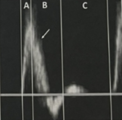
Pulsatile Flow
Early _____ (A)
Cardiac _____
Aortic valve _____
_____-velocity ejection of blood
Late _____ / Early _____ (B)
Rapid _____ and aortic valve _____
_____ _____ (arrow) represents the aortic valve _____ - variable in appearance and not always disernable
Pressure wave hits the distal peripheral _____
Possible hydraulic reflection of the pressure wave, which may result in temporary flow _____
Late _____ (C)
_____ velocity or no _____ flow
Depends upon the _____ of the vascular bed
Systole, contraction, opens, High, Systole, Diastole, deceleration, closes, Dicrotic Notch, closure, resistance, reversal, Diastole, Low, forward, resistance
Flow Characteristics of a Stenosis
Remember:
_____ = narrowing of the lumen, typically due to plaque
Changes in flow _____ at area of decreased diameter due to the disease
Velocity _____ at narrowest point
_____ at the exit (post-stenotic)
Changes in resistance _____ (downstream), depending on the severity of disease
Severe – flow and pressure remain _____ distal to the stenosis
Hemodynamically significant stenosis is defined as one that causes a notable reduction in flow and pressure (approximately _____% diameter reduction)
50% diameter reduction = _____% area reduction
Stenosis, direction, increase, Turbulence, distally, decreased, 50, 75

Normal Waveform
Rapid _____ _____
Systole flow _____
Projections _____ the baseline
Minimal _____ flow
_____phasic
acceleration time, reversal, below, diastolic, Multi

Flow Limiting Stenosis Waveform
_____ resistance
Slightly delayed _____ _____
Loss of flow _____
Forward _____ flow
_____phasic with _____ flow
Decreased, acceleration time, reversal, diastolic, Mono, diastolic

Significant Stenosis Waveform
Significantly decreased _____ _____
No _____ flow
_____ velocity
acceleration time, diastolic, Decreased
Ability of most tissues and organs to maintain constant blood flow despite changes in perfusion pressure
Sympathetic nervous system causes dilation of arterioles and capillaries when flow is decreased
Pressure is lowered, flow is increased
May be the result of:
Systemic hypotension (low blood pressure)
Stenosis
In short: if blood pressure drops, vasodilation occurs
Autoregulation
If blood pressure drops, _____ occurs.
vasodilation
Stenosis Interpretation
Velocity Ratio (Vr) – highest PSV in _____ divided by _____-_____ PSV
> 2:1 ratio increase = > _____% diameter reduction
> 4:1 ratio increase = > _____% diameter reduction
Stenotic PSV > 400 cm/s = > _____% diameter reduction
What to think: Is the stenosis PSV __x or __x greater than the pre-stenosis PSV?
stenosis, pre-stenosis, 50, 75, 75, 2, 4

What is the velocity ratio and percent diameter reduction if the stenosis PSV is 360 cm/s and the pre-stenosis PSV is 150 cm/s?
2.4, 50%

What is the velocity ratio and percent diameter reduction if the stenosis PSV is 240 cm/s and the pre-stenosis PSV is 110 cm/s?
2.2, 50%
What is the percent diameter reduction if the stenosis PSV is 409 cm/s and the pre-stenosis PSV is 183 cm/s?
75%
***Stenotic PSV > 400 cm/s = > 75% diameter reduction AUTOMATICALLY
What is the most common arterial pathology?
Atherosclerosis (plaque)
Most common arterial pathology
Thickening, hardening, and loss of elasticity of the arterial wall
Deposition of plaque between the intimal and media layers of the vessel wall
Systemic disease process
Symptomatic patients will have disease in multiple spots – CCA bifurcation, AO/Iliac, CFA bifurcation
Progression of disease results in increased severity, extension of the disease, and number of lesions
Atherosclerosis
Atherosclerosis
Most common _____ pathology
Thickening, hardening, and loss of elasticity of the arterial _____
Deposition of _____ between the _____ and _____ layers of the vessel wall
Systemic disease process
Symptomatic patients will have disease in _____ spots – CCA bifurcation, AO/Iliac, CFA bifurcation
_____ of disease results in increased severity, extension of the disease, and number of lesions
arterial, wall, plaque, intimal, media, multiple, Progression
Treatment for Atherosclerosis
Control _____ _____ – stop smoking, diet & exercise
Pharmacological – control blood _____, cholesterol, and diabetes
Beta blockers, diuretics, statins
Surgical – _____
Removal of atherosclerotic _____ from arteries
May be followed by _____ or _____ to keep lumen open
“_____ _____” – rotational atherectomy
Spinning brush that knocks off the plaque
risk factors, pressure, atherectomy, plaque, stent, angioplasty, Roto rooter
The process/condition of a blood clot forming inside a blood vessel and blocking or reducing blood flow
Result of disease progression
Stenosis eventually completely occludes
Acute occlusion can occur in less severe lesions resulting in minimal collateralization
Acute: rapid or new onset
An occlusion forms quickly meaning that the body has less time to respond by forming collaterals
Very dangerous
Thrombosis
Thrombosis
The process/condition of a _____ _____ forming inside a blood vessel and blocking or reducing blood flow
A _____ is the actual blood clot that forms
Result of disease _____
Stenosis eventually completely _____
_____ occlusion can occur in less severe lesions resulting in minimal _____
_____: rapid or new onset
An occlusion forms quickly meaning that the body has less time to respond by forming _____
Very _____
blood clot, thrombus, progression, occludes, Acute, collateralization, Acute, collaterals, dangerous
Obstruction of a vessel by a foreign substance
Most frequent cause is thrombus or plaque
Once plaque is dislodged, it is called this
Travels distally through the vessel until it lodges in an artery smaller than it is
Embolus
Embolus
_____ of a vessel by a _____ substance
Most frequent cause is _____ or _____
Once plaque is dislodged, it is called an _____
Travels distally through the vessel until it lodges in an artery _____ than the embolus
Obstruction, foreign, thrombus, plaque, embolus, smaller
Alternate pathways that may supply blood around an arterial obstruction
In most cases – small, already existing vessels that dilate over time in response to chronic vasodilation
In a chronic condition, the body already kind of knows what to do
Ex: patient with an SFA occlusion walks regularly for a year and the small vessel already present dilates in response to the perfusion need
May result in “false ok-ness,” meaning the vessel is diseased but because these are present, we see adequate flow. This is particularly tricky with physiologic testing.
Collaterals
Collaterals
Alternate pathways that may supply blood around an arterial _____
In most cases – small, already existing vessels that dilate over time in response to _____ vasodilation
In a _____ condition, the body already kind of knows what to do
Ex: patient with an SFA occlusion walks regularly for a year and the small vessel already present _____ in response to the perfusion need
May result in “false ok-ness,” meaning the vessel is diseased but because these are present, we see adequate flow. This is particularly tricky with _____ testing.
obstruction, chronic, chronic, dilates, physiologic

The image shows _____ collateralization.
Complete artery occlusion with _____ collateralization allows _____ blood flow, no significant _____, waveform remains similar or only minimally altered _____ to an occlusion.
adequate, good, adequate, vasodilation, distal

The image shows _____ collateralization.
Complete artery occlusion with _____ collateralization. _____ is decreased distally with results in _____ and therefore the waveform is altered exhibiting _____ flow.
insufficient, inadequate, Pressure, vasodilation, diastolic
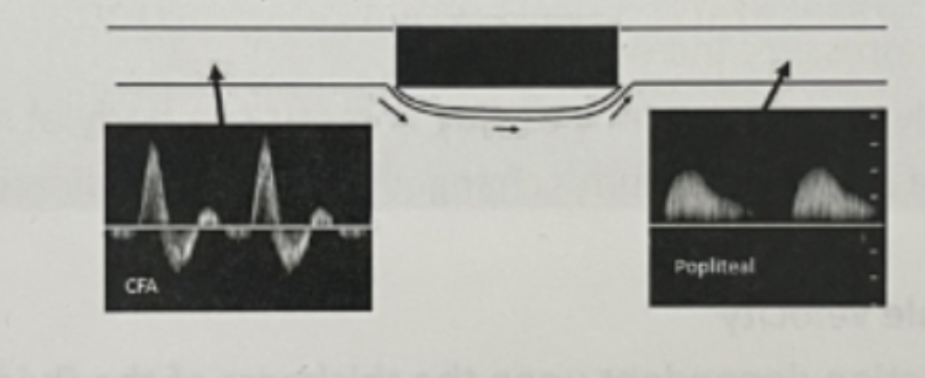
The image shows _____ collateralization.
Complete artery occlusion with very _____ collateralization. Arterioles are maximally _____ BUT _____ is insufficient to maintain flow during _____.
poor, poor, vasodilated, pressure, diastole
Aorto-Iliac Imaging
_____ – may present with exertional pain in the hips, thigh, or calf in the case of aorto-iliac disease
Aorta evaluation is often limited due to _____ _____
Evaluate the AO (proximal, mid, distal), then the internal and external iliac arteries
_____ – dilated 1.5x normal diameter
_____ – narrowing interpretation criteria is the same as peripheral arteries
_____ – segment of vessel with no detectable flow
_____ _____: aorto/iliac obstruction or occlusion, most common in males
Claudication, bowel gas, Aneurysm, Stenosis, Occlusion, Leriche Syndrome
An aneurysm occurs when the vessel is dilated __x the normal diameter
1.5
Inflammation of the arterial wall
Arteritis
Inflammatory condition of the palmar arch/digital arteries leading to small vessel obstruction; occurs primarily in men over 40 who are heavy smokers
AKA: Thromboangitis obliterans
Buerger’s disease
Rare, chronic autoimmune condition that causes inflammation of the aorta (arch) and its primary branches; most common in females
Takayasu’s arteritis
Serious inflammatory autoimmune disease that affects the larger arteries, especially in the head, eyes, and neck
Affects the temporal artery, which is a branch of the ECA
AKA: Giant cell arteritis
Temporal arteritis
Arteritis is most common in _____ vessels
Chronic inflammation may lead to _____
Arteritis is a type of _____ – group of disorders characterized by inflammation of blood vessels
smaller, thrombosis, vasculitis

Intermittent digital ischemia due to small vessel vasospasm secondary to cold exposure or emotional stress
Affected digits will turn white
Most common in females
Raynaud’s Phenomenon
Vasospastic Disorders
_____ – vasospastic disorder without underlying disease; digital and palmar arteries are without obstruction, and perfusion to the digits at rest is normal
_____ – vasospasm associated with underlying autoimmune disease or connective tissue diseases
Primary, Secondary
A sudden, abnormal tightening or constriction of an artery's muscular wall that reduces or temporarily blocks blood flow to downstream tissues
Vasospasm
Popliteal artery is compressed by the medial head of the gastrocnemius muscle
May result in stenosis, aneurysm, or occlusion
Typical in young athletes who present with exertional pain
Popliteal Artery Entrapment
Compression of the neurovascular bundle by the shoulder structures
Symptoms occur when the arm is in a certain position
Primarily neurogenic; minority of cases caused by compression of the vasculature
Thoracic Outlet Syndrome (TOS)
True or False: Patients with an aneurysm in one location are more likely to have an aneurysm elsewhere
True
The most likely complication of a peripheral artery aneurysm
Embolization

A _____ artery aneurysm seems to be the most common location of embolization (intraluminal thrombus) in the lower extremities
popliteal
Ulnar artery aneurysm due to repetitive blunt trauma
Hypothenar hammer syndrome
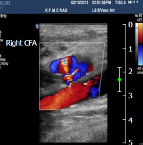
Pocket of swirling flow adjacent to an artery
Typically the result of an injury or tear to the vessel wall such as…
Percutaneous arterial catheterization
Penetrating trauma
Bypass graft anastomosis blowout
There are no walls encapsulating the structure
Treatment: thrombin injection and manual compression
Pseudoaneurysm
Option for “longer” areas of occlusion/severe disease
Basically a man made collateral
Alternative to amputation or limb loss
Bypass grafts

Bypass Graft Types:
_____ _____ _____
When the patient’s own GSV is tied to the artery to redirect blood around the area of blocked flow
_____
Man made material graft that is sewn into the proximal and distal arteries that redirects blood around the area of blocked flow
Great saphenous vein, Synthetic
Vein Graft
_____ _____ _____ is typically used
Assess during a “vein mapping” exam
Most common GSV graft: _____ to _____
Most likely the _____ _____ _____
May be left in place or _____
_____ _____ – vein left in “normal” position, valves are excised (taken out)
_____ – vein is flipped valves are left alone since they are in the direction of flow; will stay open
Great saphenous vein, femoral, popliteal, common femoral artery, reversed, In situ, Reverse
Synthetic Graft
PTFE or Dacron material
From the CFA to _____ or sometimes _____
Typically do not go below the _____ to avoid risk of kinking with motion of the patient’s leg
SFA, popliteal, knee

Graft Images
Bottom right: the double _____ lines of the vessel are very characteristic of a synthetic graft
A bypass graft runs pretty _____
Middle left: the bypass graft is the _____ vessel with _____ in it. The _____ vessel is the _____ vessel.
As you can see, the bypass graft runs _____ to the _____ vessel
When you assess a bypass graft, you will do the same protocol that you would do on a _____ vessel
The only additional images that you would obtain would be of the proximal and distal _____
This is where the bypass graft is sewn in or connected to the _____ vessel
This is shown in the bottom left image
echogenic, superficially, top, color, bottom, native, superficial, native, native, anastomosis, native
Guide wire introduced through the skin via needle through the CFA
Guide wire is advanced to the stenotic area where a catheter with a long balloon is pressed over the guidewire and into the area of plaque
Balloon is inflated to a fixed size, plaque is “smooshed” against the vessel walls, and a wire stent is opened
Balloon is withdrawn, stent is left
Complications: restenosis, neointimal hyperplasia, thrombosis, stent shift, aneurysm at stent edge
Percutaneous Transluminal Angioplasty
BPG =
Bypass graft
Which vessel is a continuation of the anterior tibial artery (ATA)?
Dorsalis pedis artery (DPA)
DISTAL to a significant stenosis →
DILATION
Your tissues always need blood flow. The demand for blood flow distal to a stenosis does _____ change, but _____ occurs so that the perfusion of blood to the tissues continues.
NOT, vasodilation