Lecture 15: Common Pediatric Orthopedic Conditions
 Call Kai
 Learn
 Practice Test
 Spaced Repetition
 Match
 Flashcards
 Knowt Play
1/127

There's no tags or description
Looks like no tags are added yet.
128 Terms
What is scoliosis?
Structural, lateral, rotated curvature of the spine; Cobb angle ≥10° (<10° = spinal asymmetry)
What age range is “adolescent” scoliosis?
Age ≥10 years at diagnosis
Early-onset scoliosis (EOS) = onset before age 10
What does idiopathic mean?
Other causes have been ruled out: congenital (vertebral malformation), neuromuscular, syndromic

Scoliosis Etiology
Probably a genetic component
Gene has not yet been identified
Neuro pathoanatomy
Chiari 1 malformation, syrinx
Abnormal paravertebral musculature
Denervation leads to thoracic scoliosis in animals
Vertebral body growth asymmetry
Unsure if cause or effect
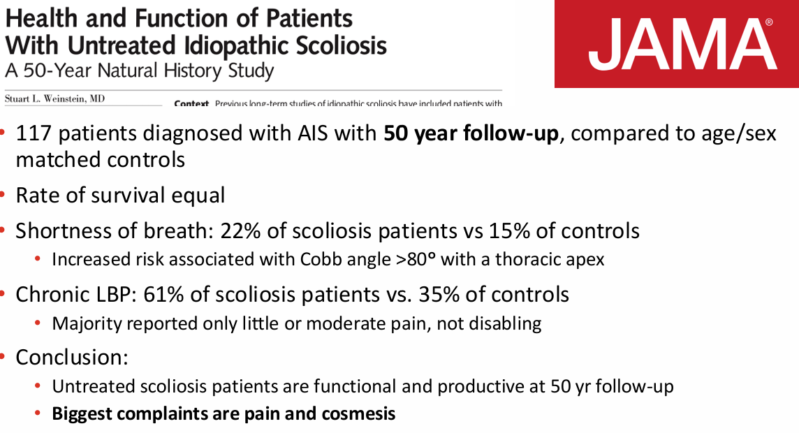
Scoliosis Natural History
Early long term studies of scoliosis led to common misconception that all types of idiopathic scoliosis lead to disabling back pain and severe cardiopulmonary compromise
Studies included non-idiopathic causes of scoliosis and early onset scoliosis
New studies have proven otherwise
Evaluation of Scoliosis Physical Exam
Shoulder height
Pelvic obliquity
Trunk shift
Waist asymmetry
Adams forward bend test
Evaluation of Scoliosis Neuro Exam and Reflexes
Neuro exam
Sensation
Motor
LE Reflexes
Patellar Tendon: L2,3,4
Achilles Tendon: S1, S2
Abdominal reflexes
T8-T12
Foot deformity
Radiology of Scoliosis
Standing spine radiographs
AP and Lat first assessment
PA subsequently
Must be able to see pelvis and proximal femur
Risser Sign, TRC, proximal femoral physis
EOS
low dose radiation system, results in 3d imaging
AP Radiology Results of Scoliosis
Curves
Right thoracic is normal
Left thoracic is abnormal
Cobb angle
End vertebra to end vertebra
5 degree variability
Risser sign
growth sign (5 means done growing, 0 means a lot of growth to go)
Tri-radiate cartilage
Growth measure
Sagittal Profile Radiology Results of Scoliosis
• Hyperlordosis at apex

Who needs an MRI?
A typical curves and/or symptoms
Left curve
Kyphosis
Abnormal appearing curves
Abnormal exam
Headaches
Rapid progression
Progression despite bracing
Early onset or juvenile
Neural axis abnormalities in ~20% of early onset (infantile/juvenile) curves; much less common in typical AIS
Treatment Determination Based on
Decision making based on age/skeletal maturity + curve magnitude
Chronological age, bone age, Risser sign, menarchal status all play a role
Sanders Stage now most widely used
Sanders Stage
Skeletal maturity scoring system that correlates more strongly with curve behavior than Risser sign or bone age
Uses hand x-ray
Stage 3 – start of Adolescent Rapid stage (peak height velocity, Risser 0, open TRC)
Curves >20 deg at Stage 2 and >30 deg at Stage 3 are at greatest risk for progression

Surgery recommended at _ degrees or more
50
Curves followed for avg 40 years
Curves <30 deg at maturity tended not to progress
Curves between 50-75 degrees at skeletal maturity with thoracic apex progressed the most
This is where the 50 deg threshold for surgery comes from
When to brace?
Curve magnitude
25-40 degrees
Age
At least 2 years of growth remaining (13-14 boys, 11-12 girls)
Risser sign (0,1,2), menarchal status
What type of brace?
TLSO
Night time bending brace (Providence, Charleston)
Conditions Effecting Brace failure
Male patients
74% of curves progressed >5 deg; 46% progressed to >50 deg or had PSF
Compliance rate 38%
Obese patients
Curve progression to >45 deg in 45% of obese patients vs. 28% normal weight
3.1 more likely to have unsuccessful result if obese
Thoracic vs. Lumbar Curves
Rate of PSF or progression >50 deg 34% in thoracic curves vs. 15% in lumbar curves
Risser stage
Rate of progression highest (70%) for Risser 0 with open TRC, especially if curve >30 deg (even with 18 hours of daily brace wear)
Risser 0, TRC open should wear brace 18 hours/day
Bracing should start with curves <30 if Risser 0, especially if TRC open
The “monitor” effect
Patients who are given feedback about their compliance with brace wear, wear their brace more
13.8hrs/day vs. 10.8 hrs/day
25% who were counseled progressed to >50 deg or surgery vs. 36% who were not
Surgical Treatment Scoliosis
When
Curve >50 degrees
Why
Weinstein data about curve progression
Cosmesis
Age
At least 10 yo
Alternative
Vertebral body tethering: skeletally immature, curves 30–65°, failed bracing (FDA HDE 2019)
Early Bracing Through History
20 years ago
SRS guidelines → brace curves 25-40° in order to stop progression to surgical magnitude
Previous studies have shown that 33% of patients with curves 25-29º braced at Risser 0 and open TRC still go on to surgery (Karol, et al. JBJS 2016)
Now want to try to brace earlier so it doesnt get to that point
Early brace treatment (curve <25º and open TRC) may reduce curve progression and the need for surgery
Only 8% of patients with fulltime brace progressed to surgery vs. 33% in previous study of patients with curves 25-29º and open TRC
Early Bracing For Scoliosis
Often results in curve improvement
Decreases risk of curve progression
Usually amenable to nighttime bracing
Now treating younger patients with smaller curves
How has Dr. Meagan’s practice changed?
Start bracing curves at 20°, especially in younger patients
I will brace curves <20° when there is clinical deformity
Mostly use nighttime braces for small curves; will change to full time if curve progression
Arthrogryposis
Non-progressive congenital disorder of unknown etiology that presents with multiple rigid joints leading to stiffness and severe limitation in motion in all 4 limbs
NOT a specific diagnosis but a clinical finding
1:3000 births
Arthrogryposis Etiology
Joint development is almost always normal during early embryogenesis
Decreased fetal movements occurring as a result of fetal or maternal abnormalities
Lack of movement = extra connective tissue around the joint = contracture
Arthrogryposis Pathophysiology
Due to failure of movement, joints that had initially formed with normal articular cartilage and joint space undergo changes
Joint capsules thickened
Fibrotic
Joint creases absent
Skin and subcutaneous tissue adherent to bone
Bursal areas poorly formed
Amyoplasia
Amyoplasia (Classic Type of Arthrogryposis)
“No Muscle Growth”
Most common type
Comprises 1/3 of all cases
Usually Symmetric in limbs with trunk spared (84% symmetric involvement)
Normal intelligence
Incidence: 1 in 10,000 live births
Amyoplasia Symptoms
Shoulders internally rotated and adducted
Elbows extended
Wrists flexed and ulnar deviated
Fingers stiff
Thumb in palm
Hips may be dislocated
Knees can be extended or flexed
Equinovarus feet
Multidisciplinary Clinics for Amyoplasia
Pediatric neurologist
Pediatric orthopedic surgeon
Hand orthopedic surgeon
Geneticist
Occupational therapist
Physiotherapist
Orthotic professionals
Goals of Treatment for Arthrogryposis
Orthopaedic goals of treatment
Independent ambulation
Plantigrade standing and walking
Increase active motion where muscle transfers are possible
Reposition stiff joints for functional advantage
Independent function of upper extremities for ADL
Treatment Arthrogryposis: Hip
Typical deformity is flexion, abduction, and external rotation
Hip Dislocations - Teratologic (14-42%)
Also…
Hip Dysplasia (65%)
Data and studies are sparse
Early reduction of both unilateral and bilateral dislocations is typically recommended
Exception is when child demonstrates little active hip movement and ambulation is unlikely; positioning for sitting is more appropriate
Knee Flexion Contractures Arthrogryposis
Knee deformities occur frequently with an incidence between 38 – 85%
Flexion contractures are reported as more common
More disabling than extension contractures
Only 50% of patients with knee flexion contractures became community ambulators
8 of 9 patients with knee extension contractures were community ambulators
Surgical Treatment of the Knees
Flexion contracture
consider ambulatory potential and quad function
Surgical options
Extensive soft tissue release
Posterolateral
Posteromedial
Anterior, including ACL/PCL
Femoral shortening
Extension osteotomy (in mature individuals)
Ilizarov
Hyperextended knee
Completely dislocated unlikely to respond to nonoperative management
Surgical options
Anterior release of knee capsule and patella, V-Y plasty of quad
Femoral shortening
Advantageous in that weakening of the quad may be avoided
Typical Foot Deformities Arthrogryposis
Clubfoot
High rate of recurrence (73%)
Avg 2.5 procedures per foot
Equinocavus
Can be extremely rigid
Seems to have a lower tendency to relapse
Vertical Talus
Low incidence (3-7%)
Arthrogryposis Foot Deformities Treatment Goals
Plantigrade foot for standing
Arthrogryposis Treatment: Clubfoot
More severe and rigid than idiopathic clubfoot
Was described as “Typically resistant to casting”
Operative treatment
Extensive posterior medial release
Residual Adductus: lateral column shortening
Residual Equinovarus: Decancellation of talus (Verebelyi-Ogston procedure)
Talectomy
Ilizarov distraction
Salvage: Triple arthrodesis
Arthrogryposis Treatment: Congenital Vertical Talus
Dobbs Method an option
Can be very stiff
Operative Treatment
Reduction of navicular on talus
Release Ant Tib Tendon
Release Tibionavicular and talonavicular ligaments
Lengthening of toe extensors
Release of equinus contracture after securing talonavicular reduction
Arthrogryposis Treatment: Scoliosis
Incidence varies greatly 20-67%
Neuromuscular type curve
Very stiff curves
May result from pelvic obliquity
Correct hip contractures first
Bracing not effective
May be used to delay surgery in young
patients
Surgical treatment for progressive curves
PSF
Instrumentation to Pelvis
Spina Bifida
Spectrum of neural tube defects
Incidental radiographic findings to significant central and peripheral nervous system dysfunction
Neurologically normal
Spina bifida occulta
Meningocele (may have mild deficits)
Neurologic deficit
Lipomeningocele
Myelomeningocele
Caudal regression

Spina Bifida Incidence
3.6 per 10,000 live births or 1 in every 2,758 live births
Result of defect in neural tube closure that occurs by 26-28 days of life
Risk factors
Folate deficiency – 400mcg folic acid daily
Maternal Hyperthermia
Maternal Diabetes
Valproic Acid
Prenatal screen – maternal serum AFP
MOMS Trial
In utero repair of myelomeningocele pioneered at Vanderbilt in 1997
Management of Myelomeningocele Study
183 patients randomized to receive in utero repair vs. post-natal closure
Stopped early due to clear benefits of in utero repair
Decreased need for shunt
Improved level of impairment
Risk of pre-term birth
School-age follow-up
More children walked independently (93% vs. 80%)
Benefit does not last as patient age
Fewer shunt placements (49% vs. 85%)
Spina Bifida Multidisciplinary Care

Spina Bifida Goals of Orthopedic Care
Correct deformities in order to allow patients to maintain mobility / ambulation and independence for ADLs
Monitor for spine balance and deformity, maintain stable posture for sitting / standing
Monitor neurologic status, look for signs of neurological deterioration
Spina Bifida Assessment of Neuro-segmental Level

Spina Bifida Classification

Spina Bifida Thoracic Level Goals
May ambulate with orthoses (RGO, HKAFO) as children, most transition to wheelchair full time
Ability to transfer, use upper limb to power wheelchair
Scoliosis
Watch for hip and knee contractures, keep feet plantigrade for positioning in wheelchair
Spina Bifida Upper Lumbar Level Goals
Household walkers with braces / forearm crutches, transition to wheelchair for longer distances
Watch for hip and knee contractures, keep feet plantigrade
Spina Bifida Lower Lumbar Level Goals
Community walkers
Below knee orthoses (AFOs)
May need forearm crutches / wheelchair for long distance
Foot deformity common
Factors affecting the potential for ambulation Spina Bifida
Neurologic level
Hip deformity
Scoliosis
Foot and ankle deformity
Age – as kids get bigger and heavier their mobility changes
Obesity
Spina Bifida Neurologic level affecting ambulation
Level of involvement:
L4 and below → mostly community walkers
L3 and above → non-functional walkers
Muscle group strength
Quadriceps (knee extension) most important
80% of patients with grade 4 or 5 strength – community or household walkers
88% with grade 0-2 strength – non-functional walkers
Iliopsoas (hip flexor)
Spina Bifida Hip Deformity affecting ambulation
Status of hips (dislocated or located) does NOT correlate with ability to ambulate
Spina Bifida Obesity affecting ambulation
60%
Increased risk with higher lesions
Interferes with walking, transfers and self-esteem
Spina Bifida - Hips
Subluxed / dislocated hips caused by muscle imbalance / paralysis around the hip joint
Emotional and complicated topic:
If the hips remain dislocated, will my child be able to walk?
Won’t the hips hurt if they aren’t put back in the socket?
Our therapist is nervous to work with our child because his / her hips are dislocated
What are the outcomes of hip reduction surgery?
76 patients – showed that there was NOT an increase in ROM or ability to walk in children who had their hips reduced
No benefit in reduction of pain or need for bracing
20 patients low-lumbar level spina bifida with hip dislocation on one side only – walking speed in children with dislocated hip was the same as those without
High rate of complications associated with hip reduction surgery
Loss of motion in 30%
Pathologic fracture in 17%
Decline in ambulatory ability in 36%
Bracing spina bifida
Many children with spina bifida (except low sacral lesions) require some type of bracing:
Prevent deformity
Correct flexible deformities
Facilitate independent mobility
Protect insensate limb
Bracing spina bifida when to start
When babies show interest in pulling to stand
May not require if intact ankle function
Started earlier if need to maintain alignment of foot / leg
Bracing needs change over time
Common Orthopedic Conditions Spina Bifida
Hip
Knees
Rotational Deformities
Foot & Ankle
Clubfoot
Calcaneus / Calcaneovalgus
Ankle Valgus
Cavus / Varus / Cavovarus
Spine
Abnormally formed vertebrae
Complete or partial absence of sacrum
Kyphosis
Lordosis – usually due to hip flexion contracture
Scoliosis
Common Orthopedic Conditions Spina Bifida - Hip and Knee
Hip
Flexion Contracture
Can lead to pelvic tilt and compensation through the spine (excessive lordosis)
Dislocation
Knee
Contractures – thoracic / high lumbar level
Flexion
If ambulatory and >20º of contracture, can interfere with brace fitting and ambulation
Non-ambulatory patients tolerate more of a contracture, but severe contracture can interfere with sitting and transferring
Extension
Maybe congenital or acquired
Makes sitting very difficult for non-ambulatory patient
Common Orthopedic Conditions Spina Bifida - Rotational Deformities
Femoral anteversion
Tibial torsion - more common in spina bifida patients
Many rotational deformities improve over time
Not always true in patients with spina bifida due to abnormal gait and activity levels
Common Orthopedic Conditions Spina Bifida - Foot and Ankle
Almost universal in patients with spina bifida
Lead to problems with walking, brace fitting, shoe wear, skin breakdown / pressure sores
Goal for ambulatory patients is a flexible, braceable, plantigrade foot
Goal for non-ambulatory patients is a foot that fits in braces and shoes and does not interfere with positioning in wheelchair
Common Orthopedic Conditions Spina Bifida - Clubfoot
Most common deformity – 30-50%
Rigid and resistant to treatment
Initial treatment is similar to non-neuromuscular clubfeet – Ponseti method
Recurrence rate 70%
May require comprehensive release around 10-12 months
Common Orthopedic Conditions Spina Bifida - Calcaneus / Calcaneovalgus
17-35%
Most common in low lumbar level
Problems with gait, pressures sores from weight-bearing on hee
Common Orthopedic Conditions Spina Bifida - Ankle Valgus
Caused by weakness of gastroc / soleus leading to abnormal gait pattern
Alters forces across the ankle joint
More stress on the lateral side → growth slows → medial side continues to grow normally
Valgus deformity of the ankle joint
Common Orthopedic Conditions Spina Bifida - Cavus / Varus / Cavovarus
8-17%
Sacral level or lipomeningocele
Elevated arch (cavus), hindfoot varus
Can lead to pressure sores on lateral foot
Common Orthopedic Conditions Spina Bifida - Spine
Variety of deformities:
Abnormally formed vertebrae
Complete or partial absence of sacrum
Kyphosis
Lordosis – usually due to hip flexion contracture
Scoliosis
Spina Bifida Kyphosis
10%
More common in thoracic level
Progressive – caused by absence of the posterior elements of the spine along with weak muscles
Sharp kyphotic deformity = gibbus
Can cause skin breakdown
Not amenable to bracing → wheelchair modifications
Surgical treatment for progressive deformity, skin breakdown
Removing some of the vertebrae and correcting the deformity with rods and screws
Spina Bifida Scoliosis
52-70% by age 6, as high as 100% for thoracic level
Paralytic type curves – long “C” curve, pelvic obliquity
Skin breakdown from uneven pressure when sitting
Not amenable to standard bracing → wheel chair modifications
Surgery for progressive deformity, pelvic tilt causing seating problems
Posterior spinal fusion
Significant risks
Decreased trunk flexibility/mobility (can affect ADLs – self-cathing, etc.)
Wound healing
Deep infection – 30-40%
Legg-Calvé-Perthes Disease Epidemiology
Incidence = 1 in 1200
Regional variability:
Low in South and East Asia
Higher in Northern Europe, USA and Canada.
Male:Female = approximately 5 : 1
Most common in ages 4-9
Bilateral 10-15%
Usually asynchronous
If same stage, consider other diseases
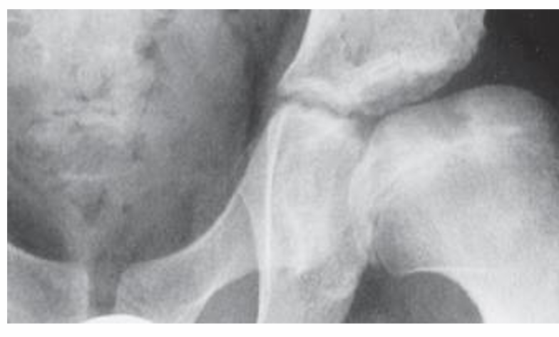
Perthes Disease Pathophysiology
blood supply of femoral head decreased
bone becomes ischemic
bone revascularizes
dead bone is resorbed

Waldenström Classification
Classification system of Perthes Disease
Initial/Ischemic
Fragmentation
Reossification
Healed/Residual

Ischemic Stage of Perthes Disease
Disruption of the blood supply to the femoral head
Deep layer of the epiphyseal cartilage undergoes cell death
There is increased mineral content (Sclerosis)

Revascularization/Resorption Stage of Perthes Disease
Increased osteoclast activity leads to bone resorption.
Mechanical strength of the femoral head decreases during revascularization.
Mechanical loading can lead to femoral head deformation.
Walking is 2.5X body weight and running is 4.5X body weight

Reossification Stage of Perthes Disease
New bone formation occurs.
There is remodeling of the femoral head with mixed osteoclastic and osteoblastic activity.
Femoral head mechanical strength increases

Healed (Residual) Stage of Perthes Disease
Bone reformed but with deformity
Clinical Presentation of Perthes Disease
Most often present with a limp.
Limp worse with activity
Insidious onset of groin pain (sometimes thigh, buttock, knee, back pain)
Stiffness
Self-limited activity
Signs of Perthes Disease
Antalgic gait and Trendelenburg gait (active phase)
Reduced hip motion during initial and early fragmentation stage
Variable presentation in late stages
Loss of extreme range of motion to adduction and flexion contractures
Lateral Pillar (The Herring Lateral Pillar Classification)
Classification of predicted outcome of Perthes Disease

Treatment Perthes Disease
Treatment options are controversial
Non-operative
NWB
Casting (Petrie)
Bracing
Traction
Operative
Femoral varus osteotomy
Salter Osteotomy
Combined
Hip joint distraction
Epiphyseal drilling
Greater trochanter epiphysiodesis
Hip Preservation
Hip arthroscopy
Surgical hip dislocation with osteochondroplasty
Greater trochanter distalization
PAO for secondary dysplasia
Valgus Osteotomy
Head Reduction Osteotomy
Perthes Disease Summary
Perthes disease involves a process of femoral head avascular necrosis with revascularization, resorption, reossification and remodeling.
Age of onset, lateral pillar classification and amount of femoral head involvement are associated with prognosis.
younger it happens better they heal
Interventions are directed towards femoral head containment to improve femoral head remodeling
Children <6 years old should be managed non-operatively.
Operative management can be beneficial for older patient.
Containment procedures during early stages may improve hip sphericity at skeletal maturity
Developmental Dysplasia of the hip
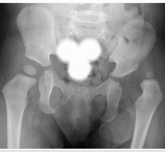
Shallow socket and/or under-covered femoral head
Malformation of anatomic structures that have developed normally during the embryologic period
Broad spectrum of disease ranging from physiologic immaturity to subluxation to frank dislocation
Epidemiology DDH Incidence
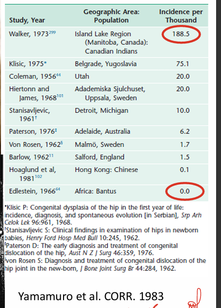
15% with sonographic findings at birth
1-2% with hip dysplasia
0.1% with frank hip dislocation
Referral rates 4-5% of births either due to risk factors or abnormal exam
4-5:1000 treated for dysplasia
Epidemiology DDH Risk Factors
Female (8 in 10 cases)
First born (6 in 10 cases)
Family Hx (12x relative risk)
Feet First/Breech (2-27% with dysplasia)
Environmental Factors
tight swaddling
Physical Exam DDH - Outcomes
Normal
Instability but not full dislocation (“subluxatable”)
Dislocatable reduced but dislocatable (“Barlow positive”)
Dislocated but reducible (“Ortolani positive”)
Fixed and dislocated (“stable, but dislocated”)
Physical Exam of DDH
The physical findings are a spectrum, which vary with the severity of the pathology and the age of the child
Asymmetric thigh fold
Limited abduction
Positive Galeazzi
Pistoning
Trendelenburg gait

Dislocatable Hip (Barlow Positive)
Reduced in the resting position
Dislocatable with the Barlow maneuver
May stabilize spontaneously or with treatment
May convert to dislocated without treatment

Dislocated But Reducible Hip (Ortolani Positive)
Dislocated in the resting position
Reduces with abduction and anteriorly directed force on trochanter
This will be lost, usually by 6-8 weeks of age
Will not resolve without treatment

Dislocated And NON-Reducible Hip
Dislocated and NON-reducible in the resting position:
Asymmetric thigh fold
Limited abduction
Positive Galeazzi
Pistoning
Trendelenburg gait

Late Presenting Dislocation
Swayback deformity
Trendelenburg gait
Contralateral hemipelvis drop
Lean towards dislocated hip

Imaging DDH
Ultrasound
Obtained 6-8 weeks of age unless clinical instability
Reduces false positive rate
Selective, not universal, screening: breech, family history, or abnormal exam (AAOS)
X-ray – AP Pelvis
Can replace ultrasound from ~4 months of age
Sufficient ossification
Treatment DDH
Pavlik Harness – 0-6 months
Maintains hip flexion/abduction to allow for remodeling
Hip Abduction brace
Second line treatment
Closed or open reduction and spica cast

Prevention DDH
Healthy Hip Swaddling
Avoid forced hip extension
Allow hips to flex and abduct
Sleep sacks
Baby carriers

Clubfoot Etiology
Congenital dysplasia of all musculoskeletal tissues distal to the knee
1-2 in 1000 live births
Higher in Hawaiians, Polynesians and Māori
2:1 M:F ratio
Bilateral 50%
4 classes of clubfoot
Postural
Idiopathic
Neurogenic
Syndromic
Club Foot Associated Conditions
Arthrogryposis
Diastrophic dysplasia
Amniotic bands
Freeman-Sheldon syndrome
Prune Belly
Möbius syndrome
Spina bifida
Down syndrome
Larsen’s syndrome
Tibial/fibular hemimelia
Initial Evaluation Club Foot
Distinguish from “postural” clubfoot
flexible will correct back on its own
Evaluate for associated anomalies/neuromuscular conditions
Determine the initial severity
Dimeglio Score
Way of grading club foot
4 components: equinus, varus, derotation of the calcaneo-forefoot block, adduction of forefoot
Assessed in terms of reducibility
4-point scale:
90-45° = 4
45-20° = 3
0-20° = 2
0- -20° = 1
< -20° = 0
4 “bonus points
Posterior crease
Medial crease
Cavus
Muscle condition
Components of club foot
equinus
sagittal plane
varus
frontal plane
derotation of the calcaneo-forefoot block
horizontal plane
adduction of forefoot
horizontal plane
Dimeglio Score Grades
Four categories of feet based on score
Grade 1 – 0-5, benign, 20%
Grade 2 – 6-10, moderate, 33%
Grade 3 – 11-15, severe, 35%
Grade 4 – 16-20, very severe, 12%
Ponseti Method of Treating Club Foot
Casting
Foot manipulated gently for 1-3 minutes
Long leg plaster cast is applied and kept in place for 5-7 days
Knee in 90° of flexion
5-6 cast changes
Percutaneous TAL if needed (85%)
Thumb is positioned over the lateral aspect of the talar head and fingers correct the forefoot
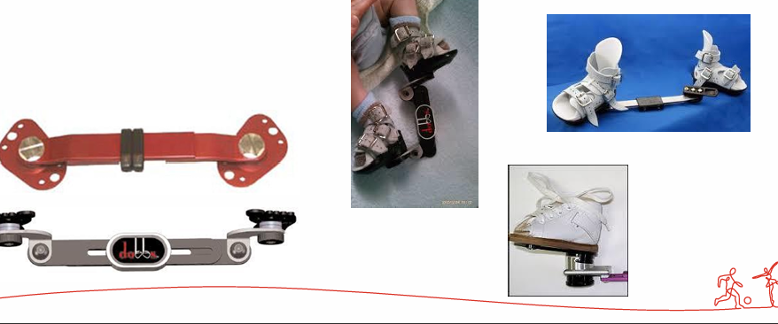
Abduction Orthosis
Feet placed shoulder width apart, 70° abduction/external rotation, 5-10° dorsiflexion
Worn 23 hrs/day for ~3 months, then nights and naps until age 4–5
French Method of Treating Club Foot
Introduced to North America by Dimeglio in 1996
Daily physical therapy sessions to stretch and relax medial tissues with gradual manipulation
Taping applied to maintain passive motion
30 min/foot, daily by PT x 3months, then by parents at home, continued until walking
CPM machine
Ponseti vs French Method Study
267 feet treated with Ponseti Method; 119 treated with French Physiotherapy Method
Initial correction obtained in 95% of patients in both groups
Perc TAL – 73% in Ponseti, 32% in French
Recurrence – 37% in Ponseti, 29% in French
Trend toward improved results using the Ponseti Method at 2 years, but not significant
Cerebral Palsy
Brain injury in period of early brain growth (<2yo)
Disordered motion, sensation, perception, cognition, communication and behavior
Epilepsy
Secondary MSK problems
Cerebral Palsy Epidemiology
Most common cause of chronic physical disability in children
2-3/1000
Causes:
Prematurity (main)
Intrauterine infection, hemorrhage, thromboembolism/stroke
Hypoxia
Brain malformations
Primary lesion in the brain – non-progressive/static encephalopathy
Progressive secondary MSK consequences
Cerebral Palsy Classification - Topography
Unilateral
Monoplegia
Hemiplegia (30%)
UE>LE
Focus on hand and foot
Bilateral
Diplegia (50%)
LE>UE
Focus on legs/walking
Triplegia
Quadriplegia (20%)
Focus on seating, hips, spine
