Rad 130 Lower Limb II
 Call Kai
 Learn
 Practice Test
 Spaced Repetition
 Match
 Flashcards
 Knowt Play
1/193

There's no tags or description
Looks like no tags are added yet.
194 Terms

Identify this projection
AP Tibia/ Fibula (Right)

What joints must be included?
Knee and ankle joints

The bottom of the IR must be just below what structure?
Lateral malleolus

The top of the IR must be just above what structure?
Apex of the patella

Identify this projection
Mediolateral Tibia/ Fibula (Right)

What structures must be superimposed for this to be lateral?
Femoral condyles

Identify this projection
AP Knee (Right)

what is the centering point for the beam?
½ inches inferior to apex of patella
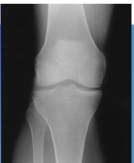
Identify this projection
AP Knee (Right)

State the acceptance criteria
Distal femur, proximal tib/fib
Femorotibial joint space open
No rotation

Identify this projection
Mediolateral knee (Right)

What structures must be superimposed?
Femoral condyles

How much is the knee flexed?
20-30 degrees

What is the angle AND direction of the CR?
5-7 degrees cephalic

Identify this projection
Mediolateral knee (Left)

State the acceptance criteria
Distal femur, proximal Tib/fib, patella in lateral profile
Patellofemoral and knee joint spaces open
Femoral condyles superimposed

Identify this projection
AP lateral oblique Knee (Right)

How much is the entire leg rotated laterally?
45 degrees

Identify this projection
AP lateral oblique Knee (Right)

State the acceptance criteria
Distal femur, proximal Tib/Fib, with patella superimposing lateral femoral condyle
Tibia superimposing the fibula

How will you recognize this projection?
The fibula is superimposed by the tibia
The patella is superimposing the lateral femoral condyle

Identify this projection
AP medial oblique knee (Right)

How much is the entire leg rotated medially?
45 degrees

Identify this projection
AP medial oblique knee (Right)

State the acceptance criteria
Distal femur, proximal Tib/Fib with the patella superimposing the medial femoral condyle
Lateral condyles are well demonstrated
The fibular head and neck are not superimposed

How will you recognize this projection?
Patella is superimposing medial condyles
Fibula is free of superimposition
Lateral condyles are well demonstrated

Identify this projection
PA Intercondylar fossa (Camp Coventry method)

What is the relationship of the CR to the lower leg?
45 degrees caudal

Where does the CR enter?
Center of knee joint (popliteal fossa)

Where does the CR exit?
Apex of the patella

Identify this projection
Knee- Intercondylar fossa (tunnel view)

State the acceptance criteria
Intercondylar fossa, articular facets, and knee joint spaces open
No rotation

How will you recognize this projection?
The large open joint space
The tibial eminences are prominent

Identify this projection
Lateraomedial patella (Left)

How much is the knee flexed?
5-10 degrees

Where is the CR directed?
Mid-patellofemoral joint space

Identify this projection (common name)
Sunrise view

Identify this projection (technical name)
Tangential sunrise/skyline patella
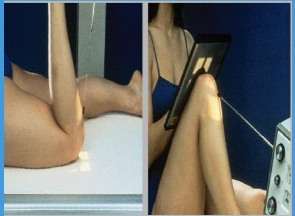
How much is the CR angled for the Settegast method?
20 degrees cephalic

Identify this projection
AP Tibia/ Fibula (Right)

Identify this projection
AP Tibia/ Fibula (Right)

Identify this projection
Mediolateral Tibia/ Fibula (Left)

Identify this projection
AP Distal Femur

How much rotation is required of the entire leg for this projection?
5 degrees internal

Identify this projection
Mediolateral Distal Femur

Identify this projection
Mediolateral proximal femur

What bony landmark is used to center for this projection?
ASIS

How many projections are performed during routine radiographs of the Femur?
4 projections

Identify this projection
Superoinferior patella (Hobbs method)

Identify this projection
Axial sunrise patella (Settegast Method)
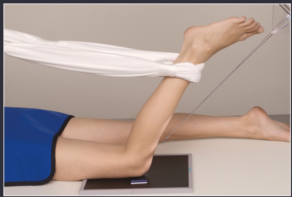
Identify this projection
Axial sunrise patella (Hughston Method)

Identify this projection
Axial sunrise patella (Inferosuperior axial projection)

Identify this projection
Axial sunrise patella (Merchant Method)

Identify this projection
Lateral Patella (Left)

Identify this projection
PA patella

Identify this projection
Knee- Intercondylar fossa (Beclere Method)

Identify this projection
Knee- Intercondylar fossa (Holmblad Method)

Identify this projection
Knee- weight bearing (Rosenberg Method)

Identify the structure in blue
Base (Superior border)

Identify the structure in blue
Apex (Inferior border)

Identify the structure in blue
Base (Superior border)

Identify the structure in blue
Posterior surface (Smooth)

Identify the structure in blue
Anterior surface (Rough)

Identify the structure in blue
Apex (Inferior border)

Identify the structure in blue
Patellofemoral joint

Identify the structure in blue
Patellar Surface
(Intercondylar sulcus/ Trochlear groove)

Identify the structure in blue
Lateral epicondyle

Identify the structure in blue
Lateral condyle

Identify the structure in blue
Medial condyle

Identify the structure in blue
Medial epicondyle

Identify the structure in blue
Intercondylar fossa (notch)
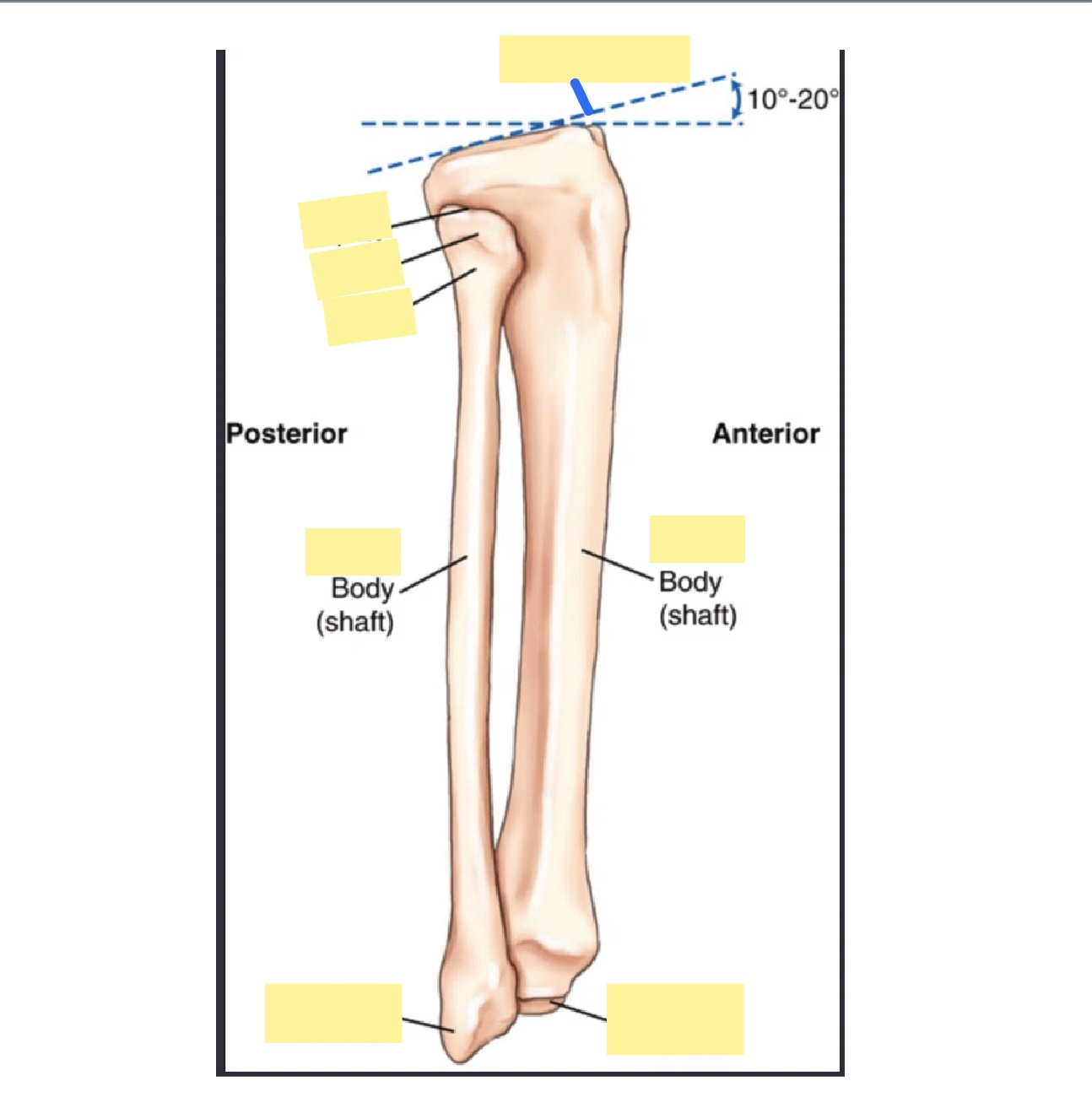
Identify the structure in blue
Tibial plateau

Identify the structure in blue
Apex of fibula

Identify the structure in blue
Head of fibula

Identify the structure in blue
Neck of fibula

Identify the structure in blue
Fibula

Identify the structure in blue
Tibia

Identify the structure in blue
Lateral malleolus

Identify the structure in blue
Medial malleolus
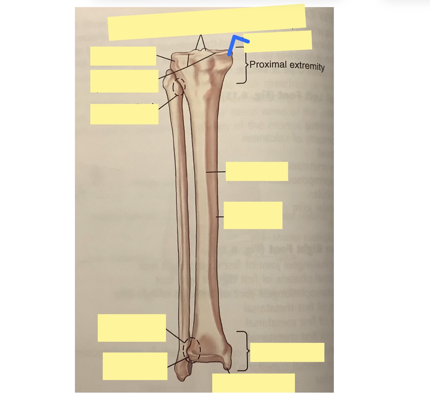
Identify the structure in blue
Medial condyle

Identify the structure in blue
Intercondyloid eminence

Identify the structure in blue
Lateral condyle

Identify the structure in blue
Articular facets (tibial plateau)

Identify the structure in blue
Proximal tibiofibular joint

Identify the structure in blue
Anterior crest

Identify the structure in blue
Body of tibia

Identify the structure in blue
Fibular notch of tibia

Identify the structure in blue
Distal tibiofibular joint

Identify the structure in blue
Medial malleolus

Identify the structure in blue
Adductor tubercle

Identify the structure in blue
Medial epicondyle

Identify the structure in blue
Medial condyle

Identify the structure in blue
Lateral condyle

Identify the structure in blue
Lateral epicondyle

Identify the structure in blue
Intercondylar fossa (Notch)

Identify the structure in blue
Popliteal surface

Identify the structure in blue
Lesser trochanter

Identify the structure in blue
Femoral neck

Identify the structure in blue
Femoral head

Identify the structure in blue
Greater trochanter