Review LOWER EXTREMITY AND HIP/SHOULDER GIRDLE
 Call Kai
 Learn
Learn Practice Test
 Spaced Repetition
 Match
Match Flashcards
 Knowt Play
1/17

There's no tags or description
Looks like no tags are added yet.
Name | Mastery | Learn | Test | Matching | Spaced | Call with Kai | Chat |
|---|
No analytics yet
Send a link to your students to track their progress
18 Terms
PA patella
Patient is prone and internal rotation the knee about 5 degrees. CR in perpendicular to IR.

Mediolateral projection
Lateral patella
Patient will be in a lateral recumbent position with the affected side down, flex knee 5-10 degrees and make sure the body and knee are in a true lateral position.
CR is perpendicular to IR.
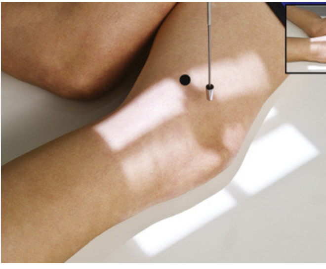
Tangential (merchant method)
Patient is supine with knees flexed 40 degrees over end of the table, place IR on edge of legs, below the knees. CR should be angled caudal 30 degrees from horizontal plane

Tangential (Hughston)
Patella
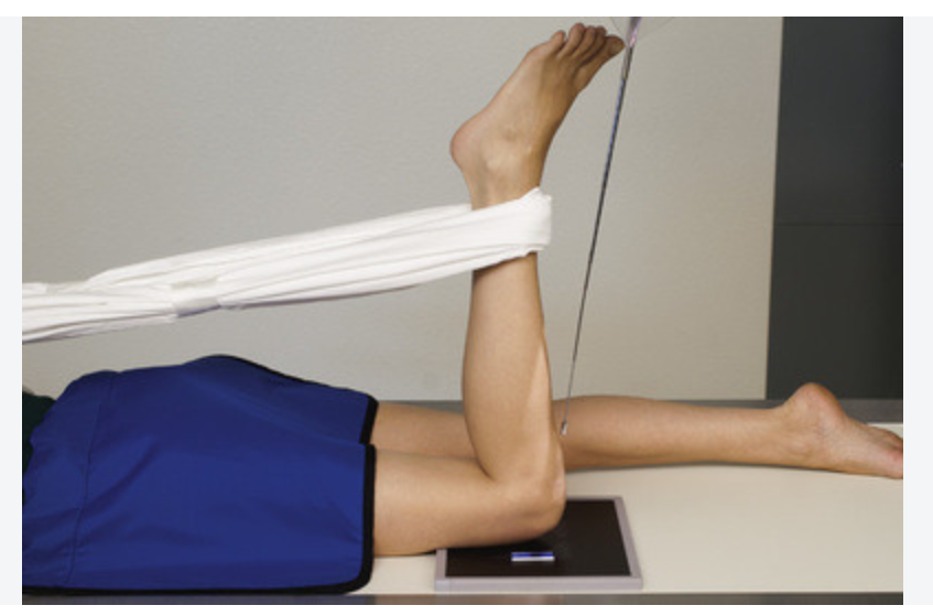
Patient in prone position with IR under knee, flex knee between 50-60 from full extension of lower leg. CR should be angled 45 degree cephalad.

Tangential (Settegast)
Patella
Patient should be prone with IR underneath knee, flex knee to a minimum of 90 degrees. CR should be tangential to the patellofemoral joint space which is about 15 to 20 degrees. SID 40 inches.
Superinferior sitting method (Hobbs)
This is with the patient sitting in a chair with the IR placed underneath their knees, knees should be flexed with feet placed slightly underneath chair. The CR should be perpendicular to IR with a minimum SID of 48-50 inches to reduce magnification

Camp Coventry
Intercondylar fossa
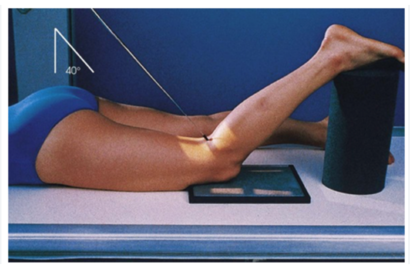
Prone position with 40-50 degree flexion and center IR at knee joint
Holmblad
Intercondylar fossa
Have the patient knee on the Xray table and ask them to lean forward 20 to 30 degrees, direct CR to midpopliteal crease

Beclere
Intercondylar fossa
Patient is supine on the table with knee flexed 40-45 degrees. Place support under the leg with the IR firmly against posterior thigh and lower leg. Direct CR perpendicular to lower leg which is 40-45 degree cephalad angle.

When do we utilize a tube angle for a knee series and why?
We use a tube angle for the lateral knee which is about a 5 to 7 degree angle. We need to angle for the lateral because the condyles are not perfectly level
-the medial condyle is lower than the lateral, therefor put a slight angle will get the femoral condyles superimposed and open the joint space.
Compare what is best visualized on a mortise oblique versus a 45 degree oblique.
How is a 15 degree mortise oblique of the ankle positioned.
A 15 degree oblique ankle will show the entire mortise joint open while the 45 degree
oblique ankle will show the tibiofibular joint open.
Internal rotation shoulder
Whats in profile & epicondylar plane?
Epicondylar plane is perpendicular, lesser tubercle in profile
External rotation shoulder
Whats in profile & epicondylar plane?
Epicondylar plane is parallel, greater tubercle in profile
AP Neutural shoulder
Whats in profile & epicondylar plane?
Epicondylar plane is 45degrees to IR, both greater and lesser tubercles are superimposed by humeral head, can see the relationship between humeral head to glenoid cavity.
Grashey
Will show glenoid cavity
35-45 degree rotation

Non-Trauma Lawrence
Coracoid process and lesser tubercle
-shows anterior dislocation: humeral head under coracoid process
-posterior dislocation: humeral head under acromion
Trauma Lawrence
Lateral view of proximal half of humerus and scapulohumeral joint visualized through the thorax without superimposition
Scapular y lateral for trauma shoulder
Lateral scapula, and scapulohumeral joint