Nervous System chp 12
 Call Kai
 Learn
 Practice Test
 Spaced Repetition
 Match
 Flashcards
 Knowt Play
1/129

There's no tags or description
Looks like no tags are added yet.
130 Terms
What organs & structures make up the nervous system?
Brain & spinal cord
Sensory receptors (eyes, ears, etc.)
Nerves that connect to other body systems
What are the two types of cells in nervous tissue?
Neurons (dendrites/cellbody (soma)/axon
Neuroglia (glial cells) pitcrew help w mainnance of neurons
What is the main function of neurons?
Intercellular communication
Transmit information between cells; release neurotransmitters
What are neuroglia (glial cells)?
- Supporting cells that help neurons survive & function.
essential to:
Support neuron survival
Help neurons work properly
Preserve structure of nervous tissue.
What are the two main anatomical divisions of the nervous system?
Central nervous system (CNS); when damaged no going back
Peripheral nervous system (PNS); MAYBE restore
What makes up the central nervous system (CNS)?
Brain & spinal cord.
Processes & coordinates sensory info (from outside of body)
Sends motor commands to control peripheral organs ex. SmoothMuscle
Responsible for higher functions: memory/learning/intelligence/emotion.
made of nervous tissue/CT/BV
What makes up the peripheral nervous system (PNS)
All nervous tissue outside the CNS & is including enteric nervous system (ENS)
Delivers sensory info to CNS.
Carries motor commands from CNS → peripheral tissues.
Includes cranial nerves & spinal nerves (nerves)
What are nerves?
Bundles of axons surrounded by connective tissue & blood vessels.
Carry sensory info & motor commands.
Cranial nerves: connect to brain.
Spinal nerves: attach to spinal cord.
the PNS
What are the two functional divisions of the PNS?
Afferent division: Carries sensory information; from receptors in peripheral tissues→CNS.
Efferent division: Carries motor commands from CNS→ muscles, glands, & adipose tissue.
What are receptors & effectors?
Receptors: Detect changes or respond to stimuli.
(neurons/specialized cells/single cells/sense organs eyes or ears)
Effectors: Target organs that respond to motor commands.
What are the two divisions of the efferent PNS?
- Somatic nervous system (SNS): Controls skeletal muscle contractions (voluntary or involuntary)
- Autonomic nervous system (ANS): Controls subconscious (can’t control) actions, smooth muscle, cardiac muscle, and gland secretions.
What are the two divisions of the autonomic nervous system (ANS)
Sympathetic:stimulating effect; prepares body for action (fight or flight)
Parasympathetic: relaxing effect; supports resting & digesting function
What is the enteric nervous system (ENS)?
abt 100 mill neurons in walls of digestive tract.
Initiates & coordinates digestive (visceral) reflexes locally.
Can function w/o instructions from CNS
Can be influenced by ANS
(BOOK KEEP: as many neurons or more than spinal cord; use same neurotransmitters as brain)
What are neurons?
Basic functional units of the nervous system.
Send & receive signals.
Responsible for communication/info processing/control.
What does the neuron's cell body (soma) house?
Nucleus & Nucleolus.
Perikaryon: Cytoplasm of the cell body.
Mitochondria: makes energy.
RER & ribosomes: Make proteins (to keep func)
(rough endoplasmic reticulum)

What are dendrites & what is their function?
Short/highly branched extensions from the cell body.
Dendritic spines get info from other neurons: (small extensions on dendrites)
abt 80–90% of a neuron's surface area.
spirit fingers

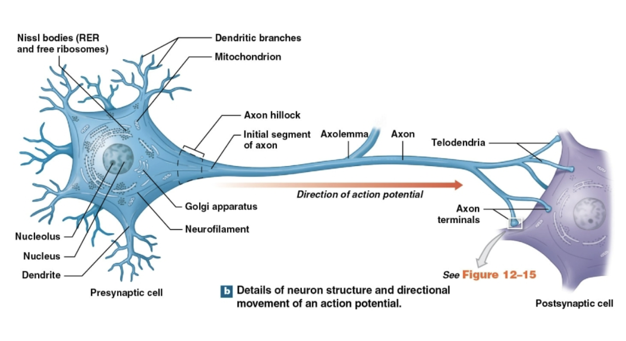
What is an axon & what is its function?
Single/long extension from the cell body.
Propagates (carries) electrical signals (action potentials) away from the cell body (to axon)
Axoplasm: Cytoplasm inside axon (BK: have neurofibrils/neurotubules/enzymes/organelles)

What are the main structures of an axon?
Axolemma: Plasma membrane of axon.
Initial segment: Beginning of axon (tgt w/ Axon hillock;look at tgt)
Axon hillock: Thick region connecting cell body to→initial segment.
Collaterals: Branches of the axon.
Telodendria: Fine extensions of distal/end of axon.
Axon terminals (synaptic terminals): Tips of the telodendria; communicate w/ other cells
What is axonal (axoplasmic) transport?
Movement of materials between cell body & axon terminals; along neurotubules in axon.
moved by mitochondria & kinesin & dynein (transport proteins) → axon
What are the four structural classifications of neurons?
Anaxonic.
Bipolar.
Unipolar.
Multipolar.
What are anaxonic neurons?
Small neurons.
more than 2 cell processes that look similar; (might all be dendrites) & axon not obvious
Found in brain & special sense organs (smell/ taste/vision/balance)

What are bipolar neurons?
Have 1 dendrite & 1 axon; separated by cell body
Small & rare.
Found in special sense organs; sight, smell, & hearing.

What are unipolar neurons?
Axon & dendrites are fused together; elongated process
Cell body sits off to side (out of way)
Most sensory neurons in the PNS are unipolar.

What are multipolar neurons?
Have 1 long axon & 2 or more dendrites.
Common in CNS.
All motor neurons that control skeletal muscles are multipolar.
most common discussed

What are the three functional classifications of neurons?
Sensory neurons (to brain)
Motor neurons. (send back down)
Interneurons.
What are sensory neurons?
(afferent neurons) afferent fibers carry sensory info from receptors to → CNS.
unipolar.
Cell bodies grouped in sensory ganglia. (group of neurons)
VISCERAL = ORGANS
2 types of sensory neurons?
Somatic sensory neurons: Monitor the external environment. (muscle)
Visceral sensory neurons: Monitor the internal environment. (organs)
What are the three types of sensory receptors?
Interoceptors: Monitor internal systems (dig & urine) & senses (stretch, deep press, pain)
Exteroceptors: Monitor external environment (temp) & complex senses sight, smell, & hearing.
Proprioceptors: Monitor position & movement of skeletal muscles & joints.
What are motor neurons and the 2 kinds?
(efferent neurons)
Carry instructions from CNS → to peripheral effectors (w/ efferent fibers)
Somatic motor neurons: Control skeletal muscles.
Visceral motor neurons: Control smooth muscle/cardiac muscle/glands/adipose tissue
VISCERAL=AUTONOMIC NS !SAMEEE!
What are preganglionic & postganglionic fibers?
Carry signals from CNS → autonomic ganglia→ visceral effectors.
Preganglionic fibers: Extend from CNS→autonomic ganglion.
Postganglionic fibers: Extend from ganglion → target.
—-O—-
What are interneurons?
most in brain & spinal cord (some in auto ganglia)
Connect sensory neurons & motor neurons (between)
Distribute sensory info to coordinate motor activity.
Help w/ memory/planning/learning
What are neuroglia & what do they do?
Also called glial cells.
Support & protect neurons.
Help maintain structure of nervous tissue.
abt 50% volume of nervous system.
What are the four types of neuroglia in the CNS?
Astrocytes.
Ependymal cells.
Oligodendrocytes.
Microglia.

What are astrocytes and what are their functions?
large cell bodies w/ many extensions.
Maintain blood-brain barrier (BBB).
repair damaged nervous tissue w/ scar tissue
Guide neuron development.
Control environment around neurons (nutrients/ions/concentration)
(give structural support for CNS.)
What are ependymal cells and what do they do?
Line ventricles of the brain & central canal of the spinal cord.
Make & monitor cerebrospinal fluid (CSF).
Have cilia that help circulate CSF.
like rocks around beaver lake
What are oligodendrocytes and what do they do?
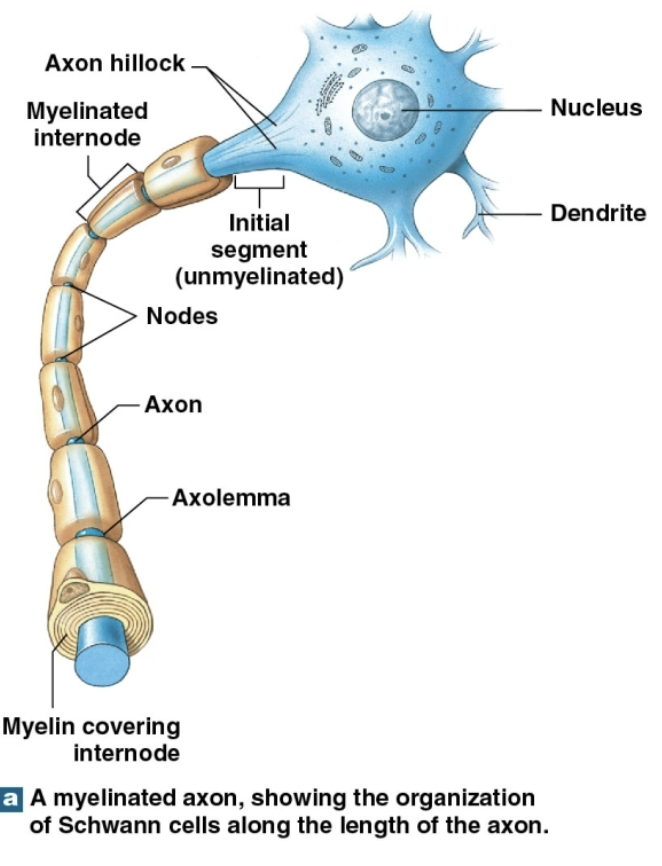
Form the myelin sheath around axons in the CNS (struct framework)
Myelin insulates axons.
Increases speed of action potentials.
make nervous tissue appear white.
What are internodes and nodes of Ranvier?
Internodes: Myelinated parts of axon.
Nodes of Ranvier: Gaps between the myelinated segments; Let action potentials move faster along myelinated axons.(MORE BOUNCING)
What is the difference between gray matter & white matter in the CNS?
BOOK KEEP
Gray matter: has neuron cell bodies/dendrites/unmyelinated axons.
White matter: has many myelinated axons.
What are microglia and what do they do?
Smallest/least numerous neuroglia.
Move through nervous tissue.
Clean up cellular debris/waste/pathogens.
phagocytosis
What are the two types of neuroglia in the PNS?
Satellite cells.
Schwann cells.
insulate CB & axons

What are satellite cells and what do they do?
Surround ganglia (groups of neuron cell bodies).
Regulate fluid environment around neurons.
(O2/CO2/nutrients/neuotrans.)
What are Schwann cells and what do they do?
Form myelin sheath around axons in the PNS.
Also called neurolemmocytes.
One Schwann cell covers only one section of one axon.
Many Schwann cells work tgt to cover an entire axon.
repair after injury

What is the neurolemma?
The outer surface of a Schwann cell.
more wrapping of burrito=more insulation faster bouncing/saltitory conduction
What happens when a peripheral nerve is injured?
Wallerian degeneration: axon distal to injury degenerates
Schwann cells help form a pathway for new axon growth;wrap around axon
axon sends growth buds→ strarts to regrow on pathway
neuron can regain some function.
Why is nerve regeneration limited in the CNS? (ACTUALLY STOPPED HERE)
Astrocytes make scar tissue and release growth blocking chemicals that block axon regrowth.
What is a membrane potential?
An electrical diff. between inside & outside of a cell.
made by movement & distribution of ions across cell membrane.
important for neurons.
What are the three main types of membrane potential changes?
Resting membrane potential: Electrical charge (mem poten) of a cell when its resting.
Graded potential: Temporary, localized change in membrane potential caused by a stimulus (crawling up)
Action potential: Electrical impulse that travels along the axon; made by grade potential (actual up spike)
What is the resting membrane potential?
electrical charge across the membrane of a resting cell.
A typical neuron resting is abt −70 mV.
inside of the cell is negative compared to outside.
How are ions distributed differently inside & outside a neuron?
Outside (ECF): High concentrations of sodium (Na⁺) & chloride (Cl⁻).
Inside (cytosol): High concentrations of potassium (K⁺) & neg. charged proteins.
What three things help create the resting membrane potential?
Different ion concentrations inside & outside the cell (cytosol is diff from EF in chemical & ionic comp)
cell (plasma) membrane is selectively permeable.
membrane has diff levels of permeability for diff ions.
What are chemical, electrical, and electrochemical gradients? LOOK AT/SEMI STUDY STARTING HERE
Chemical gradient: Diff in ion concentration across the membrane (Na⁺ & K⁺)
Electrical gradient: Diff in electrical charge across the membrane; cytosol is neg compared to extracell. fluid (membrane seperates the 2)
Electrochemical gradient: Combined chemical & electrical forces acting on an ion→move across mem. (form of Poten. energy)
What are current & resistance across a cell membrane?
Current: Movement of electrical charges; to get rid of potential diff
Resistance: How much the membrane restricts ion movement.
Higher resistance means less current.
What is an equilibrium potential?
membrane potential where there’s no net movement of a particular ion across the membrane.
K⁺ equilibrium potential = −90 mV.
Na⁺ equilibrium potential = +66 mV.
Why does potassium (K⁺) have a major effect on the resting membrane potential?
membrane is highly permeable to K⁺.
K⁺ moves out of cell.
so helps make inside of cell negative.
K⁺ equilibrium potential (−90 mV) is close to the resting potential (−70 mV).
(Na+ perm is low=small effect on RMP)
What is the sodium-potassium exchange pump? (ACTuAlLY STUDY RN)
Uses ATP to move ions across cell membrane.
Pumps 3 Na⁺ out of cell.
Brings 2 K⁺ into cell.
Maintains sodium & potassium concentration differences.
Helps stabilize resting membrane potential (-70 mV)
3:2 ratio (Na⁺ & K⁺ channels are either passive or active)
Why does the membrane potential change?
stimulus causes certain ion channels to open or close.
changes the membrane's permeability to ions.
Movement of ions changes electrical charge across the membrane.
What are passive ion channels?
Always open; let ions to move across membrane.
Help maintain resting membrane potential.
permeability changes w/ conditions
(leak channels)
What are active ion channels?
Open & close in response to specific stimuli.
Most are closed when cell is at resting membrane potential
(gated channels)
What are the three types of active (gated) ion channels?
Chemically gated.
Voltage-gated.
Mechanically gated.
What are chemically gated ion channels?
ligand-gated channels.
Open when a specific chemical binds to them.
ex: Acetylcholine (ACh).
Found on neuron cell bodies & dendrites
What are voltage-gated ion channels?
Open or close in response to changes in membrane potential.
Found in neuron axons & muscle cell membranes.
Have activation (open when stimmed) & inactivation gates (close to stop ion movement)
What are the three possible states of voltage-gated ion channels?
Closed but able to open.
Open (activated; when stimmed).
Closed and unable to open (inactivated; to stop ion movement).
What are mechanically gated ion channels?
Open in response to physical changes or distortion of the membrane.
Found in sensory receptors; respond to touch, pressure, and vibration.
What are graded potentials?
Temporary, localized changes in membrane potential.
Caused by stimuli that open gated ion channels.
Can’t travel far from stimulation site.
Can involve depolarization or hyperpolarization.
How does a graded potential occur when sodium channels open?
A stimulus opens chemically gated Na⁺ channels.
Na⁺ enters the cell.
The inside becomes less negative.
This causes depolarization (MP rises)
What is depolarization?
inside of the cell becomes less negative.
happens when positive sodium ions (Na⁺) enter the cell.
What is repolarization?
membrane returns toward its normal resting potential after a change.
inside becomes more negative again.
What is hyperpolarization?
The inside of the cell becomes more negative than its resting potential.
positive ions move out not into cell
Happens when positive potassium ions (K⁺) leave the cell.
What are the characteristics of graded potentials?
Strongest at the site of stimulation.
Become weaker as they travel farther away.
Spread through local currents.
Can be depolarizing or hyperpolarizing.
Stronger stimuli produce larger changes.
trigger cell func./ACh causes graded potential at motor end plate is neuomuscular junction
What is an action potential? (HERE?)
nerve impulses
An electrical impulse propagated along an axon;changes in mem. potential→ affect entire excitable mem.
Begins at the initial segment of the axon.
Does not weaken as it travels.
Starts when a graded potential reaches (depolarizes) axolemma to threshold.
What is the threshold for an action potential?
The membrane potential that must be reached to trigger an action potential.
In an axon, is −60 to −55 mV
What is the all-or-none principle?
An action potential either happens completely or does not happen.
If threshold is reached (by stim in change of MP), an action potential occurs.
All action potentials are the same size, regardless of stimulus strength.
What are the steps of an action potential?
Depolarization to threshold: The membrane reaches threshold.
Voltage-gated Na⁺ channels open: Sodium enters the cell, making the inside positive (depolarization)
Na⁺ channels inactivate and K⁺ channels activate: Potassium leaves the cell, beginning repolarization.
Return to resting potential: K⁺ channels close and the resting membrane potential is restored.
What happens during depolarization of an action potential?
Voltage-gated Na⁺ channels open.
Na⁺ rushes into the cell.
inside changes from negative to positive.
membrane rapidly depolarizes
to much Na+
What happens during repolarization of an action potential?
Na⁺ channels become inactivated (close at +30mV)
Voltage-gated K⁺ channels open.
K⁺ moves out of the cell.
The inside becomes negative again.
loss on K+
What happens when an action potential returns to resting potential?
Voltage-gated K⁺ channels begin closing.
K⁺ may continue leaving briefly.
The membrane may become temporarily hyperpolarized.
The resting membrane potential is restored.
Action potential is over
What is the refractory period?
time from the beginning of an action potential until the membrane returns to its resting state.
During this time, the membrane cannot respond normally to another stimulus.
What is the difference between the absolute and relative refractory periods?
Absolute refractory period: No new action potential can occur because Na⁺ channels are open or inactivated.
Relative refractory period: A new action potential can occur, but only with a stronger stimulus; till membrane potent. stabalizes; Na+ regain resting
What is the role of the sodium-potassium pump after an action potential?
Uses ATP to restore ion concentrations over time.
Moves 3 Na⁺ out of the cell.
Moves 2 K⁺ into the cell.
Maintains the concentration gradients needed for future action potentials.
What is action potential propagation? HERE TOO
The movement of an action potential along an axon.
Occurs through a series of steps.
Can be continuous or saltatory.
What is continuous propagation?
Occurs in unmyelinated axons.
The action potential moves along one segment at a time.
Local currents depolarize the next segment.
Slower than saltatory propagation.
straight line;one direction ——
What is saltatory propagation?
Occurs in myelinated axons.
The action potential appears to bounce from one node of Ranvier to another.
Faster than continuous propagation.
Requires less energy.
Depolarization occurs at the nodes.
What two factors affect the speed of action potential propagation?
Myelination: Myelinated axons conduct signals faster.
Axon diameter: Larger diameter means less resistance and faster conduction
What are the three types of axon fibers?
Type A fibers.
Type B fibers.
Type C fibers.
What are Type A fibers?
Myelinated; large diameter.
Fastest conduction speed to & from CNS (up to 120 m/sec).
Carry sensory information about position and balance.
Carry motor impulses to skeletal muscles.
critical info transmitted through these; like threat to survival/motor commands to prevent injury
What are Type B fibers?
Myelinated; Medium diameter.
Intermediate conduction speed (about 18 m/sec).
What are Type C fibers?
Unmyelinated; Small diameter.
Slowest conduction speed (about 1 m/sec).
Carry many types of sensory information.
What is a synapse?
A specialized site where a neuron communicates with another cell.
presynaptic neuron sends the message.
postsynaptic neuron receives the message.
What are the two types of synapses?
Electrical synapses: Cells communicate through direct physical connections.
Chemical synapses: Cells communicate using neurotransmitters across gap.
What are electrical synapses?
Cells connect through gap junctions.
Ions pass directly between cells.
Allow very fast communication; currents affect both cells (of AP)
Found in some areas of the brain/eye/ciliary ganglia.
What are chemical synapses?
most common type of synapse between neurons (only type between neurons & other cells)
Cells separated by small space: synaptic cleft.
Neurotransmitters carry message across gap.
presynaptic cell sends the message.
postsynaptic cell receives it.
What are the two types of chemical junctions?
Neuromuscular junction: Between a neuron & a skeletal muscle cell.
Neuroglandular junction: Between a neuron & a gland cell
What are neurotransmitters?
Chemical messengers stored in synaptic vesicles.
Released from axon terminal of presynaptic cell.
Bind to receptors on the postsynaptic membrane.
Can be broken down by enzymes or recycled.
released into synaptic cleft
How do chemical synapses work?
An axon terminal releases neurotransmitters.
Neurotransmitters cross synaptic cleft.
bind to receptors on the postsynaptic membrane.
changes membrane's permeability & creates graded potential.
action potential may/may not occur.
What determines whether a postsynaptic cell produces an action potential?
amount of neurotransmitter released.
sensitivity of the postsynaptic cell.
if the membrane reaches threshold.
What are cholinergic synapses?
Synapses that release acetylcholine (ACh).
Found at all neuromuscular junctions involving skeletal muscle.
Found at:
-many CNS synapses.
-all neuron-to-neuron synapses in PNS.
Found at parasympathetic neuromuscular & neuroglandular junctions.
What are the steps of communication at a cholinergic synapse?
An action potential reaches the axon terminal.
The terminal membrane depolarizes.
Calcium ions (Ca²⁺) enter the axon terminal.
Calcium triggers the release of ACh by exocytosis.
ACh binds to receptors on the postsynaptic membrane.
ACh is broken down by acetylcholinesterase (AChE).
THINK MUSCLE PHYSIOLogY!!!
What does acetylcholinesterase (AChE) do?
Breaks down acetylcholine (ACh) in the synaptic cleft.
Produces acetate and choline.
Helps stop the signal.
What is synaptic fatigue?
Happens when neurotransmitters can’t be recycled quickly enough.
synaptic response becomes weaker.
response improves when ACh is replenished
What are the two main functional classes of neurotransmitters?
Excitatory: Cause depolarization (of postsynaptic membranes) and promote action potentials.
Inhibitory: Cause hyperpolarization (of postsynaptic membranes) and suppress action potentials.
determined by:
properties of the receptor on the postsynaptic cell.
Not simply the type of neurotransmitter