Gram Stain
 Call Kai
 Learn
Learn Practice Test
 Spaced Repetition
 Match
Match Flashcards
 Knowt Play
1/72

There's no tags or description
Looks like no tags are added yet.
Name | Mastery | Learn | Test | Matching | Spaced | Call with Kai | Chat |
|---|
No analytics yet
Send a link to your students to track their progress
73 Terms
Steps of Gram Stain
Drop of Water
Mix Bacteria
Air Dry
Heat Fix
Apply Primary Stain (Crystal Violet) for 1 min
Rinse
Apply Mordant (Gram’s Iodine) for 1 min
Rinse
Apply Decolorizer (Acetone) for 30 Seconds
Rinse
Apply Secondary Stain (Safranin) for 1 min
Rinse
blot with bilious paper
What color will gram positive bacteria be
Purple
What color will gram negative bacteria be
Red / Pink
Is B. cereus gram positive or negative
Gram positive
Is E. Coli gram positive or negative
Negative
Difference between Gram positive and Gram negative bacteria
gram positive bacteria have a single, thick cell wall made out of peptidoglycan and a single plasma membrane
gram negative bacteria have a very thin peptidoglycan layer that is between an inner membrane and an extra outer lipid membrane
Is b. Megaterium gram positive or negative
gram positive
is M. smegmatis gram positive or negative
gram positive
is s. marcescens gram positive or negative
gram negative
is S. Epidermis gram positive or gram negative
Gram Positive
Morphology: B. megaterium
Bacillus
Singly, in pairs, or short to long chains
Morphology: e. coli
Bacillus
Singly or in pairs
Morphology: M. Smegmatis
Bacillus
Single
Morphology: m. luteus
Coccus
Tetrad & irregular clusters
Morphology: S. marcescens
Bacillus
Singly, pairs, or short chains
Morphology: s. epidermidis
Coccus
Pairs, Tetrads, or short chains
Acid Fast Stain: M. Smegmatis
Positive
Acid Fast Stain: S. epidermidis
Negative
Spore Stain: B. Megaterium
Positive
Capsule Stain: K. pneumoniae
Positive
Capsule Stain: s. epidermidis
Positive

Name these parts of the microscope
Ocular Lens
Objective Lens

Name these parts of the microscope
Slide Holder
Arm
Stage
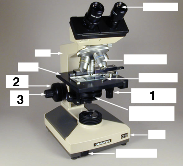
Name these parts of the microscope
Mechanical Stage Controls
Coarse Focus
Fine Focus

Name these parts of the microscope
Iris Diaphragm Lever
Base
On/Off switch
If you are viewing a specimen using the high-dry objective, what is the total magnification of the image?
400x
List IN ORDER the exact sequence of steps you should perform in order to view a prepared specimen with a compound microscope:
Clean All Lenses
Place slide on stage
rotate 10x objective into place
Move stage up (looking from side) close to objective lens
Move specimen under objective lens using stage control knobs
Move stage down with course adjustment knob wile looking in ocular lens until image is seen
Fine focus with Fine adjustment knob
Add oil and rotate directly to the 100x objective
Fine focus until image is clear
Agar Slant Terms: Abundance of Growth
None
Slight
Moderate
Large
Agar Slant Terms: Pigmentation
Used to describe the pigment of bacterial growth
Agar Slant Terms: Optical Characteristics
Opaque: No light transmission
Translucent: Partial light transmission
Transparent: full light transmission
Agar Slant Terms: Form
Filiform
Echinulate
Beaded
Effuse
Arborescent
Rhizoid
Agar Slant Form: Filiform
Continuous, threadlike growth with smooth edges
Agar Slant Form: Echinulate
Continuous, threadlike growth with irregular edges
Agar Slant Form: Beaded
Nonconfluent to semiconfluent colonies
Agar Slant Form: Effuse
Thin, spreading growth
Agar Slant Form: Arborescent
Tree like growth
Agar Slant Form: Rhizoid
Rootlike growth
Agar Slant Terms: Consistency
Dry
Buttery
Mucoid
Agar Slant Consistency: Dry
Free from moisture
Agar Slant Consistency: Buttery
Moist and shiny
Agar Slant Consistency: Mucoid
Slimy and glistening
Agar Plate Terms: Size
Pinpoint
Small
Moderate
Large
Agar Plate Terms: Pigmentation
Color of colony
Agar Plate Terms: Form / Shape of colony
Circular
Irregular
Rhizoid
Agar Plate Form: Circular
Unbroken, peripheral edge
Agar Plate Form: Irregular
Indented, peripheral edge
Agar Plate Form: Rhizoid
Rootlike, spreading growth
Agar Plate Terms: Margin
Entire
Lobate
Undulate
Serrate
Filamentous
Agar Plate Margin: Entire
Sharply defined, even
Agar Plate Margin: Lobate
Marked indentations
Agar Plate Margin: Undulate
Wavy indentations
Agar Plate Margin: Serrate
Toothlike appearance
Agar Plate Margin: Filamentous
Threadlike, spreading edge
Agar Plate Terms: Elevation
Flat
Raised
Convex
Umbonate
Agar Plate Margin: Flat
Elevation not discernible
Agar Plate Margin: Raised
Slightly elevated
Agar Plate Margin: Convex
Dome-shaped elevation
Agar Plate Margin: Umbonate
Raised, with elevated convex central region
Broth Terms
Uniform fine turbidity
Flocculent
Pellicle
Sediment
Uniform fine turbidity
Finely dispersed growth throughout
Flocculent
Flaky aggregates dispersed throughout
Pellicle
Thick, pad-like growth on surface
Sediment
Concentration of growth at the bottom of broth culture may be granular, flaky, or flocculent
List IN ORDER the exact sequence of steps you should perform in order to prepare a specimen for simple staining from a broth culture:
Flame the loop
Flame the mouth of the broth tube
Loop full of bacteria
Put bacteria on slide
Air dry
heat fix
Apply Crystal violet - 1 min
Rinse
Blot with bibulous paper
6. How does the simple stain procedure differ when preparing a specimen from an agar culture?
Will need to put a loop full of sterile water on a slide
Flame loop and get sample of bacteria
Mix bacteria into drop of sterile water
List the 3 steps of the acid-fast staining procedure
Drop of Water
Mix Bacteria
Air Dry
Heat Fix
Apply Primary Stain (Carbol Fuchsin) Over Steam - 5 min
Rinse
Apply Acid Alcohol - 30 seconds
Rinse
Apply Secondary Stain (Methylene Blue) - 1 min
Rinse
Dry with bibulous paper
What color will acid fast positive bacteria appear
Red / Pink
What color will acid fast negative bacteria appear
Blue
What is the difference in structure between an Acid-fast positive and Acid-fast negative species?
Acid Fast Positive: Have a thick, waxy outer layer rich in mycolic acids and glycolipids that prevents the carbol fuschin from being washed out by acid alcohol
Acid Fast Negative: Do not have a thick, waxy outer layer
List the steps of the spore staining procedure.
Drop of Water
Mix Bacteria
Air Dry
Heat Fix
Apply Primary Stain (Malachite Green) over steam - 5 min
Rinse
Apply Secondary Stain (Safranin) - 1 min
Rinse
Dry with bibulous paper
What are endospores?
Contains a copy of the genetic material of a cell and is surrounded by a tough spore coat; produced when the bacteria undergoes tough conditions like lack of nutrients are high heat
Describe the procedure for doing a capsule stain.
Drop of Primary Stain (Congo Red) on one end of slide
Mix in bacteria
use another slide to spread the red stain/bacteria mixture across the slide
Air dry
Flood slide with maneval’s stain - 1 min
Rinse
What is a capsule?
a thick, gelatinous outer layer that surrounds the cell wall of some bacteria that helps with evading immunity, helps adhesion, and prevents drying