axial skeleton flashcards - a&p practical 1
 Call Kai
 Learn
Learn Practice Test
 Spaced Repetition
 Match
Match Flashcards
 Knowt Play
1/48

There's no tags or description
Looks like no tags are added yet.
Name | Mastery | Learn | Test | Matching | Spaced | Call with Kai | Chat |
|---|
No analytics yet
Send a link to your students to track their progress
49 Terms
frontal bone
makes up the forehead and upper part of eye sockets

supraorbital margin
superior rim of orbit (eyes)

supraorbital foramen (notch)
opening in the supraorbital margin for the supraorbital nerves & vessels

glabella
smooth area b/w the eyebrows

frontal sinus
cavity in frontal bone above eyes

parietal bone
top of the head

coronal suture
joins frontal & parietal bones

squamous suture
joins parietal & temporal bones

lambdoid suture
joins parietal & occipital bones

sagittal suture

occipital bone
back of head

external occipital protuberance
midline bump on the back of the skull

external occipital crest
midline ridge running from protuberance toward the foramen magnum

superior nuchal line
ridge extending laterally from protuberance (attachment for neck muscle)
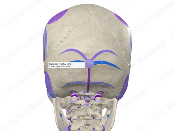
inferior nuchal line
ridge below the superior nuchal line (attachment for neck muscle)

occipital condyle
articulates w/ C1 (atlas)
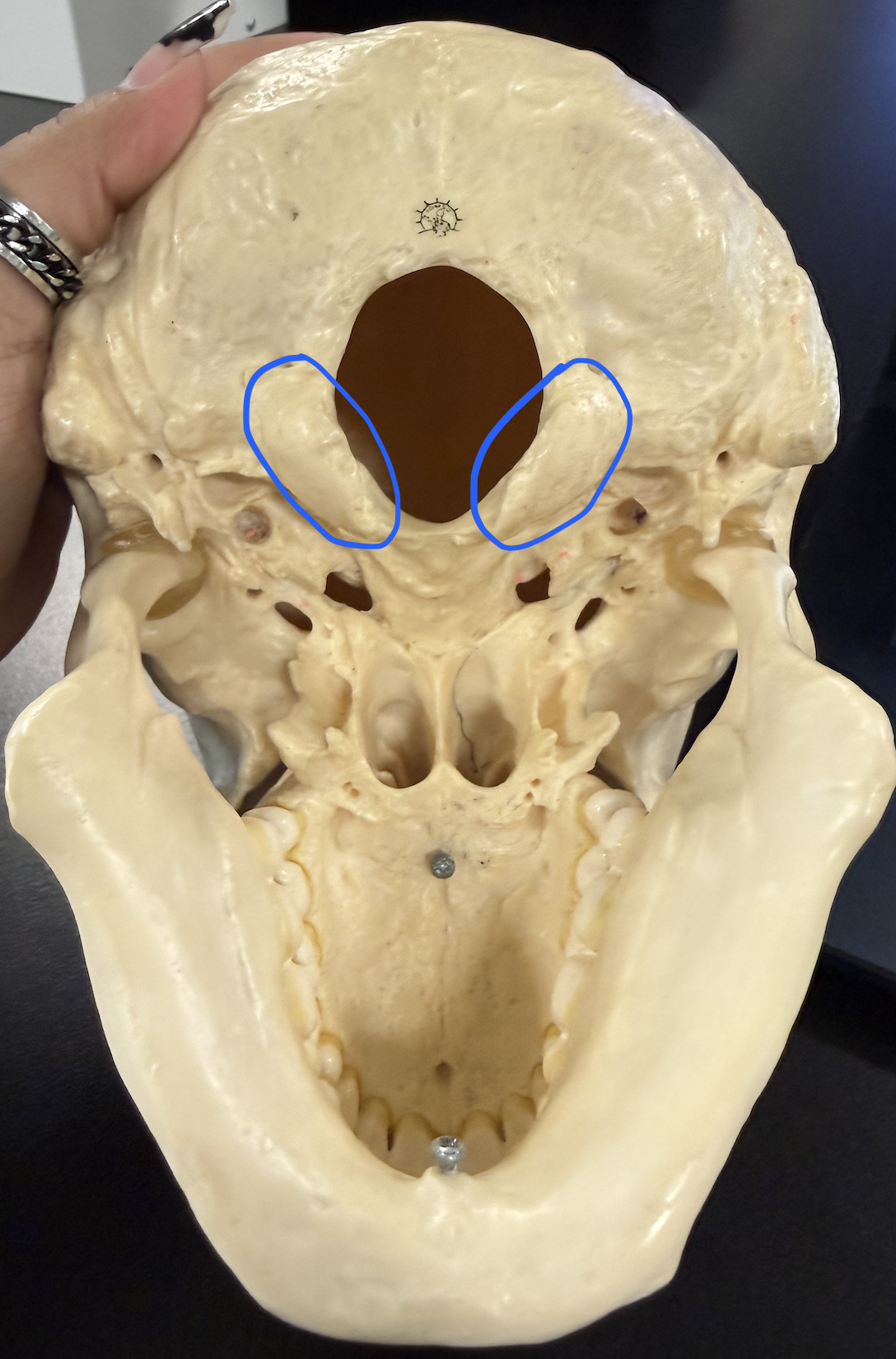
foramen magnum
large opening where the spinal cord leaves the skull

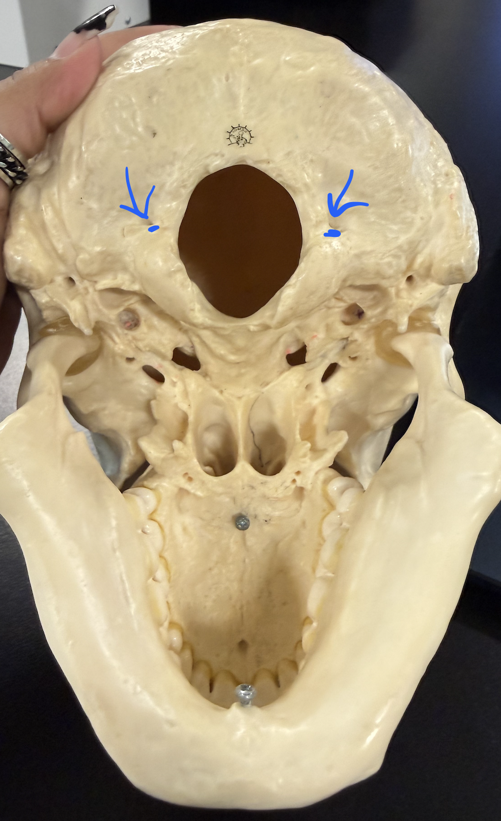
hypoglossal canal
for hypoglossal nerve; under occipital condyles (need to tilt to be visible)
temporal bone
sides of head

squamous region
thin, flat, platelike part of the temporal bone

zygomatic process
projects forward to help form zygomatic arch

mandibular fossa
articulates w/ mandibular condyle of the mandible

external acoustic meatus
ear canal opening

styloid process
slender spike below the ear canal

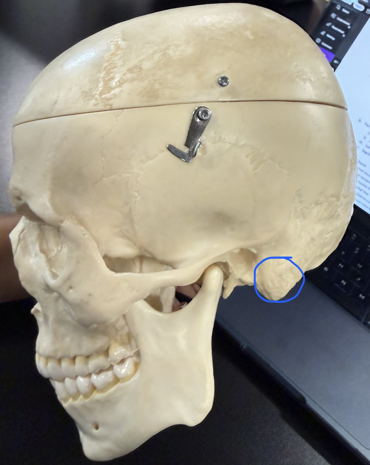
mastoid process
middle ear infection can spread here (mastoiditis), then to meninges (meningitis); sternocleidomastoid attaches here
jugular foramen
internal jugular vein, CN XI, CN X, CN IX

internal acoustic meatus
CN VIII , CN VII

foramen lacerum
jagged opening at base of the skull b/w the temporal, sphenoid and occipital bones

carotid canal
internal carotid artery

sphenoid bone
complex, butterfly-shaped bone located at the middle base of the skull

lesser wings
thin wings forming part of the anterior cranial floor and eyes

greater wings
large lateral wings forming part of the cranial floor and eyes
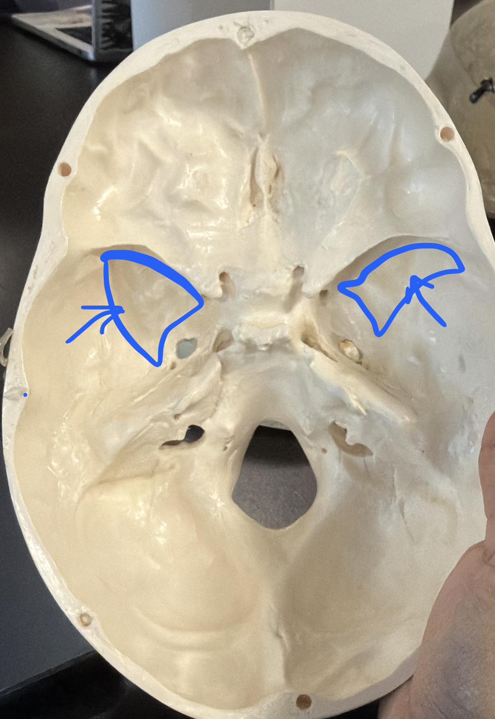
optic canal
optic nerve (CN II)

superior orbital fissure
CN III, CN IV, CN VI, ophthalmic branch of CN V

foramen rotundum
maxillary branch of trigeminal nerve (CN V)

foramen ovale
mandibular branch of trigeminal nerve (CN V)

foramen spinosum
smaller opening in the greater wing, posterior and lateral to foramen ovale

sella turcica
saddle-shaped depression; contains hypophyseal fossa
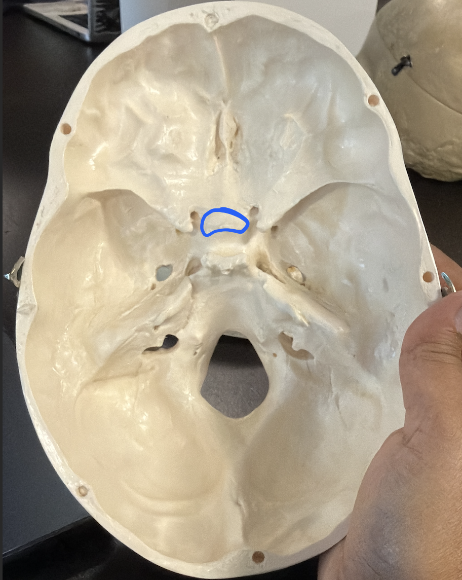
anterior clinoid processes
anterior projections of the sella turcica region
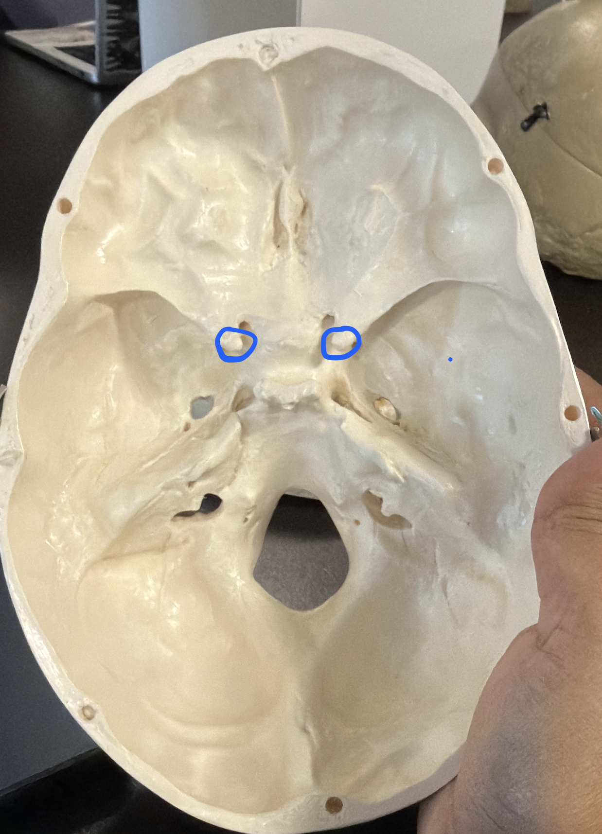
posterior clinoid processes
posterior projections flanking the sella turcica

tuberculum sellae
ridge forming the anterior edge of the sella turcica

dorsum sellae
ridge forming the posterior wall of the sella turcica

hypophyseal fossa
pituitary gland (hypophysis)

pterygoid processes
downward projections from the sphenoid; each has a lateral and medial plate

lateral plates (pterygoid)
_______ plate of the pterygoid process

medial plates (pterygoid)
______ plate of the pterygoid process

sphenoid body
central part of the sphenoid; houses the sphenoidal sinus
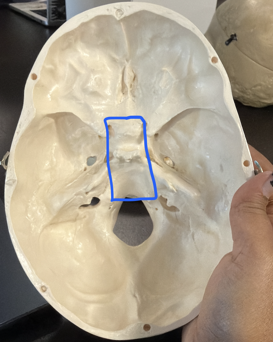
sphenoidal sinus
cavity within the sphenoid body

ethmoid bone
