heart physiology: electrical events
 Call Kai
 Learn
Learn Practice Test
 Spaced Repetition
 Match
Match Flashcards
 Knowt Play
1/55

There's no tags or description
Looks like no tags are added yet.
Name | Mastery | Learn | Test | Matching | Spaced | Call with Kai | Chat |
|---|
No analytics yet
Send a link to your students to track their progress
56 Terms
electrical events
heart depolarizes & contracts w/out nervous system stimulation; basic rhythm can be modified by autonomic NS (sympathetic/parasympathetic); Intrinsic conduction system
setting the basic rhythm
coordinated heartbeat is a function of: presence of gap junctions & intrinsic conduction system
intrinsic conduction system
network of noncontractile (autorhythmic) cells; specialized to initiate & distribute impulses > coordinated depolarization & contraction of heart
pacemaker (autorhythmic) cells (step 1)
unstable resting membrane potentials due to opening of slow Na+ channels (continuously depolarize);
pacemaker (autorhythmic) cells (step 2)
at threshold, Ca2+ channels open; explosive Ca2+ influx prosuce the rising phase of AP (depolarization)
pacemaker (autorhythmic) cells (step 3)
repolarization results from inactivation of Ca2+ channels & opening of voltage-gated K+ channels
three parts of action potential for pacemaker cells
1. pacemaker potential
2. depolarization
3. repolarization
pacemaker potential (pacemaker cells)
slow depolarization is due to both opening of Na+ channels & closing of K+ channels; membrane potential is never a flat line
depolarization (pacemaker cells)
action potential begins when the pacemaker potential reaches threshold; depolarization is die to Ca2+ influx through Ca2+ channels
repolarization (pacemaker cells)
due to Ca2+ channels inactivating & K+ channels opening; allows K+ efflux, which brings the membrane potential back to its most negative voltage
sequence of excitation
cardiac pacemaker cells pass impulses, in order, across heart in ~220 ms
Pathway of sequence of excitation
1. sinoatrial (SA) nodes
2. atrioventricular (AV) node
3. atrioventricular (AV) bundle
4. right & left bindle branches
5. subendocardial conducting network (purkinje fibers)
intrinsic conduction system sequence of electrical excitation
1. sinoatrial (SA) node (pacemaker) generates impulses
2. impulses pause (0.1s) at atrioventricular (AV) node
3. Atrioventriculat (AV) bundle connects atria to ventricles
4. bundle branches conduct impulses theough interventriculat septum
5. purkinje fibers depolarize contractile cells of both ventricles
sinoatrial (SA) node
pacemaker of heart in right atrium (primary pacemaker) depolarizes faster than rest off myocardium
sinoatrial (SA) node generation of impulse
ab 75X/min (sinus rhythm); inherent rate of 100X/min modified by extrinsic factors (parasympathetic NS)
sinoatrial (SA) node spreading impulse
through atria (via gap junctions), & to AV node (via internodal pathway
picture of intrinsic conduction system

Atrioventricular (AV) node location
in inferior interatrial septum
atrioventricular (AV) node
delays impulses approximately 0.1 sec, allows atria to finish their contraction before the ventricles begin to contract
atrioventricular (AV) node generation of impulse
inherent rate of 50X/min in absence of SA node input (secondary pacemaker)
Atrioventricular (AV) bundle (bundle of His) location
in superior interventricular septum
Atrioventricular (AV) bundle (bundle of His)
only electrical connection between atrial & ventricles; atria & ventricles not connected via gap junctions
right & left bundle branches
2 pathways in interventricular septum; carry impulses toward apex of heart
subendocardial conducting network (purkinje fibers) location
complete pathway through interventricular septum to apex & up into ventricular walls; more elaborate on left side of heart
subendocardial conducting network (purkinje fibers)
AV bindles & this depolarize 30X/min in absence of AV node input; ventricular contraction immediately follows from apex upward toward atria
timing of conduction events

extrinsic innervation (description)
modifies the basic rhythm; HR modified by ANS via cardiac centers in medulla oblongata
extrinsic innervation types
1. sympathetic NS
2. parasympathetic NS
3. cardioacceleratory center
4. cardioinhibitory center
sympathetic NS effect (extrinsic innervation)
higher heart rate & force of contraction
parasympathetic NS effect (extrinsic innervation)
lower heart rate; no effect on contraction bc it does not innervate myocardium
cardioacceleratory center
sympathetic output; to pacemaker & myocardium
cardioinhibitory center
parasympathetic output; via vagus nerves; only pacemaker cells
extrinsic innervation

Action potential of contractile cardiac muscle cells
has characteristic 'hump' or plateau
contraction differences from skeletal muscle (AP) pt.1
depolarization wave ALSO opens slow Ca2+ channels in sacrolemma > SR release its Ca2+
contraction differences from skeletal muscle (AP) pt.2
Ca2+ surge prolongs the depolarization phase (plateau)
contraction differences from skeletal muscle (AP) pt.3
Action potential & contractile phase last much longer; allow blood ejection from heart
contraction differences from skeletal muscle (AP) pt.4
repolarization result of inactivation of Ca2+ channels & opening of voltage-gated K+ channels; Ca2+ pumped back to SR & extracellularly
3 parts of AP for contractile cells
1. depolarization
2. plateau phase
3. repolarization
pacemaker cell action potential

depolarization (contractile cells)
due to Na+ influx through fast voltage-gated Na2+ channels; positive feedback cycle rapidly opens many Na+ channels reversing membrane potential; channel inactivation ends this phase
plateau phase (contractile cells)
due to Ca2+ influx through slow Ca2+ channels; keeps cell depolarized bc few K+ channels are open
repolarization (contractile cells)
due to Ca2+ channels inactivating & K+ channels opening; allows K+ efflux, which brings membrane potential back to resting voltage
AP for contractile cells

AP for contractile cells skeletal vs cardiac

electrocardiography
conduction of impulses through heart generates electrical currents; impulses can be detected on body's surface & recorded
electrocardiogram (ECG or EKG)
composite of ALL AP's generated by intrinsic conduction system & contractile cells at given time
3 waves
1. P wave
2. QRS complex
3. T wave
p wave
atrial depolarization (starts w/SA node)
QRS complex
ventricular depolarization
T wave
ventricular repolarization
What do EKGs show
electrical events ONLY; electrical events come before mechanical events
EKG
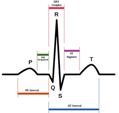
Atria AP on EKG
1. atrial depolarization - p wave
2. atrial plateau - P-Q segment
3. Atrial repolarization - not shown on EKG
ventricles AP on EKG
1. ventricular depolarization - QRS complex
2. ventricular plateau - S-T segment
3. ventricular repolarization - t wave
steps of electriocardiogram (EKG)
1. atrial depolarization, initiated by SA node, causes P wave
2. W/atrial depolarization complete (plateau of AP) impulse is delayed at AV node P-G segment
3. ventricular depolarization begins at apex causing QRS; atrial repolarization occurs
4. ventricular depolarization is complete (plateau of AP) S-T segment
5. ventricular repolarization begins at apex, T wave
6. ventricular repolarization complete T-P segment