Chapter 12 - The Cell Cycle
 Call Kai
 Learn
 Practice Test
 Spaced Repetition
 Match
 Flashcards
 Knowt Play
1/45

There's no tags or description
Looks like no tags are added yet.
46 Terms
Why does DNA need to be duplicated during cell division?
In order to make sure that each new daughter cell receives a complete and identical copy of genetic blueprint
Multicellular organisms depend on cell division for…
Development from fertilized cell
Growth
Repair
Genome
All the genetic information (DNA) present in a cell, consisting of a single DNA molecule in prokaryotes or multiple DNA molecules in eukaryotes.
Chromosomes
A thread-like structure made of DNA tightly coiled around proteins called histones, found inside the nucleus of a cell.
Chromatin
The complex of DNA and associated protein molecules that condenses to form discrete chromosomes during eukaryotic cell division.
Somatic Cells
All body cells except reproductive cells; in humans, they contain two sets of chromosomes.
Gametes
Reproductive cells (sperm and eggs) that contain half as many chromosomes as somatic cells, possessing only one set of chromosomes.
Sister Chromatids
Two identical joined copies of an original duplicated chromosome, which are attached along their lengths and separate into individual chromosomes during cell division.
Centromere
The narrow "waist" of a duplicated chromosome where two sister chromatids are most closely attached.
Mitosis
The division of the genetic material in the nucleus of a eukaryotic cell.
prophase
prometaphase
metaphase
anaphase
telophase
cytokinesis*
Meiosis
Yields nonidentifcal daughter cells that have only 1 set of chromosomes
Cytokinesis
The division of the cytoplasm to form two separate daughter cells, which overlaps with the latter stages of mitosis.
What does the cell cycle consist of?
Mitotic (M) phase
Interphase (G1, S, and G2 phases)
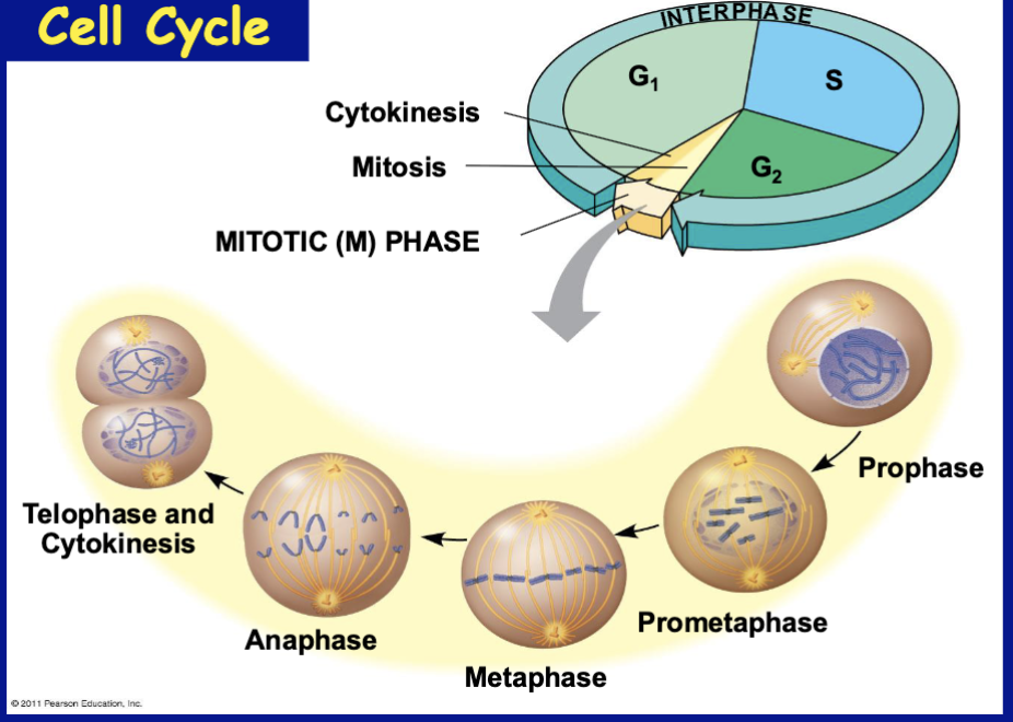
Interphase
The period of the cell cycle accounting for about 90% of the time, consisting of the G1, S, and G2 subphases, during which the cell grows and copies its chromosomes.
G1 phase
Cell duplicates or makes DNA, longest of the three
S Phase
The "synthesis" subphase of interphase during which cellular DNA is replicated and chromosomes are duplicated; chromosomes become sister chromatids
G2 phase
The cell undergoes final growth and intense preparation
Prophase
early mitotic spindles, sister chromatids coming together, nuclear envelope disappearing

Prometaphase
kinetochore assembly, nuclear envelope breakdown, spindle attachment, chromosome movement towards middle, spindles attach to kinetochores
Complete breakdown of the nuclear envelope, which fragments into small vesicles and allows the mitotic spindle microtubules to access the cell's genetic material. During this time, specialized protein complexes called kinetochores assemble on the centromere of each sister chromatid pair. Microtubules extending from opposite centrosomes (the spindle poles) invade the nuclear region and bind tightly to these kinetochores.
*some spindle microtubules attach to the kinetochores of chromosomes & begin to move the chromosomes

What is a kinetochore microtubule?
Specialized protein complexes assembled on the centromeric region of chromosomes where spindle microtubules attach during prometaphase.
Nonkinetochore microtubule
Microtubule that does not attach to chromosomes and instead push the cell poles apart
Metaphase
Condensed chromosomes align in the center of the cell along an imaginary line called the metaphase plate

Anaphase
Sister chromatids (or homologous chromosomes) separate and move toward opposite poles of the cell
Microtubules shorten by depolymerizing at their kinetochore end
Nonkinetochore microtubules from opposite poles overlap and push against each other, elongating the cell

Telophase and Cytokinesis
Nucleus/nuclear envelope reforms, mitotic spindle breakdown, chromosomes uncoil, cytoplasm splits into two new daughter cells

Mitotic Spindle
An apparatus composed of microtubules and associated proteins that controls chromosome movement during mitosis, begin at centrosome (organizes microtubules)
Centrosome
A sub-cellular structure serving as the microtubule-organizing center in animal cells, which replicates during interphase to organize the poles of the mitotic spindle.
Aster
A radial array of short microtubules that extends from each centrosome.
Metaphase Plate
An imaginary plane located midway between the two poles of the mitotic spindle along which chromosomes align at metaphase.
Cleavage Furrow
A shallow groove on the animal cell surface formed by a contractile ring of microfilaments, which pinches the cell into two daughter cells during cytokinesis.
Cell Plate
A membrane-bounded structure formed across the midline of a dividing plant cell by fusing vesicles, eventually developing into a new cell wall.
Binary Fission
A type of asexual cell division in prokaryotes (bacteria and archaea) where the single chromosome replicates starting at the origin of replication and the cell divides into two.
Origin of Replication
The specific site on a bacterial chromosome where DNA synthesis and chromosome replication initiate during binary fission.
Cell Cycle Control System
A cyclically operating set of molecules that triggers and coordinates key events in the cell cycle through internal and external regulation at specific checkpoints.
Checkpoint
A control point in the cell cycle where stop and go-ahead signals regulate cycle progression based on cellular surveillance and external signals.
3 primary checkpoints: g1, g2, and m phase
G0 Phase
A non-dividing state exited into by a cell that fails to receive a go-ahead signal at the G1 checkpoint.
Cyclin-Dependent Kinases (Cdks)
Regulatory protein involved in cell cycle control whose enzymatic activity fluctuates based on the periodic accumulation and attachment of cyclin proteins.
MPF (Maturation-Promoting Factor)
A cyclin-Cdk complex that triggers a cell passage past the G2 checkpoint into the M phase.
Platelet-Derived Growth Factor (PDGF)
An external protein for platelets, growth factor released by certain cells that stimulates the division of human fibroblast cells in culture.
Density-Dependent Inhibition
An external signal in normal animal cells where crowded cells stop dividing.
Anchorage Dependence
The requirement that a normal animal cell must be attached to a surface in order to divide.
Transformation
The process by which a normal cell is converted into a cancerous cell capable of uncontrolled division.
Benign Tumor
A mass of abnormal cells that remains localized only at its original site within otherwise normal tissue.
Malignant Tumor
A cancerous mass of abnormal cells that invades surrounding tissues and can metastasize to distant body parts.
Metastasis
The spread and export of cancer cells through blood and lymph vessels to other parts of the body, where they may establish secondary tumors.
Taxol
A chemotherapeutic drug that prevents microtubule depolymerization, thereby stopping cancer cells from progressing past metaphase during mitosis.
A normal cell is converted to a cancerous cell by…
Transformation