Biology: Chapter 3
 Call Kai
 Learn
Learn Practice Test
 Spaced Repetition
 Match
Match Flashcards
 Knowt Play
1/72

There's no tags or description
Looks like no tags are added yet.
Name | Mastery | Learn | Test | Matching | Spaced | Call with Kai | Chat |
|---|
No analytics yet
Send a link to your students to track their progress
73 Terms
List all tenets of Cell Theory
All organisms are made of one or more cells
The cell is the fundamental unit of life
All cells come from preexisting cells/every cell came from a preexisting cell
All cells have the same basic chemical composition
All Cells use energy
Cell Theory
the ideas that all living matter consists of cells, cells are the structural and functional units of life, and all cells come from preexisting cells.
Why are cells so small?
Smaller cells have more surface area relative to their volume. High surface area allows the cell to quickly exchange materials with its surroundings.
List the features common to all cells
DNA/RNA, Cell membrane, cytoplasm, and Ribosomes
DNA
Nucleic Acid that stores cell’s genetic information.
RNA
Nucleic acid that participates in the production of proteins.
Cytoplasm
the watery mixture that occupies much of a cell’s volume; in eukaryotic cells, it consists of all materials, including organelles, between the nuclear envelope and the cell membrane. (All contents except the nucleus (if the cell has one))
Cytosol
the fluid portion of the cytoplasm
Cell Membrane
the boundary of a cell, consisting of proteins embedded in a phospholipid bilayer; also called the plasma membrane. Separates the cytoplasm from the surrounding environment.
List the Major Functions of the Plasma Membrane
Separates the cytoplasm from the cell’s surroundings
Transports Substances into and out of the cell through selective permeability/Regulates passage of substances in and out of the cell
Receives and responds to external stimuli
Helps maintain homeostasis
Selectively Permeable (Plasma Membrane)
In the phospholipid bilayer, the tails are hydrophobic, so they allow lipids and nonpolar molecules to move freely in and out, but they do not allow ions or large polar molecules to move freely in and out.
How Ions and Polar molecules Move Through the Cell Membrane
they do so through the help of transport proteins, as proteins are embedded in the membrane along with the phospholipids.
Difference Between the Nucleus and a Nucleoid
The difference is that the nucleoid is the part of a prokaryotic cell where the DNA is located and is not bounded by a membrane, while a nucleus is found in eukaryotic cells and is membrane-bounded.
Cell Wall
often made up of peptidoglycan or proteins. Helps with protection and gives it a rigid shape to the bacteria cell. Prevents it from bursting if it absorbs too much water. (a rigid boundary surrounding cells of many prokaryotes, protists, plants, and fungi)
Nucleoid Region
part of a prokaryotic cell where DNA is present.
Capsule
Sticky polysaccharide surrounding cell wall. Helps it attach to surfaces and gives some protection. (Found in some bacteria)
Plasmids
some bacteria have this. It is additional DNA molecules that carry certain genes which helps them digest/adapt to unusual circumstances (Ex. being antibiotic resistant).
Flagellum
a long whiplike appendage made up of long proteins which helps it propel. Helps in the movement of the bacterial cell as many bacteria can swim in fluids.
Pili
finger-like projections. Rigid fibers made up of proteins. Helps them attach to surfaces.
List all the organelles of Eukaryotic Cells
Nucleus, Endoplasmic Reticulum, Golgi Apparatus, Cell Membrane, Lysosomes, Vacuoles, Peroxisomes, Mitochondria, Chloroplasts, and Vesicles
Nucleus
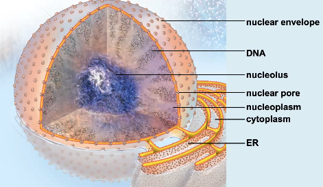
Structure: surrounded by double membrane (nuclear envelope). Has nuclear pores on outside (help move things in and out). Contains DNA, RNA, and protein. Space within nucleus is the nucleoplasm. Nucleolus at center.
Function: Separates DNA from rest of cell; site of first step in protein synthesis; nucleolus produces ribosomal subunits.
Endoplasmic Reticulum
an extension of the nuclear envelope that forms a continuous, folded compartment. A network of membranous sacs and tubules. Has rough and smooth ER.
Rough Endoplasmic Reticulum
Structure: Membrane network studded with ribosomes.
Function: Produces proteins (polypeptides) destined for secretion from the cell. Polypeptides enter the inner compartment of this, where they fold into their tertiary form. Proteins exit organelle in vesicles (then fuse to golgi apparatus).
Smooth Endoplasmic Reticulum
Structure: Membrane network without ribosomes.
Function: synthesizes lipids, breaks down carbohydrates, and detoxifies drugs and poisons.
Golgi Apparatus
Structure: Stacks of flat, membranous sacs. Folded membrane.
Function: Packages materials to be secreted; produces lysosomes. Processes polypeptides and lipids delivered by ER and makes them functional. Its enzymes add carbohydrates to proteins and lipids, making them glycoproteins and glycolipids. Packages finished products in vesicles (to plasma membrane/lysosomes/other organelles).
Lysosomes
Structure: Vesicles containing digestive enzymes manufactured in RER and processed and shipped from the Golgi apparatus; surrounded by single membrane. Enzymes contain hydrolases.
Function: Enzymatically dismantles molecules, bacteria, pathogens, and worn-out cell parts. Helps with digestion and waste processing. The enzymes inside break down the large organic molecules into smaller subunits by hydrolysis, releasing them into the cytoplasm for the cell to use.
Malfunctioning: Tay-Sachs disease
Vacuoles
Structure: Present in plant cells. Sac containing a watery solution of enzymes that degrade and recycle molecules and organelles (cellular digestion). Occupies 90% of the cell’s volume. Surrounded by a single membrane.
Function: serves similar function to lysosomes. Degrades wastes, exerts turgor pressure (keeps plant upright and rigid), and stores acids, pigments, water, etc., and helps regulate size/water balance of plant cells.

Peroxisomes
Structure: Sac containing enzymes, forming visible protein crystals; surrounded by single membrane. Originate at ER.
Function: disposes of toxins; breaks down fatty acids; eliminates hydrogen peroxide. Where oxidation occurs. Breaks down substances into cholesterol and other lipids.
Malfunction: Adrenoleukodystrophy (brain damage and death).
Mitochondria
Structure: Two membranes; inner membrane is folded into enzyme-studded cristae; contains DNA and ribosomes. The outer membrane is smooth. Has its own DNA. Inside fluid is matrix.
Function: Releases energy from food by cellular respiration to supply the cell with energy. (Energy is ATP).

Chloroplasts
Structure: present only in plant cells and some protists. Two outer membrane layers enclose an enzyme-rich fluid sac called the Stroma. Within the Stroma is a third membrane system folded into flattened sacs called thylakoids, which are stacked and interconnected into a structure called Grana. Photosynthetic pigments are embedded in thylakoids. Contains DNA and ribosomes.
Function: Produces food (sugars) by photosynthesis for the cell.

Centrosomes
Structure/Function: present only in animal cells. They are the part of the cell that organizes microtubules. Contains two centrioles, which form the basis of basal bodies that give rise to cilia and flagella (extensions that enable some cells to move)

Ribosomes
Structure: Two globular subunits composed of RNA and protein, which are synthesized in the nucleolus of eukaryotic cells.
Function: Location of protein synthesis.
2 Important Functions of the Nucleus in A Eukaryotic Cell
Is where the genetic information is stored (DNA)
Where mRNA is sent from so that other parts of the cell can receive the “recipes” that DNA has encoded in it.
Endomembrane System and Its Major Organelles
Organelles inside the cell, which work together as a unit. Major organelles: Nucleus/Nuclear Envelope, Endoplasmic Reticulum, Golgi Apparatus, Lysosomes, Vacuoles, Cell membrane, and Vesicles.
Vesicle
a sac made of membrane which helps in transport of macro molecules from one organelle to another. Formed by enoplasmic reticulum when a piece of membrane bulges outward and disconnects, forming into a tiny sphere that holds whatever molecules it is transporting. Pinches off from one organelle, travels within the cell, and fuses with another.
Endosymbiotic Theory
the theory that mitochondria and chloroplasts were once free living bacteria but were engulfed by certain ancestors and developed a mutually beneficial relationship.
Evidence Supporting the Endosymbiotic Theory
both mitochondria and chloroplasts have their own DNA, ribosomes, can divide independently, and are surrounded by double membranes. These characteristics point towards the theory that they used to be their own bacterial cells. Also, the structures and genetic sequences of today’s bacteria, mitochondria, and chloroplasts supply powerful evidence for this theory.
Tight Junctions
Found in animals. Fuses the membranes of adjacent animal cells together, preventing substances from flowing between the cells. Found in cells in inner lining of the stomach and small intestine.

Anchoring (adhering) Junctions
Found in animals. use intermediate filaments to hold cells together so there is a gap and so things can move. Connect adjacent animal cell membranes in one spot; connect cells to the extracellular matrix. Found in cells in outer skin layer.

Gap Junctions
Found in animals. connecting animal cells are analogous to plasmodesmata. Is a protein channel that links the cytoplasm of neighboring cells. Protein channel helps with the movement of a substance from one cell to another. Form channels between animal cells, allowing exchange of substances. Found in muscle cells in heart and digestive tract.

Plasmodesmata
Found in plants. the cytoplasmic connections in plant cells. Plant cells communicate through plasmodesmata. Nutrients and biochemicals travel through these channels to adjacent cells. Allow substances to move between plant cells. Found in plant cell walls.

Where are Cell Junctions Found?
Between cells, where they connect.
Cytoskeleton
a network of protein tracks and tubules in a cell (made from these protein filaments: microfilaments, intermediate filaments, and microtubules). Functions: structural support, aids in cell division, organelle transport, and cell movement.
Microfilaments
Long rod made of protein actin which helps with muscle contraction and provides strength for cells to survive stretching and compression, and they help to anchor one cell to another.

Intermediate Filaments
component of the cytoskeleton; intermediate in size between microtubule and a microfilament. Proteins that form these vary by cell type. Their function is to maintain a cell’s shape by forming an internal scaffold in the cytosol and existing mechanical stress. Also help bind some cells together.

Microtubules
component of the cytoskeleton; made of subunits of the protein tubulin. Have many functions including: can form a “trackway” along which substances move within a cell and they split a cell’s duplicated chromosomes apart during cell division. Form the internal framework of cilia and flagella. Help is movement of sperm. Helps in movement of cells and to propel food.

3 major Difference Between a Plant Cell and an Animal Cell
Outermost layer of animal cell is cell membrane. Outermost layer of plant cell is cell wall. (Plants have both a cell membrane and cell wall).
Plants have a large central vacuole. Animal cells do not.
Plants have chloroplast. Animal cells do not.
Difference Between Domain Bacteria and Archaea
Chemical composition of flagella
The molecules that compose cell membranes
Evolutionary relationship to eukaryotes. Archaea are more closely related to eukaryotes than bacteria. Bacteria are not as closely related to eukaryotes.
Eukaryotic Cell
Cell interior is divided into functional compartments, (organelles), including a nucleus. Includes: animals, plants, fungi, and protists.
Prokaryotic Cell
Small, simple cells without a nucleus. Includes Bacteria and Archaea. Smaller in size as compared to eukaryotic.
Animal Cells
eukaryotic. Have membrane bounded organelles.
Plant Cells
eukaryotic, but have cell walls and chloroplasts as compared to animal cells.
Fluid Mosaic Model
the model that explains the structure of the plasma membrane. Called mosaic because the membrane has a pattern of proteins embedded in it.
Chromatin
All DNA and its associated proteins in the nucleus
Chromosome
A single DNA molecule with its attached proteins. During cell division, chromosomes condense and become visible in micrographs. Human body cells have 46 chromosomes.
Cell Wall (plants)
rigid. Gives plants protection and shape. Interact with other molecules to help determine how a cell in a complex organism specializes. Made of polymers like cellulose pectin.
Membrane Proteins
some of the proteins extend through the phospholipid bilayer, whereas others face only the inside or outside of the cell.
Transport Proteins
proteins embedded in the phospholipid bilayer create passageways through which ions, glucose, and other polar substances pass into or out of the cell.
Enzymes
These proteins facilitate chemical reactions that otherwise would proceed too slowly to sustain life. (Not all of them, however, are associated with membranes).
Recognition proteins
Carbohydrates attached to cell surface proteins serve as “name tags” that help the body’s immune system recognize its own cells. The immune system attacks cells with unfamiliar surface molecules, which is why transplant recipients often reject donated organs. Surface proteins also distinctively mark specialized cells within an individual, so a bone cell’s surface is different from that of a nerve cell or a muscle cell.
Adhesion Proteins
These membrane proteins enable cells to stick to one another.
Receptor Proteins
Receptor proteins bind to molecules outside the cell and trigger an internal response, a process called signal transduction. (Ex. when a hormone binds to a receptor, the resulting chain reaction produces the hormone’s effects on the cell).
Nuclear Pores
a hole in the nuclear envelope. They are specialized channels composed of proteins through which regulatory proteins enter and mRNA molecules leave to send copies of directions to proteins cells can make.
Cilia
short, numerous extensions; movable protein extensions from a cell. Enables some cells to “swim” like in some protists. Present in the human respiratory tract; coordinate cilia movement allows particles to be propelled up and out. Cilia moves egg cell through female reproductive tract.
Flagella
occur singly or in pairs. Much longer than a cilium. Whiplike movement propels cells. Present in sperm cells in many species.
Eukaryote
organism composed of one or more cells containing a nucleus and other membrane-bounded organelles
Organelle
compartment of a eukaryotic cell that performs a specialized function
Prokaryote
a cell that lacks a nucleus and other membrane-bounded organelles; bacteria and archaea
Phospholipid Bilayer
double layer of phospholipids that forms in water; forms the majority of a cell’s membranes
Nuclear Envelope
the two membranes bounding a cell’s nucleus
Fluid Mosaic
two-dimensional structure of movable phospholipids and proteins that form biological membranes
Nucleolus
a structure within the nucleus where components of ribosomes are assembled
Cristae
fold of the inner mitochondrial membrane along which many of the reactions of cellular respiration occur