Pathophys Exam 2
 Call Kai
 Learn
 Practice Test
 Spaced Repetition
 Match
 Flashcards
 Knowt Play
1/413

There's no tags or description
Looks like no tags are added yet.
414 Terms
L9 - liver
macrostructure of liver
liver is the largest visceral organ in the body
high capacity for regeneration
right lobe is the largest lobe
75% - 85% of blood supply is from the portal vein

liver metabolic functions
carb metabolism
gluconeogenesis
glycogenolysis and glycogenesis
synthesis of fatty acids, lipoproteins, cholesterol
ketogenesis (breakdown of fatty acids to ketones)
protein metabolism
synthesis of plasma proteins (albumin, globulin, fibrinogen)
urea synthesis (ammonia to urea)
hormone metabolism
RBC production (in fetal livers during the first trimester of pregnancy)
liver storage functions
glycogen (broken down into glucose for E)
vitamins
A, D, E, K (lipid soluble)
B12 (water soluble)
iron
copper
liver excretory/secretory functions
bile
water
cholesterol
bile pigments (bilirubin and biliverdin)
anions of the bile acids
phospholipids (mainly lecithin)
bicarbonate and other ions
insulin-like growth factor 1 (IGF-1)
most blood proteins
cholesterol, fatty acids
liver protective functions
purification, transformation, and clearance
endogenous (hormones, ammonia)
exogenous drugs and chemicals
Kupffer cells (residential macrophages)
bacteria and other foreign materials form the (portal) blood
liver circulatory functions
antechamber of the heart
collecting portal blood from the GI tract

liver coagulation functions
production of coagulation factors
fibrinogen I
prothrombin II
factors (V, VII, IX, X, XI)
protein C
protein S
antithrombin
transaminases
from damaged/dead hepatocytes
aspartate aminotransferase (AST or SGOT)
alanine aminotransferase (ALT or SGPT)
cholestatic enzyme
from injured biliary epithelial cells
alkaline phosphotase (ALP)
gamma-glutamyl transferase (GGT)
hepatic lobule
portal triad
portal vein → brings deoxy blood + waste products from body into liver
hepatic artery → brings oxy blood from heart
bile duct → takes bile and waste products to intestine
kupffer cells → resident macrophages; 20-25% of liver cells
sinusoid → blood vessel where blood flows from portal vein to central vein
endothelial cells → line sinusoid
central vein → clean blood going to heart to get reoxygenated via interlobular vein to hepatic vein
hepatocyte → 70% of cells; make bile components
cholangiocyte → modifies bile
stellate cells → store vit A
bile canaliculi → lumen where bile is secreted

acute hepatitis
cholestasis - cells filled w pigment
fatty change/deposits - from hepatitis C
contains any immune cells besides Kupffer cells

chronic hepatitis
FIBROSIS
bridging necrosis to many central veins
fatty change - hepatitis C

viral hepatitis - HAV & HEV
HAV & HEV → direct disruption of hepatocytes
acute

viral hepatitis - HBV & HCV
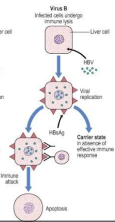
HBV & HCV → immuno-attack of hepatocytes
HCV - 75% asymptomatic
HBV - prevalence is lower due to vaccine administration
HCV - treated w direct-acting antiviral meds, which are oral drugs that cure the virus in most ppl within 8-12 weeks
pibrentasvir (Mavyret)
velpatasvir (Epclusa)
ledipasvir (Harvoni)
drug-induced liver injury
high incidence
10% adverse drug reactions
2 types
hepatocellular (tylenol) - hepatocyte death
cholestatic (methyl testosterone) - cholangiocyte injury
most common drugs
augmentin
diclofenac
azathiopurin
infliximab
nitrofurantoin
bile plug and feathering changes
central lobular necrosis
chronic liver injury; -hepatitis, fibrosis, cirrhosis, HCC etiology
hepatitis (most to least common)
obesity
alcohol
drugs
viral
autoimmune
less critical etiologies
iron overload
copper overload → Wilson’s disease (hepatolenticular degeneration)
alpha1-antitrypsin deficiency
autoimmune-biliary
PSC - progressing sclerosing cholangitis
PBC - progressing biliary cholangitis
global MASLD/NAFLD prevalence
MASLD: metabolic dysfunction-associated steatotic liver damage
NAFLD: non-alcoholic fatty liver damage
quite common in US

MASH
metabolic dysfunction associated steatohepatitis
NASH
non-alcoholic steatohepatitis
ASH
alcoholic steatohepatitis
North american incidence of MASLD, MASH ± fibrosis
MASLD (by ultrasonography) - 25.2%
MASH among MASLD patients - 21%
MASH patients who experience fibrosis (global) - 41%

North american incidence of MASLD - NAFLD and NASH ± fibrosis
N American prevalence of NASH + fibrosis - 2.1%
# of individuals in US with or will have NASH + fibrosis → 7,078,411
epidemiology
fatty liver diseases - most common cause of abnormal liver enzyme tests
liver steatosis affects 30-40% pop. in developed countries
fibrosis, cirrhosis, hepatocellular carcinoma % of children (35-50% of obese children)
affects 2-3% of children (35-50% of obese children)
ASH and MASH/NASH
histologic features - NASH
steatosis
lobular inflammation
hepatocyte ballooning
risk factors
female
obesity
dietary factors
polymorphisms
drinking pattern
smoking
comorbidity
viral hepatitis
hemochromatosis
HIV
characteristics
macrovesicular steatosis + inflammation
infiltration of both lymphocytes and Kupffer cells
ballooned hepatocytes that also contain Mallory-Denk bodies
perivenular/pericellular (“chicken wire”) fibrosis
NAFLD spectrum
fatty liver
fat accumulates in liver
NASH
fat plus inflammation and scarring
cirrhosis
scar tissue replaces liver cells
liver lipid homeostasis
HUHHH

molecular mechanism of MASH/NASH
two-hit (multi-hit) theory
first hit → fat accumulation in the liver
obesity
long-term fasting
additional/parallel hit → injury leads to inflammation
drugs
pathogens
leaky gut (LPS)
genetic abnormalities (PTEN, FXR, or other deficiency)
oxidative stress
ASH pathophysiology
hepatic fat storage
inflammation → inc hepatic uptake of gut-derived endotoxins triggering Kupffer cell activation and release of proinflammatory factors
oxidation → induction of cytochrome P4502E1 producing toxic acetaldehyde and ROS
ER stress → ethanol-mediated ER stress, protein mis-folding
alcohol inc hepatic lipid contents

ALD - inflammation mechanism
activation of innate immunity
inc pro-inflammatory cytokine production in all cells in the liver
inc adaptive immunity

liver fibrosis
persistent HSC activation → lots of connective tissue → collagen
fibrosis potentially irreversible
dec hepatic blood flow
numerous sequelae
no therapeutic agents
req liver transplantation

cirrhosis induced by MASH/NASH
NASH histologic features may no longer exist - “burnt out NASH”; the characteristic perivenular/pericellular fibrosis may still be present
HBV pathology
nodules of damaged and regenerating hepatocytes
fibrous bands w chronic inflammatory cell infiltrates (dark purple dots)
micronodular (<3 mm) vs macronodular (>3 mm) cirrhosis
leads to hepatocellular carcinoma (HCC)
ground-glass cytoplasm (light colored surface Ag particles)
bile pigment accumulation (dark brown)

Hepatitis B viruses
DNA virus structure
HBsAg - surface antigen
most abundant antigens, detectable at onset of clinical symptoms
HBcAg - core antigen
marker of viral replication
HBeAg - antigen between nucleocapsid and lipid envelope
marker of viral replication
anti-HBc IgM
acute infection or disease flare
anti-HBc IgG
chronic infection

phases of chronic HBV infection
proliferative phase
more HBeAG
virus proliferates
peak in HBV DNA causes peak in ALT → signals liver damage
dormant phase
??? dont know why
more anti-HBe → no replication
lays dormant in DNA
reactivation causes
mAb therapy
organ transplant
HIV co-infection
treatment resistance
stopping treatment

hepatitis C virus
RNA virus structure
positive-sense
single-stranded RNA virus from Flavivirdae family
each viral particle contains a single RNA strand
does not have its own proteins

HCV pathophysiology
HCV life cycle tightly linked to hepatocyte lipid metabolism
HCV assembly/release connected to fatty acid pathways and biogenesis of VLDL
spontaneous resolution of acute infection in 15-45% ppl
chronic infection if not treated//clinically undetected
predominantly immune response mediated injury
direct viral cytotoxic effect uncertain
innate immune system response to HCV infection
type I or type III interferon driven response
interferon stimulated genes and NK cells partially control viral replication but does not eliminate infection
adaptive immune system response to HCV infection
neutralizing antibodies target hypervariable envelope glycoproteins E1 and E2
short lived and nonpersistent in chronic infection
HCV specific CD8+ T lymphocytes mediate viral clearance thru cytolytic and noncytolytic functions
primed by HCV specific CD4+ T lymphocytes
vigorous, sustained cytotoxic T lymphocyte response needed for resolution of acute infection and permanent clearance
chronic Hep C infection
cord of liver parenchyma w a patchy distribution of chronic inflammation - lymphoid aggregates
prominent chronic inflammatory infiltrate of the portal tracts and the periportal region
comprised of lymphocytes w occasional plasma cells
interface hepatitis
scattered acidophil bodies - Councilman bodies
sinusoidal pattern of lymphocytic infiltrate
mild degree of steatosis

gall bladder ductal system
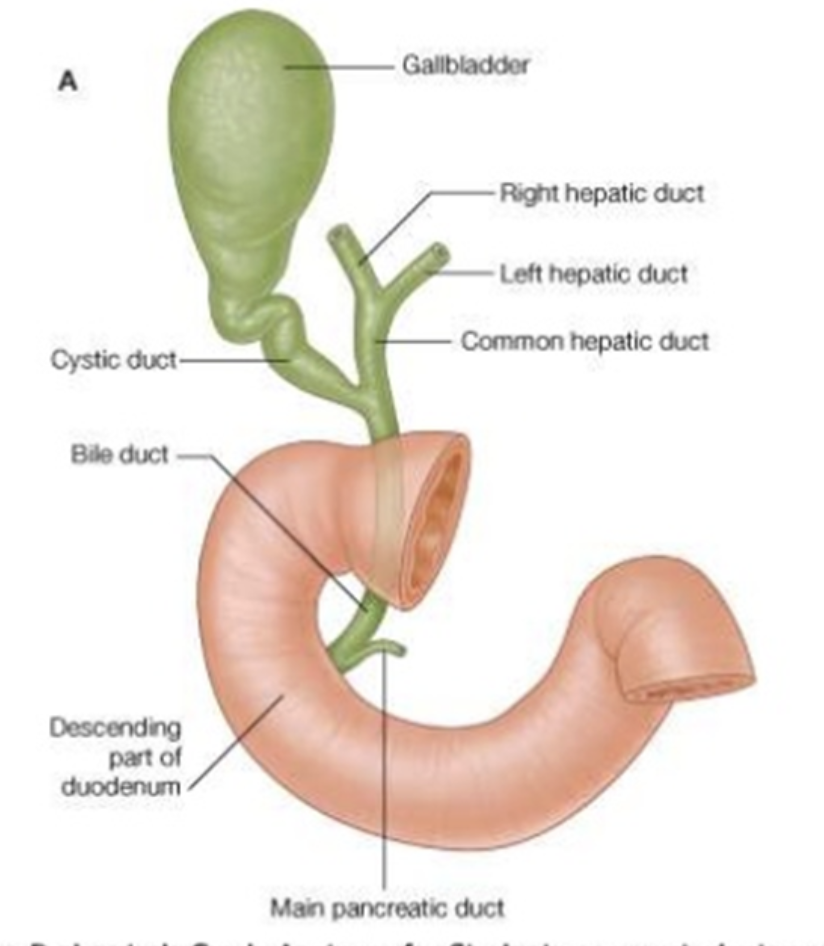
diseases of gall bladder
cholelithiasis (gallstone)
carcinoma of the bile duct
carcinoma of the gall bladder
biliary obstruction
cholelithiasis (gallstone)
gallstones can result from
excess of cholesterol
deficit of bile salts
cholesterol stones may form if
imbalance between ratio of cholesterol and bile salts
form micelles which have hydrophilic exterior enclosing and hydrophobic cholesterol

risk factors for gallstone disease
female
forties
fat
fertile
FXR mutation?
liver tumors - benign
liver cell adenoma
angioma
bile duct hamartoma
focal nodular hyperplasia
liver tumors - malignant
primary
hepatocellular carcinoma (HCC)
cholangiocarcinoma (adenocarcinoma of bile ducts)
angiosarcoma (malignant neoplasm of vascular endothelium)
hepatoblastoma (primary liver tumor in childhood)
secondary
metastases from the GI, lung, and breast cancers
colon cancers metastasize to liver
highly common in east Asia and central/south Africa
hepatocellular carcinoma mechanism
heterogenic → many diff pathways result in HCC
well differentiated
tumor cells resemble hepatocytes
form trabeculae, cords and nests
may contain bile pigment in cytoplasm
poorly differentiated
malignant epithelial cells are discohesive
pleomorphic
anaplastic
giant

NAFLD → HCC progression
fatty liver → NASH → Cirrhosis → HCC
abt 25% of patients never develop cirrhosis
cholangiocarcinoma
malignant tumors in the bile duct

L10 - GI
GI functions
ingest and prepare food for digestion
mechanical and chemical digestion
regulated absorption of nutrients
metabolism of endogenous and exogenous chemicals
immune surveillance
regulated secretion of hormones, enzymes, lubricants, and water
microbiome functions
propulsion of waste
regulated defecation
regions of GI
upper GI tract
mouth - mechanical & chemical digestion
esophagus
stomach
middle GI tract (small bowel)
duodenum - 9'“
jejunum - 2.5 m
ileum - 3 m
lower GI tract (large bowel)
cecum
colon
ascending
transverse
descending
rectum
sphincters between segments
accessory structures
salivary glands - release amylase
liver
pancrease
gall bladder - can live without it
sphincters of the GI
small bands of smooth muscle that constrict and dilate; most under involuntary control
upper esophageal sphincter (pharyngoesophageal)
moves food down esophagus
lower esophageal sphincter (gastroesophageal)
regulates food entry into stomach
pyloric sphincter (gastroduodenal)
regulates food going from stomach to small intestine (duodenum)
ileocecal valve/sphincter
ileum to cecum
internal anal sphincter
external anal sphincter
can be controlled to a certain point
gross anatomy of the GI tract

stomach
has extramuscular layer to aid in lots of mechanical digestion
first place of protein digestion
acidity helps denature
layers of the GI tract
for protection of tissues
serosa
longitudinal muscle
myenteric (Auerbach’s) nerve plexus
circular muscle
submucosa
submucosal (Meissner’s) nerve plexus
muscularis mucosae
mucosa
epithelial lining
GI cross section
muscles move food up & down, side to side, twisty
enteric nervous system “second brain of body”
myenteric nerve plexus
meissner’s nerve plexus
submucosal gland
muscles - prevent food from going backward; keep propelling bolus forward
longitudinal muscle - accordion; pinches and helps move by shortening & lengthening
circular muscle - rings; regulates diameter of lumen

enteric nervous system (ENS)
connects gut to brain via vagus nerve
location - gut wall from esophagus to anus
composition - cell bodies, axons, dendrites, nerve endings
innervation - gut cells, sensory nerves, other neurons
integration - can occur entirely within ENS; can function independent of ANS
transmitters - many excitatory and inhibitory
intrinsic control
ENS
myenteric (Auerbach’s) plexus
muscle
coordination of smooth muscle
submucosal (Meissner’s) plexus
regulates secretion and in turn absorption
extrinsic control
autonomic NS
in gut specifically; kinda opposite in rest of body
parasympathetic - mainly stimulates (ACh)
sympathetic - mainly inhibits (NE)
gastric mucosal barrier
protects the stomach from low pH
affect gut microbiome and happiness of gut bacteria
goblet cells → make mucus
NO and prostaglandins → control blood flow to stomach and help protect lining
sensory nerves → help chyme move around via peristalsis

basic major diseases of GI tract
constipation
diarrhea
gastroesophageal reflux disease
nausea and vomiting
acute viral gastroenteritis
overindulgence
motion sickness
constipation
infrequent passage of hard stools
causes
git (gastrointestinal tract) disorders
lack of dietary fiber and/or fluid intake
fiber → OTC (osmotic-based and stimulant based laxatives) and fruits and veggies (beans)
lack of motility
structural → carcinoma colon, diverticular disease
non git disorders
drugs: opioides, anticholinergics, aluminium salts
neurological: MS, Parkinsonism
metabolic/endocrine: DM, hypothyroidism
diarrhea
passage of loose or watery stools; greater 200 grams per day
causes deaths of children mostly in areas of poor water quality
common symptoms of many intestinal diseases including those due to infection, inflammation, ischemia, malabsorption, and nutritional deficiency
4 major categories of diarrhea
secretory diarrhea
isotonic stool and persists during fasting
problems w intestinal wall due to inc contractions
osmotic diarrhea
occurring w lactase deficiency
due to osmotic force exerted by unabsorbed luminal solutes
diarrheal fluid is at least 50 mOsm more conc than plasma
condition resolves with fasting
malabsorptive diarrhea
inadequate nutrient absorption is associated w steatorrhea (stinky stool cuz it pulls fat) and relieved by fasting
exudative diarrhea
due to inflammatory disease
characterized by purulent, bloody stools that continue during fasting
GERD (gastroesophageal reflux disease)
when acid comes up the relaxed lower esophageal sphincter
lower esophageal sphincter
dec esophageal clearance of acid - lying flat, alcohol, coffee
impaired mucosal defense - can erode lining of esophagus
impaired LES
transient LES relaxaations
hypotensive LES
hiatus hernia - upper part of stomach goes above diaphragm
stomach
inc acid output - smoking, coffee
H. pylori - protective effect
inc intragastric pressure - obesity, lying flat
dec gastric emptying - fat
inc bile reflux
meds for GERD
antacids → neutralizes acids as it comes down; ex: Tums
histamine blocker → 50% effective
PPI → very effective; inhibits H+ pumps
`
nausea and vomiting
higher centers:
hypothalamus
cerebellum
labyrinth
area postrema
vomiting center causes
closure of epiglottis - closes trachea & stops respiration
LES relaxation - releases acidity
somatomotor signals → inc abdominal P → wrenching of stomach; stomach squeeze; antiperistalsis
medications for nausea and vomiting
neurokinin1 antagonist
ex: serotonin, histamine, anti-emetics
complex major diseases of the GI tract
gastritis
inflammatory bowel disease (Crohn disease, ulcerative colitis)
irritable bowel syndrome
nausea & vomiting → complex; postoperative, chemotherapy-induced
peptic ulcer disease - including stress-related mucosal injury, GI bleeding
clinical manifestations of gastric disease
pain and dyspepsia
loss of appetite
bloating and distension
bleeding/vomiting
slow → black ‘coffee grounds’ vomit
rapid → bright red blood
‘mass’ effect mimicking satiety
outlet obstruction (pyloric stenosis) w profuse vomiting
hypokalemic alkalosis
change in pH
chemotherapy-induced emesis
chemotherapy → stimulates enterochromaffin cells of GI tract which release serotonin → attach to 5HT3 receptors and stimulate chemoreceptor trigger zone → stimulates vomiting center → controls vomiting
chemoreceptor trigger zone
5HT3 receptors
NK1 receptors
dopamine receptors
vomiting center
5HT3 receptors
NK1 receptors
dopamine receptors

gastric inflammations
gastritis (acute and chronic)
associated w enhanced gastric secretion and/or impaired gastric defenses
alcohol, smoking, infections (Helicobacter pylori, Herpes, Salmonella), corticosteroids, NSAIDs, stress, CNS trauma, shock, serious illnesses
chronic gastritis associated w anemia; common in elderly patients
H. pylori is most common cause
90% of patients w chronic gastritis are infected
autoimmunity
anemia → stomach produces factors for vit B absoprtion; B vit-linked anemia
degradation of wall which may cause bleeding
H. pylori
produces urease, inc ammonia production; basis of urea breath test
toxic to mucosal cells
hides in mucous layer
diagnosed thru biopsy
stimulates gastric emptying - overwhelming bicarbonate buffering
mechanisms of gastritis
healthy
damaging forces
gastric acidity
peptic enzymes
defensive forces
surface mucus secretion by goblet cells
bicarb secretion into mucus
mucosal blood flow
apical surface membrane transport
epithelial regenerative capacity
prostaglandin synthesis
injury
H. pylori infection
NSAIDs
aspirin
tobacco
alcohol
gastric hyperacidity
duodenal-gastric reflux
delayed gastric emptying
ischemia
shock
ulcer
accumulation of dead tissue
hole in stomach
fibrosis
acute inflammatory cells

peptic ulcer disease
occurs anywhere mucosa is exposed to acid gastric juice
often chronic, frequently reccurent
most common site → duodenum
extremely common
duodenal ulcers most common age 20-40
most common in patients w blood type O
resemble carcinomas
signs/symptoms
burning or gnawing epigastric pain w transient reduction of pain upon eating, bleeding, belching
pain worse 1-3 hrs after eating
at night
infection w H. pylori
genetic factors can contribute
clinical manifestations of intestinal disease
malabsorption
obstruction/constipation
‘paralytic’ bowel
ischemia
perforation
hemorrhage → tears and bleeds
diarrhea-rapid movement, often high volume, extremely dangerous in young and old
dysentery-blood and mucous
steatorrhea
IBS - inflammatory bowel syndrome
very varied
brain-gut dysregulation
altered motility
causes
neuronal plasticity
altered endocrine metabolism (serotonin)
transient infection
altered GI microbiome
genetic polymorphisms
inc mucosal permeability
bile acid malabsorption
altered mucosal immune function
food hypersensitivity
IBD - inflammatory bowel disease
idiopathic colitis
crohn disease
common in female teens
ulcerative colitis
common in female teens
GI issues are common among GLP-1 users
IBDs are usually idiopathic w multi-factorial origins
arise from mucosal immune activation and interaction between gut microbiome
both Crohn and UC
more common in females
often presenting in teens and 20s
inc long-term risk of Ca??
hygiene hypothesis for IBD
70% of lymphoid tissue is in GI → GALT
tm hygiene is skipping away normal bacteria needed to keep our immune system well
inc risk for more disease

Crohns disease
autoimmune related disease; US has highest IBDs per capita; lesions may occur anywhere (mouth to anus), but most common in terminal ileum
F > M; rare in young children; common in whites
etiology → unknown; can be triggered by stress
clinical signs → highly variable
acute → pain, diarrhea, fever
chronic → relapse and remission, weight loss, obstruction, malabsorption
anatomic → full thickness inflammation, ‘skip’ lesions, luminal narrowing (‘lead-pipe’ bowel, string-sign on X-ray)
multiple surgeries req leading to ‘short bowel syndrome’
suppression of inflammatory response used but has side-effects, particularly activation of Herpes virus
Ulcerative Colitis
inflammation of mucosa in the rectum and moving up the large intestine
most common in F>M, 20-30 years, US and Western Europe
etiology → unknown; episodes may be precipitated by psychological stress
‘sloughing’ of contiguous sheets of mucosa w bleeding
clinical presentation → pain, bloody diarrhea, pain and fever, exacerbations, remissions
inc risk of aggressive rectal adenocarcinoma after 8-10 years of disease
antacids
neutralizes gastric acids, inc serum bicarbonate (raises pH), or binds to intestinal phosphates
raises pH in GI tract by either inc HCO3 from kidneys or dec/neutralizing HCl acid released by stomach
ex: Al(OH)3, CaCO3, tums, sodium bicarbonate
PPI (proton pump inhibitors)
inhibits gastric acid production and secretion at the level of parietal cells in the stomach
reduces acid secretion in the stomach therefore helps ulcers heal and prevents further gastric ulcer formation
ex: omeprazole, pantoprazole
antisecretory agent prostaglanding E analog
inhibits gastric acid secretion and protects gastric mucosa
eliminates gastric acid and protects mucosa from damage
ex: misoprostol
gastric mucosal protectant
adhered to site of ulcerations in order to prevent further breakdown
helps to heal and protect gastric mucosa by adhering to surface
ex: sucralfate
histamine blockers (H2)
selectively antagonizes histamine H2 receptors to block gastric acid secretions
reduces gastric acids by blocking histamine receptors (histamine is normally released to block unwanted invaders, and inc acidity)
ex: ranitidine, famotidine
aminosalicylates
reduces prostaglandin synthesis to reduce inflammation
helps reduce inflammation in teh GI tract and elsewhere, thereby helping prevent flare-ups in diseases such as Crohn’s and UC
ex: sulfasalazine, mesalamine, olsalazine
emetics
stimulates the CNS to induce vomiting
induces vomiting for therapeutic benefit
ex: sorbitol, ipecac
L11 - diabetes
discovery of pancreatic diseases
Oskar Minkowski
found that pancreas is impt for glucose regulation
took pancreas out of dog and within 15 hrs it died of hyperglycemia
timeline of diabetes
1921 - discovery of insulin
1922 - first person treated w insulin - not recombinant & derived from dogs or cattle
1936 - type 1 and type 2 diabetes
1969 - structure of insulin found
1974 - monogenic forms of diabetes - immune-modulated
1982 - recombinant human insulin - more available for patients
1992-2018 - better understanding of diabetes thru genes
normal vs defective beta cells
T2DM - have insulin that is defective
dec islet number of beta-cell mass

islet of langerhans
reduced beta cell number
naturally smaller beta cell mass
defective beta cell proliferation
beta cell death
dysfunction
ER stress
oxidative stress
no insulin secretion → hyperglycemia
compromised identity
dedifferentiation (Ngn3+) - abnormal proportions
loss of identity
transdifferentiation

beta cells
in islet of Langerhans
secrete insulin
alpha cells
in islet of Langerhans
secrete glucagon
insulin structure
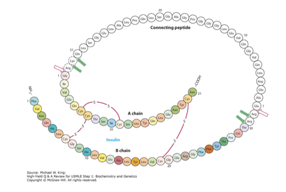
synthesized by INS gene in beta cells of islets of Langerhans
insulin and c-peptide (connecting peptide) are connected at 2 sites via dipeptide links (green dashes in pic)
half-life of abt 5 mins
1:1 ratio of breaking
1 - insulin
1 - c-peptide
can be used to measure both
tm insulin → insulin poisoning
insulin release
glucose enters cell via GLUT2 - passive movement of glucose
glucose is oxidized to generate ATP via mitochondrial oxidative phosphorylation
ATP inhibits the KATP channel which leads to membrane depolarization
membrane depolarization causes opening of Ca channels
Ca triggers the fusion of insulin-containing secretory vesicles w plasma membrane and release of insulin to circulation

insulin functions
transport glucose and amino acids
glycogen formation in liver and skeletal muscle
glucose transformation to triglycerides
nucleic acid syntheis
protein synthesis
dec degradation of glycogen, lipid, and protein
major anabolic hormone