Serine Proteases
 Call Kai
 Learn
 Practice Test
 Spaced Repetition
 Match
 Flashcards
 Knowt Play
1/19

There's no tags or description
Looks like no tags are added yet.
20 Terms
Serine Proteases (Overview)
This video will highlight important features of serine proteases as an example of how the structure of how enzyme active sites enable catalysis of a chemical reaction.
proteases, what do they
what is needed for proteolytic cleavage
products of proteolytic cleavage
Proteases cut or cleave peptide bonds.
Here is a diagram of a substrate showing the N terminus on the left, one peptide bond in the middle and the C terminus of the protein on the right.
With addition of water and a protease peptide bond can be cleaved resulting in n-terminal product and c-terminal product.
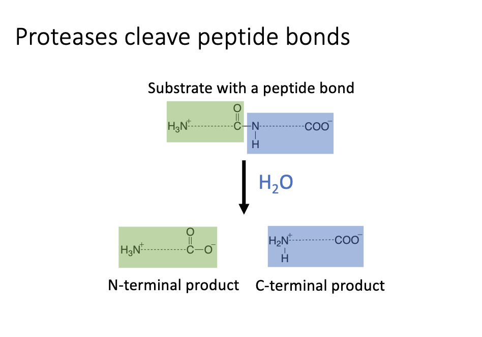
N terminal product has original N terminus and new C terminus
C terminal product has original C terminus and new N terminus
protease family names
Serine proteases are a family of proteases that include chymotrypsin, trypsin and elastase along with many others.
Key Features of Serine Proteases
serine proteases typically have a catalytic triad consisting of the amino acids aspartic acid, histidine and serine that are important for catalysis.
serine proteases have an oxyanion hole which is a space with a chemical characteristics that allow transition state stabilization.
The specificity pocket of serine proteases is the
space that binds to part of the substrate to enable substrate specificity
select which peptide bond will be cleaved in the reaction by positioning it in the right place within the active site.
Catalytic Triad
interactions between catalytic triad
The catalytic triad is made up of aspartic acid, histidine and serine.
In this image of the structure of the chymotrypsin active site, you can see that the three amino acids of the catalytic triad are close together in three dimensional space.

Here is a simplified representation of the catalytic triad of chymotrypsin. The rest of the protein is not shown and represented by the black and gray lines surrounding the catalytic triad.
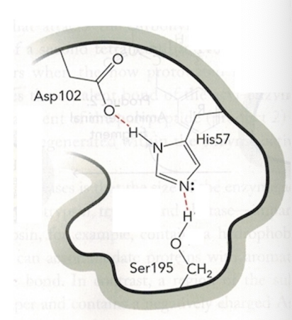
one electrostatic interaction, one H bond
A red dashed line represents an electrostatic interaction between the negatively charged oxygen of aspartic acid and the hydrogen atom of histidine.
In addition, there is a hydrogen bond formed between the lone pair of electrons of nitrogen atom on histidine, and the hydrogen atom of serine hydroxyl group, which is also represented by a red dotted line.
Oxyanion Hole
what forms the oxyanion hole
The Oxyanion Hole is an empty space in the enzyme active site that can accommodate an Oxyanion formed during the reaction.
The Oxyanion Hole has nearby chemical groups with positive or partial positive charges that can interact with the oxygen iron that enters the oxygen ion hole.
This representation of the Oxyanion Hole of Chymotrypsin shows hydrogens with partial positive charges that are part of the backbone amino groups. The partially positively charged hydrogen atoms are positioned so that they can interact with an Oxyanion located in the Oxyanion hole.

Glycine 193 and serine 195
Specificity Pocket
specificity pocket of chymotrypsin and trypsin
what amino acids
what do they bind to
The specificity pocket refers to a space that binds to specific substrates through non covalent interactions and positions the substrates within the active site for catalysis to occur.
The nature of the specificity pocket determines which peptide substrates bind and which peptide bonds are cleaved in the enzyme catalyzed reaction.
These diagrams represent the specificity pockets of chymotrypsin and trypsin

the chymotrypsin specificity pocket binds to aromatic amino acid side chains, including tyrosine represented here.
The trypsin specificity pocket contains a negatively charged aspartic acid, which binds well to long, positively charged amino acids, arginine and lysine.
4 amino acids
Gly193
catalytic triad
Ser195
His57
Asp102
diagram of the chymotrypsin serine protease active site
This diagram of the chymotrypsin serine protease active site shows the approximate locations of the catalytic triad specificity pocket and oxianion hole relative to each other.

Mechanism of Serine Protease Catalysis (Step-by-Step)
The next slides will walk through what happens in the active site of chymotrypsin during a reaction to show how each of these elements contributes to catalysis.
the substrate represented here binds to the enzyme.
In this diagram of the substrate bound to the enzyme active site, you can see that
the specificity pocket contains the pheneylalanine sidechain
the peptide bond that will be cleaved is positioned right near the serine of the catalytic triad.
The histidine of the catalytic triad deprotonates the serine Hydroxyl group, which is an example of base catalysis. The deprotononation of serine leads to a negatively charged oxygen.
negatively charged oxygen of serine attacks, the carbon atom of the peptide bond, which is an example of a nucleophilic attack. The double bond of the carbonyl group is converted to a single bond and the electron pairs move to the oxygen, creating a negatively charged oxygen also called an oxyanion.
Nucleophilic attack of the substrate by the enzyme forms the tetrahedral intermediate, which is a carbon atom with four bonds to the enzyme, the n-terminal side of the substrate, the c-terminal side of the substrate and the oxyanion. The high energy oxyanion is positioned in the oxyanion hole and is stabilized by electrostatic interactions with a partial positive charge from the hydrogen of backbone amino groups. The stabilization of the oxyanion in the tetrahedral intermediate is a main contributing factor to catalysis.
4A. Electrons from the oxyanion, reform the double bond with the carbon atom of the substrate backbone.
4B This causes movement of electrons that break the peptide bond and the c-terminal part of the original substrate is the leaving group.
4C A proton from histidine is transferred to the amino group, completing the formation of a new and terminus of the first product.
The first product, the C-terminal part of the original protein is completed and released from the active site.
The first product contains the new N terminus formed by cleavage of the peptide bond and the original C terminus of the substrate.
The active site now contains an Acyl enzyme Intermediate, referring to the carbon chain that came from the substrate, which is now COVALENTLY, linked to the serine of the enzyme.
5A Next a molecule of water shown in blue enters into the enzyme active site. The water is deprotonated by histidine, forming a negatively charged oxygen.
5B The negatively charged oxygen attacks the carbon that came from the substrate. The double bond of the carbonyl group is converted to a single bond and the electron pairs move to the oxygen, creating another oxyanion.
Nucleophilic attack of the substrate by the hydroxyl group from water forms a tetrahedral intermediate with a carbon atom connected to the hydroxyl group from water, the enzyme, the n-terminal part from the substrate and the oxyanion. The high energy oxyanion is positioned in the oxyanion hole and stabilized by electrostatic interactions with a partial positive charge from the hydrogens of the backbone amino groups. the stabilization of the oxyanion that is part of the tetrahedral Intermediate is a main contributing factor to catalysis.
6A Electrons from the oxygen ion reform the double bond with a carbon atom of the substrate backbone.
6B This causes movement of the electrons that break the bond (between substrate and serine of enzyme) with the enzyme, which is the leaving group. A proton from histidine is transferred to the oxygen of serine, restoring serine to its original state.
The second product, the N-terminal part of the original protein is now complete and released from the active site. The second product contains a new C terminus formed by the cleavage of the peptide bond and the original N terminus of the substrate.
The enzyme active site is reset to the original state and ready to catalyze another reaction
Catalytic mechanism step 1
the substrate represented here binds to the enzyme

Catalytic mechanism step 2
The histidine of the catalytic triad deprotonates the serine (of catalytic triad) hydroxyl group
which is an example of base catalysis.
The deprotononation of serine leads to a negatively charged oxygen.

Catalytic mechanism step 3
negatively charged oxygen of serine attacks the carbon atom of the substrate peptide bond
an example of a nucleophilic attack.
The double bond of the carbonyl group is converted to a single bond
the electron pairs move to the oxygen of substrate creating a negatively charged oxygen also called an oxyanion.

after 3
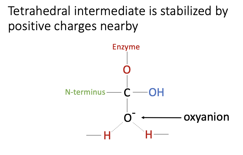
Nucleophilic attack of the substrate by the enzyme forms the tetrahedral intermediate, which is a carbon atom with four bonds to the enzyme, the n-terminal side of the substrate, the c-terminal side of the substrate and the oxyanion.
The high energy oxyanion is positioned in the oxyanion hole and is stabilized by electrostatic interactions with a partial positive charge from the hydrogen of backbone amino groups. The stabilization of the oxyanion in the tetrahedral intermediate is a main contributing factor to catalysis.

Catalytic mechanism step 4
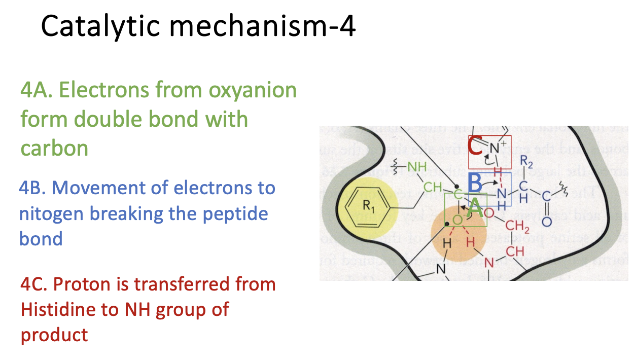
4A. Electrons from the oxyanion reform the double bond with the carbon atom of the substrate backbone.
4B. This causes movement of electrons to nitrogen (of substrate) that break the peptide bond and the C-terminal part of the original substrate is the leaving group.
4C A proton from histidine is transferred to the amino (NH) group of product, completing the formation of a new N terminus of the first product.
after 4
The first product, the C-terminal part of the original protein is completed and released from the active site.
The first product contains the new N terminus formed by cleavage of the peptide bond and the original C terminus of the substrate.
The active site now contains an Acyl enzyme Intermediate, referring to the carbon chain that came from the substrate, which is now COVALENTLY, linked to the serine of the enzyme.


Catalytic mechanism step 5
5A Next a molecule of water shown in blue enters into the enzyme active site. The water is deprotonated by histidine, forming a negatively charged oxygen.
5B The negatively charged oxygen (from water) attacks the carbon that came from the substrate. The double bond of the carbonyl group is converted to a single bond and the electron pairs move to the oxygen of substrate, creating another oxyanion.

after 5
Nucleophilic attack of the substrate by the hydroxyl group from water forms a tetrahedral intermediate with a carbon atom connected to the hydroxyl group from water, the enzyme, the n-terminal part from the substrate and the oxyanion. The high energy oxyanion is positioned in the oxyanion hole and stabilized by electrostatic interactions with a partial positive charge from the hydrogens of the backbone amino groups. the stabilization of the oxyanion that is part of the tetrahedral Intermediate is a main contributing factor to catalysis.
Catalytic mechanism step 6
6A Electrons from the oxyanion reform the double bond with a carbon atom of the substrate backbone.
6B This causes movement of the electrons that break the bond (between substrate and serine of enzyme) with the enzyme, which is the leaving group.
A proton from histidine is transferred to the oxygen of serine, restoring serine to its original state.

end
The second product, the N-terminal part of the original protein is now complete and released from the active site. The second product contains a new C terminus formed by the cleavage of the peptide bond and the original N terminus of the substrate.
The enzyme active site is reset to the original state and ready to catalyze another reaction

