Myology 1 & 2
 Call Kai
 Learn
Learn Practice Test
 Spaced Repetition
 Match
Match Flashcards
 Knowt Play
1/104

There's no tags or description
Looks like no tags are added yet.
Name | Mastery | Learn | Test | Matching | Spaced | Call with Kai | Chat |
|---|
No analytics yet
Send a link to your students to track their progress
105 Terms
latin term for musculus?
little mouse
The piriformis muscle is associated with which gluteal muscle?
middle gluteal
explain the musculus origin:
Shape and movement of some muscles (biceps) thought to resemble mice
Generation of body heat
shivering
Storage is paired with:
glycogen
list the muscle functions:
Movement of the body
Maintenance of posture and body position
Movement of substances inside the body
Generation of body heat
Storage
Provide support
list the muscle types:
smooth, cardiac, skeletal
smooth muscle characteristics
aka - visceral, unstriated, or involuntary muscle
no striations
Specialized for slow and steady contractions
Muscle cells are spindle shaped, mono-nucleated
innervated GVE neurons of ANS
smooth muscle locations
Hair follicles of skin, walls of hollow organs, walls of blood vessels, capsule of spleen, eyeball
cardiac muscle characteristics
make up the bulk of the heart
branched cells
central nuclei
joined by intercalated discs
striations
innervated by GVE neurons of ANS
characteristics of skm
40% of body weight, most attached to bones
striated, voluntary, or somatic
long, cylindrical, multinucleated (peripherally) arranged into bundles with c.t envelopes
innervated by GSE nerves
skm organization
enclosed in epimysium
epimysium- is continuous with fascia and the tendon binds muscle to bone
perimysium- wraps several fascicles in thin CT layer
endomysium- individual muscle fiber (cells) in fascicles that are surrounded by delicate c.t layer
skm unit organization
each muscle fiber is made of myofibrils
within myofibrils there are myosin + actin filaments organized in sarcomeres
sarcomeres separated by z discs
tendon
attach muscles to bone with CT
APONEUROSES
Flat sheets of densely arranged collagen fibers
used to denote any connective tissue sheet
apo- edge ; neuron= tendon?
where do muscles attach directly to?
the fleshy periosteum
ex. the attachment of the serratus ventralis m. to the serrated face of the scapula
other CT attachments
fascia
thin sheath of CT layer enclosing muscle or other organs
Superficial fascia
site for fat storage, closely associated with skin, often contains cutaneous muscles
deep fascia
fascial compartments: Covers and separates muscles enclosing them in a ‘fascial envelope or sleeve’
consisting of mostly collagen fibers
Fibers oriented in the same direction as tension and stress forces acting on the muscle
serve as origin or attachment sites
clinical use of deep fascia:
holds sutures well, allows for bloodless separation of muscle
cons- direct drainage of fluids or spread of infections + inflammation/swelling of muscle or tendon affects other structures
what are the regional thickenings in deep fascia?
retinacula (retinaculum) and annular ligaments (ring shaped)
Retinacula
aka retinaculum- that which holds or binds; band
ex. flexor retinaculum + extensor retinaculum
tendon structure
similar to skm
largely composed of collagen fibers
subfascicle- primary bundle; collagen fibers form
secondary bundles of fascicles
group of primary bundles in tendon
tertiary bundle- group of secondary bundles
tendon- group of tertiary bundles
CT layers of tendons
endotenon- surrounding individual tendon fibers
epitenon- surrounds the entire tendon with CT sheath
paratenon- epitenon surrounded by a loose areolar CT layer
tendon protection
tensile strength
can be damaged by excessive pressure or friction; when they change direction over bony prominences or hard tissues
places that provide tendon protection
synovial bursae, tendon sheath, sesamoid bones
Synovial bursae
bursa is a flattened sac containing synovial fluid to facilitate the gliding of a tendon over bone
types of synovial bursae
congenital bursa- present at birth (ex. Calcanean bursa)
induced bursa- develop SQ due to chronic outside pressure
burtitis
inflammation of bursa
tendon sheath
develop as an elongated bursa applied to one surface of a tendon
visceral layer (synovial sheath)
where the tendon sinks into the synovial bursa and is surrounded by it
different forms of tendon sheaths
parietal layer, mesotendons, visceral layer (synovial sheath)
parietal layer
External layer connected to the neighboring CT or periosteum
mesotendons
elongated mesentery that connects visceral and parietal layers
provide vascular and sensory nerve supply to tendon
Synovial Structures on the Thoracic Limb
Intertubercular (bicipital) bursa- not a true bursa/tendon sheath but extension of the shoulder joint capsule
define a muscle origin:
denotes the more fixed point of muscle attachment
define a muscle insertion:
denotes the more movable point of muscle attachment
insertion moves with the contraction
insertion of a muscle is always considered to be distal to the origin
hypertrophy
enlargement of the muscle fibers (increase in contractile proteins) resulting in increase in total mass of a muscle
Atrophy
decrease in size or shrinking of muscle fibers (loss of contractile proteins) and thus the muscle
disuse atrophy
neurogenic atrophy (denervation)
Extrinsic muscles
Muscles that attach the limb to the body/trunk
ex. trapezius
Intrinsic muscles
Muscles that are on the limb
deltoideus
muscle actions
when a muscle or its tendon crosses a joint and the muscle contracts, it causes movement
flexion and extension
agonist
prime mover
any muscle capable of producing a designated action on a joint
Antagonist
any muscle that has the opposite action on that joint
agonist for extension of elbow joint
ex. triceps brachii m.
agonist for flexion of the elbow joint
ex. biceps brachii + brachialis m.
Triceps brachii m. agonist vs antagonist:
agonist for elbow joint extension BUT an antagonist to elbow joint flexion
Biceps brachii & brachialis m. agonist vs antagonist:
Both agonists for elbow joint flexion BUT antagonists to elbow joint extension
Fixators
any muscle that stabilizes the origin of the agonist and the joint that the origin spans
Synergists
muscles that work together to create a movement
Strap-like
fibers run parallel to the long axis and throughout the length of the muscle
ex. gracilis and biceps femoris m.
Spindle-shaped
2 headed muscles- biceps (one in dogs)
3 headed muscles- triceps (4 in dogs)
2 bellied muscles- digastricus
Pennate muscles
Possess tendons throughout their length
unipennate- muscle with tendon
bipennate- muscle with a tendon on each side
multipennate- muscle with tendons distributed throughout its volume
ex of bipennate:
Infraspinatus m.
ex of multipennate:
Humeral head of DDF
ex of Unipennate:
Ulnar and radial head of DDF (deep digital flexor)
sphincter
ex of external + sphincter muscle shape (circular); anus
6 pack muscle is ex of what muscle shape/form?
wide muscle with tendinous tissue
rectus abdominus
rectus refers to what?
anything straight
ex of circular m.:
orbicularis orbis + orbicularis oculi
what muscle is in the category of wide muscle with aponeuroses?
serratus dorsalis
EAO- external abdominal oblique
IAO- internal abdominal oblique
define motor unit
One motor neuron and all the muscle fibers it innervates
stimulated to contract by action potentials produced in alpha lower motor neurons
motor unit innervation:
Every muscle fiber (cell) is innervated by a LMN nerve cell, originating in the CNS
single neuron branches to innervate more than one muscle fiber
explain contraction
Contraction of a muscle fiber is an all or none event
Force of contraction determined by the number and size
of motor units activated
sizes of motor units:
Small motor unit : a single neuron innervating a few muscle fibers
Large motor units : a single neuron innervating several hundred muscle fibers
Order of activation or recruitment of motor units is from small to large
muscle spindles
provide feedback on muscle length
protective function
golgi tendon organs
provide feedback on muscle tension
protective function
patellar reflex
Tapping on the patellar ligament results in stretching at musculotendinous junction
stimulates muscle spindle
reflexive contraction
muscle groups on the limb
Grouped based on their location and action
Often the origins or insertions within a group are
very similar
Muscles in a group are often innervated by the
same nerve
ex of location and action grouped muscles:
cranial and lateral muscles of the forearm
extensors of carpal joints or carpal and digital joints
ex of muscles grouped by origins or insertion
extensors all originate from lateral epicondylar region of the humerus
ex of muscles grouped due to innervation by the same nerve
all innervated by the radial nerve
Caudal muscles of the brachium
extensors of the elbow joint ; all insert on the olecranon
tensor fasciae antebrachii(11), triceps
brachii(6,6”,6”), anconeus mm
Cranial muscles of the brachium
flexors of the elbow joint ; both insert on the radial and ulnar tuberosities
biceps brachii(12), brachialis(8) mm.

Craniolateral forearm muscles
extensors ; carpal and digit joints ; all originate from the lateral epicondylar region of the humerus
extensor carpi radialis, common & lateral digital extensors, abductor digiti I longus

Caudomedial forearm muscles
flexors ; carpal and digital jts ; originate from the medial epicondyle of the humerus
flexor carpi radialis/ulnaris,
superficial & deep digital flexors
Lateral muscles of the rump
hip ; includes all gluteal muscles & tensor fascia latae m.
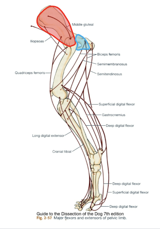
Caudal muscles of the hip
stabilizers
counter the rotation caused by the gluteals during hip joint extension
caudal muscles of the thigh
all originate from the ischiatic tuberosity ; hamstring
extend the hip variable action on the genual joint

cranial muscles of the thigh
flex the hip and extend the genual joint
Quadriceps femoris m. and iliopsoas
medial muscles of the thigh
Most are adductors of the limb
craniolateral muscles of the crus
flex tarsal joints and extend digital joints
Caudal muscles of the crus
extend tarsal joints and flex digital joints

what are muscles named based on?
direction of fibers
size & shape
muscle action
#of origins
location
origin and insertion
define direction
orientation of muscle fascicles relative to the body’s midline
rectus - parallel to rectus - rectus abdominis
transverse - perpendicular to midline - Transversus abdominis
oblique - diagonal to midline - external abdominal oblique
size greek abbreviations (brevis-, latissimus, longissimus, magnus, vastus, longus)
longus = long
brevis = short
latissimus = widest
longissimus = longest
magnus = large
vastus = huge
match the shape greek root meanings

Sphincter ; Tensor
Decreases size of an opening ; Makes body part rigid
define location
structure near which a muscle is found
temporalis, muscle near temporal bone
orgin and insertion for Sternocleidomastoid
originating on sternum and clavicle and inserting on mastoid process of temporal bone