PT-728 Differential Diagnosis Exam 1
 Call Kai
 Learn
Learn Practice Test
 Spaced Repetition
 Match
Match Flashcards
 Knowt Play
1/191

There's no tags or description
Looks like no tags are added yet.
Name | Mastery | Learn | Test | Matching | Spaced | Call with Kai | Chat |
|---|
No analytics yet
Send a link to your students to track their progress
192 Terms
Factors for Screening
Side effects of medications
Comorbidities
Visceral pain mechanisms
Reasons to Screen
Direct Access
Quicker and sicker patients
Signed prescription
Medical specialization
Disease progression
Patient disclosure
Inaccuracy of patient report to MD
Primary prevention
Stopping the process that lead to development of disease
Secondary prevention
Regular screening for early detection
Screening for Referral
Determining if individual has a condition that can be addressed in PT or if a referral is necessary
Red Flags: looking for patterns during examination
-Past Medical & Family History
-Risk Factors
-Clinical Presentation
-Pain Pattern
-Signs and Symptoms
•Medical Screening
method for detecting disease or dysfunction before medical care
Physical therapy diagnosis
the comparison of NMS symptoms to identify impairment classifications so that we can treat movement dysfunction
Patient/Client management
Examination
-Patient Interview
-Medical Screening
-Systems Review
-Physical Examination
Evaluation
Diagnosis
Prognosis
Intervention
Examination
Tailored process of:
-Obtaining the history
-Completing a medical screening
-Conducting a systems review
-Performing tests and measures
Purpose of Thorough Examination
Limit doubt
Practice according to the diagnostic evidence
Allow provider to proceed:
-With confidence
-With reasonable doubt
-With concurrent referral
Referral in lieu of treatment
PQRST of patient interview
-Palliating and Provoking/ Previous Symptoms
-Quality
-Region
-Severity
-Timing
Pain referral chart

Presence of serious pathology is suggested by
1) Pain that is worse during rest vs activity
2) Pain that is worsened at night or not relieved by any position
3) Poor response to conservative care including a lack of pain relief with prescribed bed rest
4) Poor success with comparable treatments
Red Flags - Category 1
Blood in sputum
Loss of consciousness or altered mental status
Neurological deficits not explained by monoradiculopathy
Numbness or paresthesia in perianal region
Pathological changes in bowel or bladder
Pattern of symptoms not compatible with mechanical pain
Progressive neurological deficit
Pulsatile abdominal masses
Red Flags - Category 2
Age > 50
Clonus
Fever
Elevated sedimentation rate
Gait deficits
History of disorder with predisposition for infection or hemorrhage
History of metabolic bone cancer
History of cancer
Impairment of precipitated by recent trauma
Long term corticosteroid use
Long term workers compensation
Nonhealing wounds or sores
Recent history of unexplained weight loss
Writhing pain
Red Flags - Category 3
Abnormal reflexes
Bilateral or unilateral radiculopathy or paresthesia
Unexplained referred pain
Unexplained significant upper or lower limb weakness
Referral flow chart

Constitutional Symptoms
Nausea/Vomiting
Change in bowel habits
Night Sweats/Day Sweats
Fatigue
Sleep pattern
Fever
Weight Loss
Syncope/Dizziness
Shortness of breath
Cholesterol levels
HDL
Women: >50
Men: > 40
LDL < 100
Total: < 200
Cardiovascular System: Upper Quarter
Difficulty breathing/SOB
Nausea
Syncope/Dizziness
Fatigue
Angina
Palpitations
Symptoms related to activity
Medications
(Nitroglycerin and oxygen)
Cardiovascular System: Lower Quarter
Arterial claudication
Deep Venous Thrombosis
Swelling
Varicose veins
Dry, shiny skin
Loss of hair growth
Poor Nail Condition
Rubor on Dependency
Esophagus pain referral

Large intestine pain referral

Small intestine pain referral

Stomach pain referral

Pancreas pain referral

Endocrine system
Fatigue
Weight Loss
Increased urinary frequency
Anxiety
Changes in hair growth/non-healing wounds
Diabetic changes in vascularization
Fasting blood sugar prior to therapy sessions to determine appropriateness
Hepatic/Biliary system
Nausea
Jaundice
GI Bleeding/Symptoms
Ascites
Fatigue
Chills
Vomiting
Weight loss
Pain
5th Vital Sign
-HR, BP, RR, Temp.
Understand how and when a diseased organs can refer pain to the neuromusculoskeletal system helps us identify suspicious pain patterns
-Chest, back, shoulder, scapula, pelvis hip, groin, and SI joint are the most common sites of referred pain from a systemic disease process
Mechanisms of Referred Visceral Pain
Visceral pain (pain from internal organs) is not well understood
Viscerosenseory fibers ascend the anterolateral system to the thalamus, with fibers projecting to several regions of the brain
The regions encode the site of origin of the viscera pain, but unfortunately they do it poorly
Embryologic Development
Pain is referred to a site where the organ is located in fetal development.
-Think of heart and kidney
-Ear & kidney come from same embryonic tissue
Multi-Segmental Innervation
Viscera have multisegmental innervations
Visceral-organ cross-sensitization
-Referred visceral pain to somatic tissues based on overlapping or same segmental projections of spinal afferent neurons to the spinal dorsal horn
Think of Cardiac Pain
-C5 goes to heart = have C5 (shoulder) pain during MI
-Can occur in any structure innervated by C3-T4 including jaw, neck, upper trap, shoulder, and arm
Direct Pressure and Shared Pathways
Ganglions from each neural system gathering and sharing information through the cord to the plexuses where the response is decided
Anything that impinges the central diaphragm can refer pain to the shoulder
Anything that impinges the peripheral diaphragm can refer pain to the ipsilateral costal margins and/or lumbar region
Head of pancreas impinges on central diaphragm
Tail of pancreas impinges on peripheral diaphragm
Assessment of Pain and Symptoms
Interview techniques and specific questions for pain assessment
-Need more than just a number on pain scale
Include detailed health history, physical examination, medication history, assessment of functional status, consideration of psycholosocial-spiritual factors
Elderly
-An accepted part of the aging process
-OTC analgesic meds for more than 6 months
-Loss of Independence?
-Early Stages of Dementia
Young
-Never underestimate that a young client is unable to answer questions about pain.
-Various scales
-Lack of cooperation, withdrawal, acting out, distractibility or seeing comfort, altered sleep patterns vocalizations and eating patterns.
Characteristics of Pain
Location
Description of Sensation
Intensity
Duration
Frequency
-constant vs. intermittent
-want symptoms to change w/ provocation
Pattern/Source
-MSK vs. emotional vs. visceral
Cutaneous
Somatic
visceral
neuropathic
referred
Cutaneous pain
Related to the skin: superficial somatic structures located in the skin and subcutaneous tissue
Able to point to pain
organs can refer pain to skin
Somatic pain types
Deep somatic:
-poorly localized and may be associated with an autonomic phenomenon
Superficial somatic
-involve the skin, superficial fascia, tendon sheaths and periosteum
Somato-visceral
-myalgia causes functional disturbance of the underlying viscera
Somato-emotional (psychosomatic)
-sources of pain occur when emotional or psychologic distress produces physical symptoms
Viscero-somatic
-visceral structures affect the somatic musculature
Visceral pain
Sources include the internal organs and the heart muscle
Not well localized
Visceral fibers synapse at the level of the spinal cord close to fibers supplying specific somatic structures
-When they converge, the nervous system has trouble interpreting the input which can lead to unclear impulses
Viscero-Viscero Reflex
-occurs when pain or dysfunction in one organ causes symptoms in another organ
Characteristics of Viscerogenic Pain
Gradual, progressive and cyclical pain patterns
Constant pain
-red flag
Physical Therapy Intervention "Fails"
Bone pain and aspirin
Pain does not fit expected pattern
-
How Do We Differentiate Between Somatic and Visceral Pain??
Superficial Somatic:
sharp and more localized
Deep Somatic:
dull, deep ache
responds to rest or non weight bearing
Visceral:
dull and ache but not feel better after rest
It is less likely that the client with back, hip, SI or shoulder pain that has been present for the last 5-10 years is demonstrating a viscerogenic cause of symptomsNeuropathic
Neuropathic pain
Damage or pathophysiologic changes of peripheral or central nervous system
Malfunction of nervous system
Usually described as sharp, shooting, burning, tingling, or electric shock sensation
Not alleviated by opiates or narcotics
-gabapentin
-lyrica
Referred pain
Felt in an area far from site of the lesion but supplied by the same or adjacent neural segments
Occurs by way of shared central pathways for afferent neurons and can originate from any somatic or visceral source
Questions to ask...
-Are you having any pain anywhere else in your body?
-Are you having symptoms of any other kind that may or may not be related to your main problem or concern
Tension pain
difficult to find a comfortable position
inflammatory pain
constitutional signs and symptoms
visceral peritoneum is less localized than parietal
Ischemic
sudden, intense, constant and progressive
Myofascial pain
occurs when prolonged muscular contraction or cocontraction results in
-local ischemia
-increased cellular metabolites
-subsequent pain
Faulty ergonomic, prolonged work position, or repetitive motion
increased release of substance p
Joint Pain
morning pain
-systemic
no morning pain
-MSK
Muscle Spasm
sudden involuntary contraction of muscle or group of muscles:
Somatic-somatic response: -painful musculoskeletal problem may also have a varying degree of reflex muscle spasms to protect joint
viscero-somatic: painful visceral disease can have muscle spasm of the overlying musculature
Muscle Trauma
acute trauma, burns, crush, or unaccustomed intensity or duration of muscle contraction
Broken fibers leak potassium into interstitial fluid setting off cascade of chemical reaction within the muscle
Rhabdomyolysis symptoms
-Red/brown urine
-Decreased urine output
-Fever
-Chills
-Tachycardia
-Nause/vomiting
-Confusion
Muscle Deficiency
weakness and stiffness
connective tissue changes may occur as small amounts of fibrinogen leak from vasculature into intracellular spaces, adhering to cellular structure causing muscular stiffness
Trigger Points
Hyperirritable spots within a taut band of skeletal muscle or in fascia
tender to palpation, local tenderness:
history of immobility, prolonged or vigorous activity:
reproduced w/ palpation or resisted motion:
Referred pain from a trigger point is characteristic and specific to every muscle
Active trigger point
pain when pushed
may refer pain to other site
pain at rest
Latent trigger point
pain when pushed
may refer pain to other site
NOT have pain at rest
Screening For Emotional and Psychologic Overlay
Anxiety: amplifies physical symptoms
Depression: identified factor that delays recovery for clients
Panic Disorder: sudden, unprovoked feelings of terror or impending doom with associated physical symptoms
-mimic MI
-muscle soreness afterwards
Illness Behavior Syndrome
Illness behavior is what people say and do to show they are ill and perceive themselves as sick or in pain:
-expresses and communicates the severity of pain and physical impairment
Most identified in people with chronic pain: expression depends on what and how the client thinks of their symptoms/illness
Conscious symptoms magnification is malingering
Unconscious symptoms magnification is illness behavior
Malingering
is a self-destructive, socially reinforced behavioral response pattern consisting of reports or displays of symptoms that function to control the life of the sufferer
-acts as if the future cannot be controlled because of the presence of the symptoms: "my back pain won't let me..."
-exaggerate limitations beyond those that seem reasonable in relation to the injury, apply minimal effort on maximal performance test, and overreact to physical loading during objective examination
Waddell's Nonorganic Signs
Five nonorganic signs and seven nonanatomic/ behavior descriptions of symptoms to differentiate between physical and behavior causes of back pain
3 or more positive signs: nonmovement dysfunction
A positive findings does not suggest an absence of pain but rather a behavior response:
does not confirm malingering or symptom magnification nor does it imply the nonexistence of physical pathology
Waddell's Nonorganic Signs defined
1) Superficial & Nonorganic Tenderness
-overreaction
2) Axial Loading & Acetablular Rotation
-axial loading thru head = reaction
-arms locked next to hips = reaction
3) Distraction
-PSLR test = reaction
-Quad test = no pain
4) Regional Sensory distribution & weakness
-weakness doesn't make sense
5) Overreastion
-Exaggerated response
3 or more positives on Waddell's Sign
Potential non-movement dysfunction
Nonmovement dysfunction
clinical pattern of nonmechanical, pain-focused behavior:
predictive of poor outcome and associated with delayed return to work or not working
Classification of Blood Pressure table
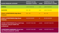
Orientation
Client's ability to answer questions about the person place, time and situation
Fruity (Sweet) Breath
could be sign of diabetic ketoacidosis
Bad Breath
could be a sign of
dental decay, lung abscess, throat or sinus infection GI disturbance, Bowel obstruction
Vitals
Vitals
Pulse
Blood Pressure
Temperature
Oxygen Saturation
Pain
(now called the 5th vital sign)
Walking speed
(the 6th vital sign)
Blood pressure and temperature are two most valuable vitals signs for screening for systemic diseases
Vital signs are the single easiest, most economic, and fastest ways to screen for many systemic illnesses
Pulse Rate
Normal resting pulse rate 60-100 beats per minute
Carotid and radial are preferred sites, but also can assess brachial, ulnar, femoral, popliteal, dorsalis pedis, posterior tibial
Pulse rate vs Heart rate
Pulse strength (amplitude)
gives an indication of the circulation blood volume and strength of left ventricle ejection
0 Absent, not palpable
1+ Pulse diminished, barely palpable
2+ Easily palpable, normal
3+ Full pulse, increased strength
4+ Bounding, too strong
Pulse Abnormalities
Weak pulse beats alternating with strong beats
Weak, thready pulse
Bounding pulse
(throbbing pulse followed by suddent collapse or decrease in force of pulse)
Two quick beats followed by a pause
Irregular rhythm
Pulse amplitude decreases with inspiration/increase with expiration
Pulse too fast
(Tachycardia)
Pulse too slow (Bradycardia)
Pulse should increase w/ inspiration and decrease w/ expiration
Respirations
Try to assess without drawing attention
Normal rate is 12-20 breaths per minute
Observe for use of accessory muscles, silent or nosiy breathing, puffed cheeks, pursed lips, nasal flaring
Pulse Oximetry
Oxygen Saturation on Hemoglobin (SaO2)
Normal range at rest and during exercise is 95% to 100%
Referral for medical evaluation advised when resting levels fall below 90%
Blood Pressure
Measurement of pressure in artery at the peak of systole (left ventricular contraction) and during diastole
(heart rest after closure of aortic valve)
Do not apply cuff above an intravenous line where fluids are infusing
It is recommended that patients who have undergone axillary node dissection avoid BP measurements on affected side
If a repeat measure is needed, wait 1 minute between measurements to avoid damaged blood vessel and inflammatory response
Yellow flags for BP
SBP greater than 120 and or DBP greater than 80 in the presence of risk factors (age, medications, personal or family history)
Decrease in DBP below 70 in adults age 75 or older
Persistent rise or drop in BP over time
Steady fall in BP over several years in and adult over 70
Lower standing SBP (less than 140) in adults over 65 with history of falls
Difference in pulse pressure greater than 40 mmHg
(60-80 is yellow flag)
More than 10mmHg difference in SBP or DBP from side to side in UEs
More than 40mmHg difference in SBP or DBP from side to side in LEs
BP in LEs is lower than in the UEs
DBP increases more than 10mmHg during activity or exercise
SBP does not rise as workload increases
SBP exceeds 200 during exercise, DBP exceeds 100 during exercise
BP changes in presence of other signs such as first-time onset angina, dizziness, nausea, pallor, or extreme diaphoresis
Sudden fall in BP
(more than 20 mmHg SBP or 10 mmHg DBP)
with
concomitant rise (10-20% rise) in pulse
Pulse Pressure
Difference between systolic and diastolic BP readings
Normally around 40mmHg
60-80mmHg difference is a yellow flag
Increase in pulse pressure is linked to higher risk of stroke and heart failure
Decrease in pulse pressure could indicate possible congestive heart failure or a blood loss
Pulse pressure does increase normally during exercises as we expect SBP to increase and DBP to remain about the same
•Systolic Blood Pressure increases with increased activity in a linear fashion
In a normal healthy adult:
Minimal to Moderate Exercise:
Expect increase in SBP 20mmHg or more
Intense Exercise:
Expect increase in SBP 40-50mmHg or more
ACSM suggests response to exercise is a progressive rise 10mmHg + 2 mmHg for each metabolic equivalent (MET)
Diastolic blood pressure should remain stable, may even see a small decrease
ACSM recommends reducing or stopping exercise if:
-SBP exceeds 200mmHg
-DBP exceeds 100mmHg
Lower standing SBP (less than 140) is and independent predictor of loss of balance and falls in adults over 65
In older adults decrease in BP may be an early warning sign of Alzheimer's disease
Body Temperature
Oral body temperature ranges from 96.8 to 99.5 (average 98.6)
Fever is likely indicator of infection
Call doctor if temperature rises above 101
Walking Speed: The 6th Vital Sign
General indication of function
Reliable, valid, sensitive measure of functional ability with predictive values in assessing future health, functional decline, hospitalization, or mortality
Timed 10 meter walk test
Low gait speed (at risk for falls/admissions/adverse events):
Pressure Ulcers table

Arterial vs. Venous Insufficiency table

Systolic Blood Pressure with doppler
Check SBP using dorsalis pedis and posterior tib
Divide the higher of each lower extremity by the highest brachial pressure
0.9 or less indicative of PVD and requires possible referral
1.0-1.3 is considered normal
Well's Criteria for DVT

Nail Bed Assessment
Systemic changes affect fingernails and toenails, but signs are typically more prominent in the faster growing fingernails.
Normal individual you can press or blanch the nail bed of a finger or toe and have a whitening effect.
Refill time should occur within 3 seconds
If the capillary refill time exceeds 3 seconds, the lack of circulation could be due to arterial insufficiency
Lymph Node Palpation
Look for obvious areas of swelling or redness
"Normal" lymph nodes usually are not visible or eaily palpable
Enlarged, visible, or palpable lymph nodes could indicate cancer, infection, virus, bacteria, allergies, thyroid conditions, or even food intolerances.
Neurological Screen Examination
Six major areas to assess
-Mental and Emotional Status
-Cranial Nerves
-Motor Function
-Sensory Function
-Reflexes
-Neural Tension
Cranial Nerve table
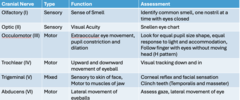
Cranial Nerve table

Motor Assessment - UQ

Motor Assessment - LQ

Reflex scale
0 No response, absent
+1 Low normal, decreased, slight contraction
+2 Normal, visible muscle twitch
+3 More brisk than normal, increased or exaggerated
+4 Hyperactive, very brisk, clonus
Deep tendon relfexes
Biceps (C5)
Brachioradialis (C6)
Triceps (C7)
Patellar (L3-4)
Achilles (S1-2)
Neural Tension
-Inability of nerve to glide in its protective sheath
-Clinical result can be numbness, tingling, pain
-Could be cause by disc protrusion, scar tissue, space occupying lesion (cyst, bone spur, tumor, cancer, disc)
-Does not tell etiology, but that peripheral nerve is involved