Learn: CH 3 Reproductive A&P & Ch 4 Hereditary and Environmental influences on childbearing | Quizlet
 Call Kai
 Learn
Learn Practice Test
 Spaced Repetition
 Match
Match Flashcards
 Knowt Play
1/81

There's no tags or description
Looks like no tags are added yet.
Name | Mastery | Learn | Test | Matching | Spaced | Call with Kai | Chat |
|---|
No analytics yet
Send a link to your students to track their progress
82 Terms
Sexual development: prenatal
• Genetic sex is determined at conception.
• Reproductive systems of males and females are similar for the first 6 weeks.
• Differentiation of the external genitalia is complete by about 12 weeks.
• Both ovaries and testes, gonads- analogous organs, secrete their primary hormones.
When can you discover your baby's gender?
Early as 6 weeks of pregnancy through at-home DNA blood tests and late as 18-22 weeks during a standard mid-pregnancy ultrasound.
What chromosomes do mom and dad carry?
- Moms egg carries a sing x chromosome.
- Dad's sperm carries either an x or and y, if the sperm is an x then the baby will become female.
Ovaries screte estrogen
By 30 weeks of gestation, the female fetus has all the ovum she will ever have,
Do males secrete testosterone in fetal life?
Yes, the testes secrete testosterone in fetal life
External Primary female sex characteristics
- Vuvla
- Labia
- Clitoris
Internal Primary female sex characteristics
- Ovaries
- Uterus
- Vagina
External Primary male sex characteristics
- Penis
- Scrotum
Internal Primary male sex characteristics
- Testes
- Prostate
- Vas deferens
When does puberty occurs and which cells? what hormones are released?
- Sexual maturation between age 9-12 years.
- Puberty occurs in the somatic cells rather than the gametes
- Main glands invovled: hypothalamus, anterior pituitary gland, and gonads (ovaries and testes).
- GnRH is not released until late childhood even if the ovaries/testes tries to secrete hormones, it is blocked by the hypothalamus
- Theory that areas of the brain mature and alert the hypothalamus to start puberty.
External female reproductive organs
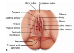
Mons pubis
Rounded, fleshing prominence over the symphysis, anterior border of external reproductive organs.
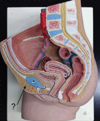
Bartholin's glands
Lubricate the vaginal opening during sex.
Skene's glands
Help lubricate the urethra
labia majora
Rounded, fleshy folds of tissue that extend from the mons pubis to the perineum, slightly darker than the surrounding skin, protect the more fragile tissues of the external genitalia.
Labia minora
Run parallel to and within the labia majora. It extends from the clitoris anteriorly and merges posteriorly to form the fourchette, which is the posterior rim of the vaginal introitus. The labia minora does not have pubic hair. It is highly vascular and responds to stimulation by becoming engorged with blood.
Clitoris
A small projection at the anterior junction of the two labia minora. It is composed of highly sensitive erectile tissue which is similar to the penis. The labia majora merges to form a prepuce over it.
Vestibule of the vagina
- Refers to the structures enclosed by the labia minora
- Contains the urinary meatus
- vagina opening/intoitus
- Ducts of Skene
- Bartholin glands
Vagina introitus/opening
- Surrounded by erectile tissue
- During sexual stimulation, blood flows into the erecticle tissue, allowing the opening to tighten around the penis. This adds a massaging feeling that heightens the male's sexual sensations and encourages ejaculation.
Hymen
A thin fold of mucosa partially separating the vagina and the vestibule. The intactness, or lack of, the hymen is not a criterion of virginity. The hymen may be broken because of injury, tampon use, intercourse, or childbirth.
Perineum (female)
Most posterior part of the external female reproductive organs. It extends from the fourchette anteriorly to the anus posteriorly. It is composed of fibrous and muscular tissues that provide support for pelvic structures
Internal female reproductive organs: Vagina, and what are the major functions?
- It is a tube of muscular and membranous tissue.
- Allows discharge of menstrual fluid flow
- Female organ of coitus
- Allows passage of the fetus to outside the mother's body during childbirth.
Female Puberty Changes
- Breast changes
- Body contours: pelvis widens
- Body Hair
- Skeletal growth
- Reproductive organs
- Menarche
What are the first changes for puberty for a female?
Breast changes are the first thing of changes during puberty and about 1 and (or) 1/2 year, the menstrual cycle should begin.
Female secondary sex characteristics
- Breasts
- More fat distribution in buttocks and thighs
- Pubic and axillary hair
- Higher-pitched voice
Male secondary sex characteristics
- Increased muscle mass
- Growth of hair on the face and body
- Growth of pubic and axillary hair
- Deeper voice
Internal female reproductive organs: Uterus
Divisions: Corpus, isthmus, cervix.
Layers: perimetrium, myometrium, and endometrium
What is the uterus, and the function of the uterus?
- The uterus is a hollow, thick-walled, muscular organ shaped like a flat, upside-down pear.
- The uterus houses and nourishes the fetus until birth and then contracts rhythmically during labor to expel the fetus.
- Each month the uterus is prepared for a pregnancy, regardless of whether conception occurs.
Corpus also known as fundus
Upper division of the uterus. The uppermost part of the uterine corpus, above the area where the fallopian tubes enter the uterus, it is the fundus of the uterus.

Isthmus
a narrower transition zone, located between the corpus of the uterus and the cervix. During late pregnancy, the isthmus elongates and is known as the lower uterine segment.

Cervix
- Is the tubular "neck" of the lower uterus is about 2-3cm length.
- During labor the cervix effaces which thins and dilates to allow passage of the fetus.
- The os is the opening in the cervix between the uterus and vagina.
- The upper and lower cervix are marked by the internal and external os.
- External os of a childless female is round and smooth.
- After vaginal birth the external os has an irregular, slit-like shape and may have tags of scar tissue.

Perimetrium layer of the uterus
- Outer peritoneal layer of the serous membrane that covers most of the uterus.
- Laterally, the perimetrium is continuous with the broad ligaments on both sides of the uterus.

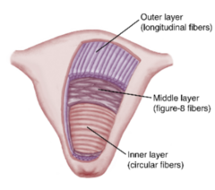
Myometrium of uterus
- Middle layer of thick muscle
- Most muscle fibers are concentrated in the upper uterus, and their number diminishes progressively toward the cervix. The myometrium contains 2 types of smooth muscle fibers, each with specific functions in childbearing:
- Longitudinal
- Interlacing figure-8 fibers
- Circular fibers
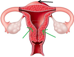
Longitudinal fibers (myometrium)
Found mostly in the fundus and designed to expel the fetus efficiently toward the pelvic outlet during birth

Interlacing figure-8 fibers (myometrium)
- Constitute the middle layer.
- These fibers contact each other after birth to compress the blood vessels that pass between them to limit blood loss.

Circular fibers (myometrium)
- From constrictions where the fallopian tubes enter the uterus and surround the internal cervical os.
- They prevent reflux of menstrual blood and tissue into the fallopian tubes.
- Promotes normal implantation of the fertilized ovum by controlling its entry into the uterus
- Retain the fetus until the appropriate time of birth
Endometrium of the uterus
- Inner layer of the uterus
- Responds to the cyclic variations of estrogen and progesterone during the female reproductive cycle
- Has two layers basal layer and functional layer
Basal layer of endometrium
The area nearest the myometrium that regenerates the functional layer of the endometrium after each menstrual period and after birth.

Functional layer of endometrium
- Lies above the layer.
- Endometrial arteries, veins, and glands extend into the functional layer and are shed during each menstrual period and after childbirth in the lochia, vaginal drainage after childbirth
Lochia
Vaginal drainage after childbirth
Internal female reproductive organs: ovaries
- Sex hormone production
- Maturation of an egg/ovum during each cycle
- At birth, we have all the eggs we will ever have

Internal female reproductive organs: fallopian tubes
- Also called oviducts, are 8 to 14 cm long and quite narrow (2 to 3 mm at their narrowest and 5 to 8 mm at their widest). They form a pathway for the ovum between the ovary and uterus.
- Fertilization occurs in the fallopian tubes. Each fallopian tube enters the upper uterus at the cornu, or horn, of the uterus.

What are the fallopian tubes lined inside?
- Lined with folded epithelium containing hairlike processes called cilia that beat rhythmically toward the uterine cavity to propel the ovum through the tube. The
Tubal blockage
May result in sterility or a tubal ectopic pregnancy because the fertilized ovum cannot enter the uterus for proper implantation.
What are the fallopian tube's four divisions
1. Interstitial portion that runs into the uterine cavity and lies within the uterine wall.
2. The isthmus is the narrow part adjacent to the uterus.
3. The ampulla is the wider area of the tube lateral to the isthmus, where fertilization occurs.
4. The infundibulum is the wide, funnel-shaped terminal end of the tube. Fimbriae are finger-like processes that surround the infundibulum
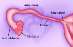
Are fallopian tubes connected to ovaries? Where is the ovum after fertilization?
- The fallopian tubes are not directly connected to the ovary.
- The ovum is expelled into the abdominal cavity near the fimbriae at ovulation.
- Wavelike motions of the fimbriae draw the ovum into the tube.
- Tubal isthmus remains contracted until 3 days after conception to allow the fertilized ovum to develop within the tube.
- Initial growth of the fertilized ovum within the fallopian tube promotes its normal implantation in the upper uterus
Female Reproductive support structures: Bony pelvis
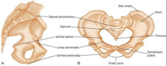
Gynecoid pelvis
The pelvis is thought to be the most favorable type for vaginal birth. This is because the wide, open shape gives the baby plenty of room during delivery. It is ideal for delivery.
Android pelvis
The narrower shape of the android pelvis can make labor difficult because the baby might move more slowly through the birth canal. Some pregnant women with this pelvis type may require a C-section.

Anthropoid pelvis
The elongated shape of this pelvis makes it roomier from front to back than an android pelvis. It is still narrower than the gynecoid pelvis. Some pregnant women with this pelvis type might be able to have a vaginal birth, but their labor might last longer.

Platypelloid pelvis
The shape of this pelvis type can make a vaginal birth difficult because the baby may have trouble passing through the pelvic inlet. Many pregnant women with this pelvis type need to have a C-section; the shape is not ideal.
Where is estrogen made?
- Ovaries, corpus luteum
- Placenta (pregnancy)
- Testosterone in sertoli cells of testes (male)
- Adipose, liver (etc)
Where is/are the target organs for estrogen?
- Internal and external reproductive organs
- Breasts (female)
- Testes (male)
What does estrogen do for females?
- Reproductive organs: maturation of puberty, stimulation of the endometrium before ovulation.
- Breasts: Induce growth of glandular and ductal tissue, initiate deposition of fat at piberty; stimulates growth of long bone but cause closure of epiphyses, limiting mature height.
- Pregnancy stimulates growth of the uterus and breast tissue; inhbits active milk production, and relaxes pelvic ligaments.
Why is estrogen necessary for males?
For normal sperm formation.
Where is progesterone made?
Ovary, corpus luteum, placenta
What is/are the target organs for progesterone?
- Uterus
- Breasts (female)
What does progesterone do for females?
- Stimulates secretion of endometrial glands; causes endometrial vessels to become dilated and toruous in preparation for possible embryo implanation.
- Pregnancy: induces growth of cells of fallopian tubes and uterine lining to nourish embryo; decreases contractions of uterus; prepares breasts for lactation but inhibits prolactin secretion.
Where is oxytocin made?
Posterior pituitary
Where is oxytocin's target organs?
- Uterus
- Breasts
What does oxytocin do for females?
Uterus: stimulates contractions during birth and stimulates postpartum contractions to compress uterine vessels and control bleeding.
- Stimulates and lets down or milk ejection reflex during breastfeeding.
Ovarian cycle
- Follicular phase
- Ovulatory phase
- Luteal phase
Endometrial cycle
- Proliferative phase
- Secretory phase
- Menstrual phase
Changes in cervical mucus
- Cervical mucus is scant, thick, and sticky.
- Just before ovulation, cervical mucus becomes thin, clear, and elastic
- Spinnbarkeit: the elasticity of cervical mucus
Breasts function
- Responsible for lactation
- Montgomery's glands: lubricate the nipples during pregnancy and lactation.
- Alveoli: Secrete colostrum/breast milk
- Mammary glands/lactiferous ducts.
Fertilization of the ovum most likely takes place in which part of the fallopian tube?
Ampulla
Environmental Influences on Childbearing causative agents:
- Maternal infectious agents
- Drugs
- Pollutants
- Ionizing radiation
- Maternal hyperthermia
- Maternal medical disorders
Genetic Screening, Testing, and Counseling
- Availability: facilities that provide maternal-fetal medicine services
- State agencies
- Agencies that focus on a specific birth defect (March of Dimes)
- Focus on the family
- The process of genetic counseling can be slow, and a diagnosis may never be established.
What is Menstrual cycle
- Regular and reccuring changes in the AP secrections, ovaries, and uterus, endometrium.
- Called because menstruation is a marker for the start and end if pregnancy does not occur.
- Duration about 28 days and can range 20-45 days.
- Significant deviation from 28-day cycle are associated with reduced fertility
- Day 1 = first day of period
Ovarian decreases and signals the anterior pituitary to produce? Where does it produce?
- FSH and LH.
- FSH and LH are secreted from the anterior pituiary.
- tells the ovaries to release an ovum and get the endometrium ready.
Follicular (Ovarian cycle
- Egg matures begins from day 1 of period and lasts about 14 days
- Estrogen and progesterone drop, stimulates ecretion of FSH and LH by the AP
- FSH and LH start to rise, 6-12 Graafian follicles begin to grow.
- each follicle secretes fluid containing high levels of estrogen, which speeds up the growth as it makes the follice more sensitive to FSH, which is a positive feedback.
- 1 follicle will outgrow the others and become known as the mature follicle.
- The reason the other follicles don't grow is that FSH decreases during ovulation
- If there is more than 1 "winner" can lead to multifetal pregnancy
Ovulatory phase of the ovarian cycle
- Middle of 28-day cycle, 2 days before ovulation
- LH increases, FSH also increases, but not as much as LH
- Less follicular estrogen is produced, more progesterone is secreted which stimulates the maturation of a single follicle and release of egg.
- Ovulation is the beginning of the luteal phase
- Mature follicle forms a stigma on the walls, ruptures, and the egg is picked up by the fallopian tube to the uterus.
Luteal phase of the ovarian cycle
- Starts with ovulation, LH is prominent, remaining cells of the old follicle remain for about 12 days as a corpus luteum.
- Estrogen and progesterone increase, secreted by the corpus luteum.
- FSH and LH decrease in response to higher levels of estrogen and progesterone.
- If the egg is fertilized, chorionic gonadotropin (hCG) maintains the corpus luteum to assist with pregnancy
- If the egg is not fertilized, FSH and LH decrease and the corpus luteum regresses; the uterine lining breaks down, which leads to your period.
What happens to estrogen and progesterone near the end of the luteal phase of the ovarian cycle?
- The loss of estrogen and progesterone from the corpus luteum at the end of the cycle stimulates the AP to secrete more FSH and LH, and the cycle starts again.
- The old corpus lutuem is replaced by the corpus albicans.
Proliferative of the endometrial cycle
- (6-14) Occurs as the ovum matures & is released during the first half of the ovarian cycle.
- After the completion of a menstrual period, endometrium is very thin.
- Basal layer of endometrial cells remain after MC & cells multiply to form new endometrial epithelium & endometrial glands under the stimulation of estrogen secreted by maturing ovarian follicles.
- Endometrium spiral arteries & endometrial veins elongates to accompany thickening of the functional endometrial layer & nourish the proliferating cells.
- As ovulation approaches, the endometrial glands secrete thin, stringy mucus that aids entry of sperm into the uterus.
Secretory phase of the endometrial cycle
- Occurs during the last half of the ovarian cycle as the uterus is prepared to receive a fertilized ovum.
- Endometrium continues to thicken due to estrogen & progesterone from corpus luteum, reaching maximum thickness 5- 6 mm.
- Blood vessels twist & dilate. Progesterone from corpus luteum causes the thick endometrium to secrete substances to nourish a fertilized ovum.
- Large quantities of glycogen, protein, lipids, & minerals are stored within the endometrium, awaiting arrival of the ovum.
Ischemic phase of the endometrial cycle
- If fertilization does not occur, the corpus luteum regresses about 2 days before the onset of menstruation, causing vasospasms of the endometrium to become ischemic and necrotic.
- The necrotic areas of the endometrium separate from the basal layers, resulting in menstrual flow.
Menstrual phase of the endometrial cycle
- The duration of the menstrual phase, from the first day of flow to the last is about 5 days.
- During the cycle, woman lose about 40 mL of blood, and some become mildly anemic during their reproductive years, and esppecially if their diets are low in iron
Lining of the uterus?
Progesterone
What is a teratogen?
An agent that causes a birth defect or is difficult to establish tertogenic potential
What are factors that make it harder to establish teratogens>
- Study in retrospect
- Timing of exposure and affects pregnancy at different times
- Placental transfer
- Individual variations
Ensure mom avoids rubella
Avoid immunization 28 days before pregnancy