17.3 - Cardiac Muscle Tissue + Electrophysiology
 Call Kai
 Learn
Learn Practice Test
 Spaced Repetition
 Match
Match Flashcards
 Knowt Play
1/33

There's no tags or description
Looks like no tags are added yet.
Name | Mastery | Learn | Test | Matching | Spaced | Call with Kai | Chat |
|---|
No analytics yet
Send a link to your students to track their progress
34 Terms
What are the characteristics of Cardiac Muscle Cells?
Branched
Intercalated Disks
Striated
Single Nucleus
Increased mitochondria = increased myoglobin for energy
Makes ATP with fatty acids + glucose
What are the characteristics of Intercalated Disks?
Connects pacemaker cells to contractile cell and connects contractile cells to each other.
Contains desmosomes to hold cardiac muscle cells together
Contains gap junctions to allow ions to pass from cell-cell.
They are electrical synapses; allows electrical activity to spread to al cardiac muscle cell.s
Allows heart to contract as a unit.
Contractile Cells:
99% of all cardiac cells
Has an RMP
Has a plateau phase
Responsible for the contractions
Autorhythmic (Pacemaker) Cells
1% of all cardiac cells
No RMP
Has a prepotential phase
Makes up conduction system
Generates spontaneous action potentials that spread to contractile cells
Voltage Gated Na+ Channels
Recall what it does
Where are they found in this case?
Found in all cells except certain pacemaker cells
Voltage Gated Ca++ Channels
Function
Which cells contain them
Voltage-gated Opening
Time-gated Closing; closes after period of time regardless of the voltage.
Present in all cardiac muscle cells.
K+ Ion Channels
In all cardiac muscle cells
Ligand or voltage gated
HCN Channel
Unique to pacemaker cells
Voltage-gated channel
Activates upon hyperpolarization
Causes Na+ to enter the cell while K+ still exists upon opening
Voltage
Difference in electrical potential between 2 points
Membrane Potential
Voltage/Charge difference that exists across membranes of all cells
Resting Membrane Potential
Membrane potential of a cell at rest
What is Pacemaker Potential?
Action Potential in pacemaker cells
AP results from reversal in membrane potential = from -60 mV to 10 mV
Characteristics of pacemaker cells electrically:
Depolarization occurs slowly; lacks Na+ channels in the sarcolemma.
Naturally hyperpolarize
Contains HCN channels; causes cell to depolarize slowly to threshold upon opening of the channel
Steps to Pacemaker Potential:
Slow Initial Depolarization
Pacemaker potential is hyperpolarized = min. potential @ -60 mV
Hyperpolarization triggers voltage-gated HCN channels to open.
Na+ leak into cell faster than K+ leaks out = slow depolarization.
Full Depolarization
Membrane reaches threshold @ -40 mV.
Voltage-gated Ca++ open and enter the cell.
Membrane fully depolarizes.
Repolarization
Ca++ are time gated to close
Voltage-gated K+ channels open as Ca++ close = K+ exits the cell.
Membrane repolarizes
Minimum Potential Phase
K+ channels remain open until reaching min. potential.
Hyperpolarization triggers K+ to close.
loops again
What is the Cardiac Conduction System?
Group of interconnected pacemaker cells that generate action potential and spread it to other parts of the cardiac conduction system
What are the 3 populations of pacemaker (autorhythmic) cells that make up the cardiac conduction system?
Sinoatrial Node (SA Node) = Primary pacemaker
Atrioventricular (AV Node) = Secondary pacemaker
Purkinje Fiber System = Slowest group of pacemaker
What is the Sinoatrial Node (SA Node)?
Location
Rate of depolarization
Influence by
Permeability
Connected to what?
The Primary Pacemaker
Located in right atrium
Cells of SA node have fastest rate of depolarization = 60-70 AP/min
Influenced by PNS and SNS
More permeable to Na+ = easier to increase MP and reach threshold = AP
Connects do internodal pathways and the AV node; not directly connected to contractile cells
What is the Atrioventricular Node (AV Node)?
Location
Rate of depolarization
Secondary Pacemaker
Located between right atrium and ventricle.
Rate of depolarization = 40-50 AP/min
Takes over is generating AP if SA Node is nonfunctional
Purkinje Fiber System:
Rate of depol.
How are they different than the SA and AV Node
Components
Slowest pacemaker cells
Rate of depolarization = 20 AP/min
Atypical Pacemakers = AP rely on different ion channels and function differently.
3 components:
AV Bundle (Bundle of His)
Left and Right Bundle Branches
Terminal Branches
What is the AV Bundle (Bundle of His) of the Purkinje Fiber System?
Component of the Purkinje Fiber System that penetrates the heart's skeleton in the inferior interatrial and interventricular septum.
Traveling across the cardiac skeleton causes the signal to slow down
What are the Left and Right Bundle Branches of the Purkinje Fiber System?
Component that courses along left and right sides of the interventricular septum
Carries signal from interventricular septum to the apex of the heart
What are the Terminal Branches of the Purkinje Fiber System?
Component of the Purkinje Fiber System that penetrates the ventricles and contacts the contractile cells
What are Internodal Pathways?
Pathways that connect directly to contractile cells.
Spreads the AP that originated from the SA node --> contractile cells of the atria
What are Purkinje Cells?
Cells that connect directly to contractile cells.
Moves AP from apex --> atria.
What are the steps of the Conduction Pathway through the Heart?
Uses the Cardiac Conduction System
SA Node generates AP = 60-70 per minute
AP spreads to atrial cells.
Impulses conduct to the AV Node.
AP conduction slows down at AV Node = 40-50 per minute.
Allows atria to depolarize and contract before the ventricles; gives ventricles time to fill.
Prevents backflow into atria.
AP conducts to R + L bundle branches of the Purkinje Fiber System = 20 AP/min.
AP conducts to Purkinje fibers = spreads to contractile cells of the ventricles
Characteristics of Contractile Cell AP:
Pacemaker potential conducts down and triggers action potentials of contractile cells to occur
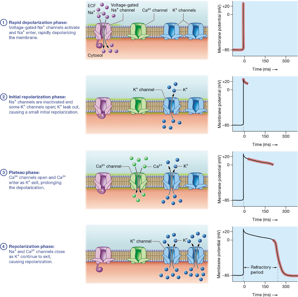
Phases of Contractile Cell Action Potential:
Rapid Depolarization Phase
Pacemaker AP conducts down to contractile cells, activating Na+ in sarcolemma.
Influx of Na+ cause rapid depol. of membrane from its RMP of -85 mV.
Initial Repolarization Phase
Small, initial repolarization immediately after depolarization.
Caused by abrupt inactivation of Na+ and small outflow of K+ through channels that were briefly opened.
Plateau Phase
Depol. sustained at about 0 mV.
Caused by opening of Ca++ channels and influx of Ca++.
As Ca++ enter, K+ leave = very little net change in MP.
Ca+ close slowly and timely, allowing the plateau phase to last longer.
Repolarization Phase
Na+ and Ca++ channels close
K+ channels open = K+ efflux
MP returns to RMP of -85 mV
Why is there a plateau phase in contractile cell AP?
Lengthens the cardiac AP = slows heart down to provide the time required for the heart to fill with blood.
Prolonged AP makes the twitch last long = develops more force (strength)
Prolonged AP allows Ca++ to enter, which is needed for the sliding-filament mech. of sarcomere.
What is the Effective Refractory Period?
The phase during an AP in which the cell cannot generate more AP.
Plateau phase lengthens during this period in contractile cell AP.
Prevents cells from entering a tetany state = sustained contraction.
Allows heart to relax = ventricles fill with blood.
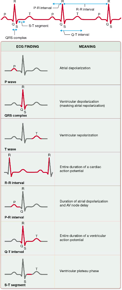
What is an Electrocardiogram (ECG/EKG)?
Function
Different Waves
Graphic depiction of electrical activity in cardiac muscle cells over a period of time.
Does NOT display mechanical events.
Waves:
P Wave
QRS Wave
T Wave
What is the P Wave of the ECG?
Atrial Depolarization; except the SA node.
= Atria are contracting; but is not depicted in ECG.
What is the QRS Complex of the ECG?
Ventricular Depolarization depicted in 3 separate waves.
Q = 1st downward deflection
R = Large upward deflection
S = 2nd downward deflection
QRS waves are represented with a large magnitude due to the ventricles being much larger than the atria.
= ventricles are contracting mechanically
Atrial Repolarization
Produces small electrical signal, but is hidden by the QRS complex.
= atria are relaxing mechanically
What is the T Wave of the ECG?
Ventricular Repolarization
Upward Deflection; sometimes inverted
= ventricles are relaxing mechanically
ECG Waves