Ortho 3 - Shoulder + Upper Arm
 Call Kai
 Learn
 Practice Test
 Spaced Repetition
 Match
 Flashcards
 Knowt Play
1/257

There's no tags or description
Looks like no tags are added yet.
Name | Mastery | Learn | Test | Matching | Spaced | Call with Kai | Chat |
|---|
No analytics yet
Send a link to your students to track their progress
258 Terms
Sternoclavicular joint
Articulation between the clavicle and the sternum
Clavicle
what bone is this?

Acromion

Spine of the scapula

acromioclavicular joint

Coracoid process

subacromial bursa
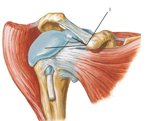
Greater tubercle of the humerus
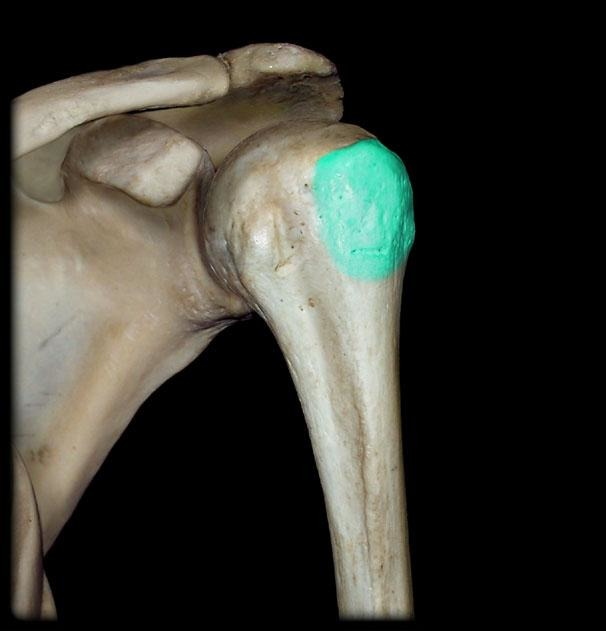
Lesser tubercle of the humerus

Glenoid Fossa

Glenohumeral ligaments

injury to a ligament - more difficult to heal d/t decreased blood supply
What is a sprain?
injury to muscle or tendon - easier to heal d/t higher vascularization
What is a strain?

Compression of neurologic and/or vascular structures between the clavicle and first rib.
What is thoracic outlet syndrome (TOS)?
95%.
What percentage of TOS is neurologic?
3-4%.
What percentage of TOS is venous?
1%.
What percentage of TOS is arterial?
presence of a cervical rib -> an extra rib above the first rib.
What structural anomaly can cause TOS?
Scalene muscle variation because the brachial plexus passes through the scalene triangle.
What muscle variation can predispose to TOS?
1. whiplash
2. falls
3. repetitive overhead activity
4. swimming
5. fractured clavicle or first rib.
What injuries/activities can cause TOS?
Brachial plexus, subclavian artery, and subclavian vein.
What structures pass through the thoracic outlet?
- Nonspecific upper-extremity paresthesia
- numbness
- weakness
- pain w/ overhead or repetitive use.
Neurologic TOS symptoms?
- head rotation or tilting
- shoulder abduction
- external rotation toward the affected side.
What clinical maneuvers can reproduce TOS symptoms in office?
- shoulder external rotation and slight aBduction
- head extension and rotation toward the affected side
What exam posture is used for the Adson test?
Over the scalene muscles.
Where is point tenderness found in TOS?
Muscular atrophy and further weakness (rare)
What can chronic neurologic TOS rarely cause?
It can mimic DVT with positional changes of the extremity.
How can venous TOS present?
It can mimic arterial thrombus with positional changes of the extremity.
How can arterial TOS present?
NCV/EMG and MR neurogram (MRI)
How to test for neurologic causes of TOS?
Ultrasound, MRA, CT angiography, and venography.
How to test for vascular causes of TOS?
PT and intrascalene steroid or anesthetic injections.
Treatment of neurologic TOS?
Anticoagulation; thrombectomy if the extremity is compromised.
Treatment of vascular TOS?
Thoracic outlet decompression.
What surgery can benefit all TOS types?
- cervical rib removal
- first-rib resection
- scalene muscle resection
Examples of thoracic outlet decompression procedures?
It is the first bone in the body to ossify (harden)
What is notable about clavicle ossification?
It is the only direct connection of the trunk to the upper extremity.
What is the clavicle's connection role?
69%
What % of clavicle fractures occur at the middle aspect of the bone and are classified as group 1?
28%.
What % of clavicle fractures occur at the distal aspect of the bone and are classified as group 2?
Acromion-clavicular (AC) separation.
What injury can accompany a distal/group 2 clavicle fracture?
3%.
What % of clavicle fractures occur at the proximal aspect of the bone and are classified as group 3?
with significant trauma.
When are proximal/group 3 clavicle fractures most common?
Fall onto the shoulder, 87%.
Most common mechanism of clavicle fracture?
7%.
What percentage of clavicle fractures result from direct blow?
6%.
What percentage of clavicle fractures result from FOOSH?
- point tenderness
- crepitus with arm movement
- possible skin tenting
- swelling
- ecchymosis
Clavicle fracture physical exam findings?
Pneumothorax.
What pulmonary complication must be assessed with every clavicle fracture?
upper extremity function w/ brachial plexus and subclavian vessels
What neurovascular structures must be assessed with every clavicle fracture?
X-ray.
Initial diagnostic imaging for clavicle fracture?
Concern for ligamentous disruption.
When is MRI considered in a group 2 clavicle fracture?
Upward displacement of the proximal fragment due to pull on it from the sternocleidomastoid
What displacement pattern is typical of a displaced midshaft clavicle fracture?
Over 4 cm.
What amount of shortening in a midshaft clavicle fracture is shown as severe?
Ice and short-term pain medication.
Initial symptomatic treatment of clavicle fracture?
Less than 1 bone-width displacement.
How is a minimally displaced clavicle fracture defined?
Figure 8 bandage or sling
Treatment of minimally displaced clavicle fracture?
To support damaged ligaments.
Why may group 2 clavicle fractures need extra arm support?
Surgical fixation/correction (it is 1 or more widths of displacement)
What is the treatment method of a significantly displaced clavicle fracture?
After bracing/sling use is no longer needed.
When does PT begin after clavicle fracture?
Flexion, extension, pronation, and supination so pt does not lose mobility
What elbow motion should begin within the first week of clavicle fracture?
High-energy trauma.
What type of trauma causes scapular fractures?
No, they are uncommon.
Are scapular fractures common?
ALWAYS child abuse
If a young child presents with a scapular fracture, how do we classify the method of obtaining the fracture?
Body, glenoid, or acromion.
What parts of the scapula are typically fractured?
The injury usually results from high-energy trauma -> neck and rib com-injuries common
Why are associated injuries common with scapular fracture?
AP shoulder, Y view, axillary view, and chest x-ray.
Diagnostic imaging for scapular fracture?
Nonoperative; sling if necessary.
Usual treatment of scapular fracture?
Significant trauma with instability, multiple fractures, or severe glenoid trauma.
When is surgery considered for scapular fracture?
Strong ligaments.
What holds the AC joint together?
Direct blow to the AC joint, especially in contact sports, or fall directly onto the shoulder.
Common mechanisms of AC joint sprain?
Six.
How many Rockwood AC injury classification types are there?
Type I.
Which Rockwood type is mildest?
AC ligament sprain with AC joint intact.
Rockwood type I pathology?
AC ligament torn, CC ligament intact, AC joint subluxed.
Rockwood type II pathology?
AC and CC ligaments torn with complete joint dislocation.
Rockwood type III pathology?
Point tenderness over AC joint, reluctance to use extremity, and positive cross-body adduction test.
AC sprain physical findings?
Types II and III.
Which AC injury classifications can show a step-off between clavicle and acromion?
Pain from compression of the AC joint, suggesting AC pathology.
What does a positive cross-body adduction test indicate?
Clavicle position relative to the acromion and coracoid processes.
What can plain x-rays assess in AC injury?
Zanca view - upward angle of the x-ray beam
Recommended x-ray view for AC joint injury?
Types II and III.
Which AC injury classes may need MRI?
Conservative - ice, NSAIDs, sling, and physical therapy.
Treatment of type I and II AC sprains?
Surgical repair may be used, but conservative treatment like types I/II may be tried.
Treatment of type III AC sprain?
6-12 weeks.
How long can conservative AC sprain treatment take to heal?
6 weeks rest, 6 weeks ROM work, then strengthening to baseline.
Postoperative AC injury rehabilitation sequence?
The humeral head/labrum is not inherently stable and needs reinforcement from the capsule, ligaments, and rotator cuff.
Why is the glenohumeral joint dependent on stabilizing structures?
Glenohumeral and scapulothoracic articulations.
What articulations provide major shoulder movement freedom?
Superior translation of the humeral head into the acromion pinches structures between them.
How can weakened shoulder stabilizers cause impingement?
Sulcus sign.
What instability sign may be positive in shoulder impingement syndrome (SIS)?
Increased scapular movement on the thoracic cage with scapular winging - causes thoracic nerve entrapment
What scapular abnormality can contribute to SIS?
- supraspinatus tendon
- subacromial bursa
- long head of the biceps tendon
Which structures become inflamed in SIS?
Examiner applies downward pressure on the antecubital fossa while the patient has a relaxed shoulder. If the humeral head is readily displaced by 2 cm or more inferiorly, sulcus sign is +
Which maneuvers test for a sulcus sign?
Repetitive activity at or above shoulder level.
Common activity risk for SIS?
Difficulty sleeping on the affected side.
What sleep complaint is common in SIS?
Risk increases with age d/t arthritic changes
How does age affect SIS risk?
Osteophytes on the underside of the AC joint/acromion.
What degenerative change can worsen SIS?
Type 1
Which acromion morphology is flat, the normal morphology that easily allows nerves to pass through the space between the acromion and clavicle

Type 2
Which acromion morphology is curved, the space where the nerves are able to travel is smaller

Type 3
Which acromion morphology is hooked -> puts pt more at risk of impingement of subacromial space d/t less space for nerves to travel

Upper-extremity inflexibility.
What flexibility issue can contribute to SIS?
Deltoid region or lateral arm.
Typical SIS pain location?
Overhead activity and lying on the affected side.
When is SIS pain commonly provoked?