MCB 2000 Chapter 12
 Call Kai
 Learn
 Practice Test
 Spaced Repetition
 Match
 Flashcards
 Knowt Play
1/32

There's no tags or description
Looks like no tags are added yet.
33 Terms
amphipathic lipid
the head and tail behave like different molecules
the phosphate head is polar and charged. it hydrogen-bonds with water and sits comfortably on the surface
often with a charged amine attached
the fatty acid tails are pure hydrocarbons. no polar bonds, no H-bond capacity. water can’t accommodate them
that structural split is what makes everything else in this module possible

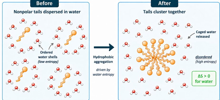
the hydrophobic effect
entropy is the driver and the entropy belongs to water
nonpolar tails cluster together to minimize ordered water, increasing the entropy (ΔS) of water
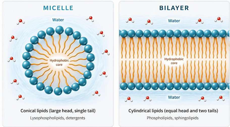
micelle vs bilayer
same thermodynamics
the lipid’s shape picks the structure
micelles shaped from lipids shaped like a cone (big head, usually one tail)
detergents, lysophospholipids
bilayer shaped from cylindrical lipids
phosphatidylcholine
pack into sheets — two layers, tails inward, head facing water
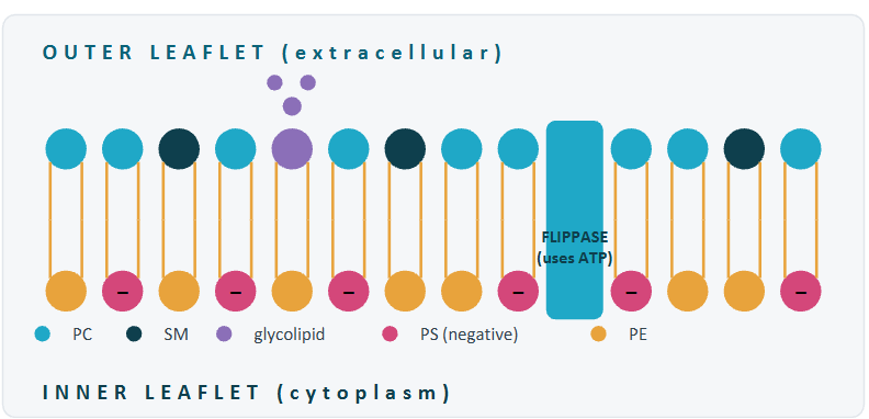
bilayer asymmetry
two leaflets carry lipids, and the cell maintains that difference
two faces, two compositions
the outer leaflet is rich in phosphatidylcholine and sphingomyelin, with neutral head groups and the occasional glycolipid for cell recognition
the inner leaflet is rich in phosphatidylserine and phosphatidylethanolamine. PS carries a negative charge that helps recruit cytoplasmic proteins
flippases use ATP to maintain the difference. It doesn’t happen on its own
asymmetry is maintained because lipids don’t naturally stay on one side and diffusion would scramble the asymmetry
what the bilayer makes possible
selective barrier
what crosses: water and small nonpolar gases (O2 and CO2) passes freely
what’s blocked: ions and large polar molecules. the hydration shell is too costly to strip
effect: foundation of cell chemistry
self sealing
why: an exposed tail is the worst possible state (thermodynamically). the bilayer rearranges within microseconds
results: sealed vesicles form spontaneously. tear close without machinery
effect: robust, self-repairing
dynamic, not static
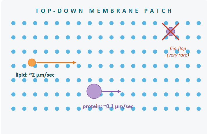
lateral diffusion: lipids slide sideways within a leaflet at ≈2 μm per second
flip-flop: crossing between leaflets is much slower because it requires dragging a hydrophilic head group through hydrophobic region: minutes to days without enzymes
effects: fluid but ordered
three properties, one cause: tails dislike water, heads loves it
lipid rafts
the membrane is more organized than a uniform sea
fluid mosaic, revised
proteins float in a fluid sea has structure
Singer and Nicolson (1972) got the broad picture right. The membrane is a 2-D fluid with proteins floating in it
rafts: cholesterol-rich patches of tightly-packed saturated lipid form more-ordered microdomains. Specific membrane proteins, especially signaling receptors and GPO-anchored proteins, concentrate there

the Goldilocks principle
fluidity has a sweet spot, and crossing into either extreme is bad news
low temp — lipid tails packed tightly side by side, membrane= ordered and viscous, almost crystalline
gel- state (solid-like)
molecules can’t pass and membrane cracks under mechanical stress
high temp — gain enough thermal energy to bend and rotate, packing starts breaking down and membrane becomes a true 2-D fluid
liquid state (fluid-like)
ions leak across, gradient collapse, cell loses membrane potential
Tm — flip from too low too high = melting temperature
cooperative, so it is sharp rather than a gradual change
narrow sweet spot where membrane is flexible enough for proteins to function, but stable enough to hold the barrier

gel vs liquid-crystalline
same lipid, two states. the difference is the order of the tails

Knob 1: fatty acid saturation
straight chains pack tight
Cis double bonds create kinds that don’t
each cis double bond bends the chain by about 30 degrees. neighbors can’t pack as tightly and Tm drops
the more cis double bonds, the lower the melting temps and the more fluid at any given temp

Knob 2: Acyl chain length
more carbons mean more van der Waals contact, which means higher Tm
why it matters:
each additional CH2 adds another point of van der Waals contact with neighboring tails. long chains have more total contact area, so they stay packed at higher temps
cells pair longer chains with unsaturation to keep Tm near physiological temps. the two knobs work together
most mammalian membrane lipids land in the C16 to C18 range for this reason

Knob 3: Cholesterol structure
three parts and each does a different job in the bilayer
polar head — a small hydroxyl group at C3. hydrogen bonds with phospholipid head groups at the bilayer surface
rigid steroid core — four fused rings, planar and inflexible. no rotation possible. this rigidity is what lets cholesterol restrain neighboring tails
flexible tail — a short, branched chains with 8 carbons. nestles in the hydrophobic core alongside the acyl tails. cholesterol is roughly as long as a 16-carbon chain
only in animals, but = hopanoids in bacteria

cholesterol as a composition buffer
same molecules, opposite effects, depending in what’s already there
the composition question: at a fixed temp, cholesterol pushes whichever composition is at hand toward intermediate fluidity

cholesterol as a temp buffer
hold composition fixed. lets temp change. cholesterol still tunes both ways
cholesterol buffers both directions: composition and temp

2 questions to determine category of membrane proteins
GO BACK TO VIDEO — M3 CH. 12

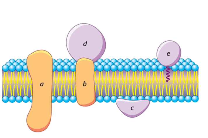
5 different protein placements

positions a and b
how integral proteins survive inside the lipid tail region: the alpha-helical transmembrane domain
why helices work: the helix hides it backbone
an alpha helix uses every backbone N-H and C=O in internal hydrogen bonds. the polar backbone is completely satisfied inside the helix
that lets the helix expose only its side chains. if those side chains are hydrophobic, the whole helix can sit comfortably in the lipid tail region
about 20 hydrophobic residues are enough to span a bilayer
multi-pass (a) and single pass (b) both use this trick. the bilayer never sees the polar peptide backbone

inside positions c and d
how peripheral proteins stay attached without ever entering the lipid core
peripheral logic: they never touch the tails
peripheral proteins (C,D) hold to the membrane by interacting with the polar head groups, not the hydrophobic tails. hydrogen bonds, salt bridges and electrostatic contacts do the work
some bind directly to phospholipid head (C). others, like D, hold onto the exposed surface of an integral protein (B)
change the pH or raise the salt and these interactions break. the protein comes off without disturbing the bilayer
if a salt wash takes a protein off the membrane, you were looking at a peripheral protein, but if it takes a detergent, it was an integral protein
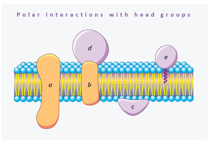
inside position e: lipid-anchored
the protein body stays out of the bilayer. a covalent lipid tail does the tethering
how it works: the lipid is the anchor
a fatty acid or isoprenoid chain is covalently attached to the protein after translation. the chain inserts into one leaflet and the protein body sits in the aqueous space on that side
which leaflet? depends on the anchor. GPI anchors face out, into the extracellular space, like alkaline phosphatase. acyl and prenyl anchors face in, into the cytoplasm, like Ras
the protein never enters the bilayer. a single fatty chain or isoprenoid tethers it to one leaflet
the fluid mosaic in action
proteins float in the bilayer, but they don’t all move at the same rate
two rules of motion:
lateral diffusion is fast. flip-flop isn’t
lipids and proteins both diffuse sideways within their leaflets. lipids move quickly because they’re small. proteins move more slowly, scaling roughly with the size of their transmembrane footprint
crossing between leaflets requires dragging polar groups through the hydrophobic core (flip-flopping) . for lipids that takes minutes to days with enzymes. for proteins it doesn’t happen (dragging a folded protein through a tail region of the membrane with all its hydrophilic surfaces exposed is not energetically feasible)
that asymmetry is what lets the cell maintain leaflet-specific protein placement
sideways yes, across no. the cell uses that to keep its leaflet distinct.
permeability scale
coefficient P spans 14 orders of magnitude

why ions can’t cross
Na+ and K+ carry a shell of 6-8 water molecules ]
stripping it to cross the hydrophobic core costs ~150 kJ/mol — prohibitive without a protein
why O2 and CO2 cross freely
small, nonpolar, uncharged = no hydration shell
the hydrocarbon core acts as a solvent so these gases dissolve in and diffuse across freely
simple diffusion
down the gradient, through the bilayer, no protein
rate law: rate is proportional to the gradient
more gradient, more flux
Fick’s law: rate scales linearly with the concentration difference across the membrane
there is no saturation. the bilayer itself is the medium and it doesn’t run out of capacity
double the gradient, double the rate
works for anything that crosses the bare bilayer, but only at modest rates

facilitated diffusion
still down the gradient, but now through a protein. the kinetics give it away
channels: open pore, fast flow
substrate flows through without binding
up to 10^8 ions per second
gated open or closed by signals
carriers: bind, switch, release
substrate binds, the protein changes conformation, the substrate is released on the other side
slower (10³ per seconds) but more specific
GLUT transporters move glucose this way


Ion channels
selective, fast, gated. The K+ channels shows how all three work together
selective:
the filter is sized for K+ and lined with carbonyl oxygens that mimic the hydration shell
Na+ is smaller and is rejected because it cannot afford to leave its hydration shell
fast:
ions pass at up to 10^8 per second, near the diffusion limit
mutual repulsion in the file keeps them moving
gated:
conformational changes open or close the channel in response to voltage, ligands, or mechanical force

aquaporins
a specialized channel that does one thing very well: move water fast
water, only water:
10^9 molecules per second per channel
the pore is narrow enough that water passes single file
a precise electrostatic arrangement passes neutral H2O but blocks even a single proton
found wherever water flux is high; kidney tubules, red blood cells, plant root cells

drug absorption: you computed the charge before, membranes explain why it decides crossing
stomach: pH 1.5
Aspirin pKa = 3.5 → mostly protonated (uncharged)
neutral and small: no hydration shell. it sits in the “crosses freely” zone and is absorbed here
small intestine: pH = 6.5
Aspirin pKa = 3.5 → mostly deprotonated (charged)
now charged: a hydration shell forms. shedding it costs ~150 kJ/mol, so the bilayer rejects it
the charge was the easy half. the membrane is the point; only the neutral form sheds its water shell and crosses

why active transport exists
cells must move things in the wrong direction, which passive transport can’t do
gradients are useful but costly:
the cells uses gradients of Na+, K+, Ca2+, and H+ to drive nerve impulses, muscle contraction, secondary transport and signaling
those gradients dissipate as ions leak back across. without pumps, replenishing them, every gradient in the cell would equilibrate within minutes
pumps don’t move bulk material. they maintain the difference that everything else uses

Na+/K+ pump
primary active transport
3 Na+ out, 2K+ in, 1 ATP — repeated billions of times per cell per second
why it matters: the cell’s most expensive habit
use this pump constantly, using roughly 30% of all the ATP the cell produces
the gradient it builds is the energy source for nerve impulses, muscle contraction, secondary transporters and Ca2+ regulations
almost everything electrical or absorptive in animal physiology runs on this pump
P-type ATPase: the protein phosphorylates itself on asp residue with each cycle

Na+/K+ pump’s conformational cycle
E1 binds Na+
pump open to cytoplasm. 3 Na+ bind
phosphorylation
ATP transfers phosphate to as aspartate
conformational switch
E2 releases Na+, binds K+
now open to outside. Na+ out, 2 K+ in
low affinity for Na, so Na leaves easily
dephosphorylation
phosphate hydrolyzes off
protein flips back, K+ releases inside
K affinity drops
the cycle is irreversible. each step has a built-in direction: bind on one side, release on the other
secondary active transport
use on ion’s downhill flow to drag another molecule uphill
primary pumps build the Na+ gradient
secondary transporters spend it to do other work

clinicals: digitalis and the heart
inhibits one pump and strengthens heart contractions. a 200-year-old drug is still in use
therapeutic uses: treats heart failure by increasing contractile force
narrow therapeutic window: too much causes arrhythmia. from the foxglove plant, in use since the 1700s
same pump biology from S4-S6 in reverse: breaks the gradient deliberately and downstream transport flips

Chapter Overview
