VTP 3
 Call Kai
 Learn
 Practice Test
 Spaced Repetition
 Match
 Flashcards
1/271

There's no tags or description
Looks like no tags are added yet.
272 Terms
What are the Dosing practices for helminths?
• Interval: Synchronized treatment at intervals, strong selective pressure
• Strategic: Seasonal treatment: before winter (May) and after winter (Oct)
• Tactical: Treatment adapted to changing situation
• Selective: Treatment of selected animals based on clinical signs, FEC, body weight, BCS, milk yield
• Continuous in-feed: Year-round daily treatment, very high selective pressure
What are refugia and why are they of importance?
Helminth population not exposed to anthelmintics, like larva and eggs in the environment, hypobiotic larvae, or helminths in untreated animals. They serve to reduce anthelmintic resistance.
What contributes to high anthelmintic resistance?
• High treatment frequency
• Single-drug regimens
• Long-acting drugs (drugs w residual effect) + Drugs effective for multiple developmental stages
• Targeting and timing of mass treatment
• Underdosing
How is anthelmintic resistance tested?
FECRT: Faecal Egg Count Reduction Test (<95% reduction means resistant)
Differentiate between tolerance and resistance
Tolerance: Innate unresponsiveness of a drug to a developmental stage of a parasite (stage-specific tolerance) or a particular parasite (total tolerance)
Resistance: Ability to cope with the effects of infection (without suffering severe morbidity or mortality)
What are species in Fasciola and their hosts
Fasciola heptica - DH: Cattle, sheep, goat, humans. IH: Galba truncatula, Pseudosuccinea columella
Fasciola gigantica - DH: Cattle, sheep, goat, humans. IH: Radix natalensis, Pseudosuccinea columella

What is the predilection site for Fasciola?
Bile ducts
How do Fasciola develop?
IH: The egg is shed in the faeces, and forms miracidium and infects the IH. Then sporocyts and redia are formed through asexual reproduction. The cercaria leaves the snail, encysts, and forms the metacercaria
DH: Metacercaria infects the DH. Excysts in the GIT and penetrates the intestinal wall. Juvenile flukes migrate to the peritonium, invades the liver parenchyma, and grow into mature flukes in the bile ducts. Aberrant migration common in cattle and aberrant DHs (encapsulated flukes in Lungs and other sites.)

Identify the following images and give characteristics
Fasciola egg, large, yellowish-brown, thin shelled, oval egg, containing fine granules and an indistinct opperculum.

Identify the following images and give characteristics
Fasciola metacercaria, circular, free in water, attached to plants.

Give characteristics of fasciola
▪ Dorsoventrally flattened
▪ Leaf-shaped
▪ Colour: Brown-greyish
▪ Conical anterior end
How is Fasciolosis diagnosed?
Demonstration of eggs in faecal samples by means of sedimentation after Benedek or Visser filter technique
F. hepatica: Antibody-detection ELISAs, + Coproantigen detection ELISA
What are reservoir hosts of Fasciola?
Domestic ruminants.
Discuss the pathogenesis and pathology of Fasciola
Traumatic hepatitis as juvenile flukes migrate through the liver parenchyma. Haemorrhagic tracts and petechia. Liver capsule can rupture, haemoperitoneum. Hepatic fibrosis. Hyperplastic cholangitis, inflammation, calcification. Adult flukes are haematophagic, anaemia, Hypoalbuminaemia
What are Clinical signs seen in Fasciolosis?
▪ Acute form (infection with large numbers of metacercariae)
- Sheep (F. hepatica + F. gigantica)
- Sudden death + Haemorrhagic anaemia + Dyspnoea + Hepatomegaly
- No fluke eggs present
▪ Subacute form (infection with large numbers of metacercariae over extended period of time)
- Sheep (F. hepatica + F. gigantica) + Calves (F. hepatica)
- Rapid loss of condition + Anaemia + Submandibular oedema (‘bottle jaw’) + Hepatomegaly (Sheep) + High mortality
- Fluke eggs present
▪ Chronic form (infection with moderate numbers of metacercariae)
- Most common form in cattle, sheep, horse and man
- Sheep: Anaemia + Submandibular oedema (‘bottle jaw’) + Inappetence + Unthriftiness + Chronic weight-loss
- Cattle: Production losses (e.g. milk)
How is Fasciolosis treated?
Benzimidazoles: Triclabendazole best for immature phases
Salicylanilides: Closantel
Halogenated nitrophenols: Nitroxynil
Sulphonamides: Clorsulon
How are Fasciola controlled?
Strategic application of anthelmintics
Farm and grazing management:
- Limit access to water sources
- Avoid wet pastures, and vlei areas
- Animals should be watered with fresh water (water from bore holes or streams)
- Regular cleaning of water troughs
Reduction of intermediate hosts
- Biological control (e.g. ducks)
What is the importance of Fasciolosis?
Man can act as an accidental DH
One of the economically most important helminth infections of ruminants
Name species in Calicophoron and their hosts
Calicophoron microbothrium: DH - sheep, goat, cattle. IH - Bulinus tropicus
What is the predilection site of Calicophoron?
Fore stomachs
Discuss Calicophoron development
Intermediate host:
- Miracidium Metacercaria (minimum 4 w)
Definitive host:
- Infection by ingestion of metacercariae
- Duodenal phase: Excystment of metacercaria in duodenum and attachment of juvenile flukes to duodenal mucosa
- Gastric phase: Migration of juvenile flukes to the fore stomachs and attachment to the epithelium

Identify the following images and give characteristics
Calicophoron adults, conical, circular in cross section, pink/red

Identify the following images and give characteristics
Calicophoron egg; large, oval, large yolk cells, colourless, thin shelled, distinct operculum
How is Calciphorosis diagnosed?
Demonstration of immature flukes by rinsing faeces
Demonstration of eggs in faecal samples by means of sedimentation after Benedek or Visser filter technique
Discuss the pathogenesis and pathology of Calciphoron
Duodenal phase (intestinal paramphistomosis): Immature stages pathogenic (plug feeders) Ulceration + Haemorrhage (pin-point haemorrhages, haemorrhagic duodenitis) + Large numbers of pinkish immature flukes attached to mucosa and in intestinal contents
Gastric phase: Adult stages - largely non-pathogenic
What are clinical signs of Calciphorosis?
▪ Animals become listless
▪ Progressive decrease in appetite, eventually complete anorexia
▪ Acute diarrhoea: Faeces extremely fluid, foetid, dark green, sometimes with blood and immature flukes + Great thirst
▪ High mortality
How is Calciphorosis treated?
Salicylanilides: Closantel1 + Niclosamide2 + Oxyclozanide3 + Resorantel3
1 Immature flukes in sheep
2 Immature flukes in sheep and cattle
3 Immature and mature flukes in sheep and cattle
What ist he importance of Calciphorosis?
▪ Seasonal and often serious problem
▪ Large numbers of adults occur in the fore stomachs without any clinical signs, however, during the early duodenal phase a severe and often fatal diarrhoea can occur
Name species in Gastrodiscus and their hosts.
Gastrodiscus aegyptiacus - DH: Horses, other equids, suids. IH - Bulinus forskalii
What are the predilection sites for Gastrodiscus?
Caecum + Colon
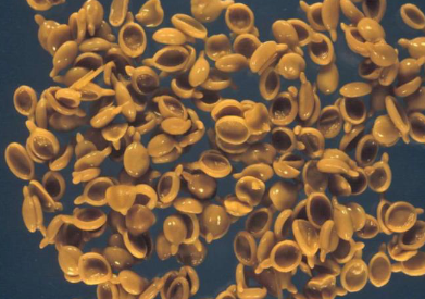
Identify the following images and give characteristics
Gastrodiscus aegytiacus adults - dorsoventrally flattened pink ‘tennis-racket’


Identify the following images and give characteristics
Gastrodiscus aegyptiacus egg - Large oval egg, with coarse granular material, colourless, thin-shelled, distinct operculum.
How is Gastrodiscosis diagnosed?
Demonstration of eggs in faecal samples by means of sedimentation after Benedek or Visser filter technique
Discuss the pathology and pathogenesis of Gastrodiscus
Pathogenicity proportional to worm burden
Severe infections: Oedematous and haemorrhagic colitis
What are clinical signs of Gastrodiscosis?
Mild infections: Clinically inapparent
Severe infections: Weight-loss + Chronic diarrhoea + Recurrent colic attacks + Collapse + Incoordination
How is Gastrodiscosis treated and controlled?
Oxyclozanide
Farm and grazing management
Avoid wet pastures and swamps
Horses should be watered with fresh water
Treatment of infected horses
What is the Importance of Gastrodiscosis?
Mild infections go always clinically unnoticed and are mostly undetectable
Large worm burdens can cause haemorrhagic and oedematous colitis as well as colic
Name species in Schistosoma and their hosts.
Schistosoma mattheei - DH: Cattle, sheep, goats, humans
Schistosoma mansoni - DH: Humans
Schistosoma haematobium - DH: Humans
IH: Bulinus africanus + Bulinus globosus
What is the predilection site for Schistosoma
Portal and mesenteric veins + Veins of urinary bladder (only in cattle)
How does Schistosoma develop?
Infective stage: Cercariae
Infection: Percutaneous infection + Ingestion of cercariae
Cercaria penetrates skin, sheds tail and transforms into a schistosomulum. Enters lymphatic or venous system. Transported to right heart and lungs. Schistosomules pass to the portal vein of the liver where they grow to maturity. Paired sexually mature worms migrate to the mesenteric veins and bladder veins (cattle). Eggs escape from the veins largely into the wall of the bowel, pass through the mucosa into the gut lumen and are shed in faeces; some eggs remain trapped in tissues

Identify the following images and give characteristics
Schistosoma adults (male inside female groove). Elongate, Unisexual and dimorphic. Always encountered in pairs

Identify the following images and give characteristics
Schistosoma egg - Large, Spindle-shaped, Thin-shelled, Terminal spine, Embryonated when laid
How is Schistosomosis diagnosed?
Demonstration of eggs in 10 % formalin-fixed faecal samples by means of sedimentation or Visser filter technique
Demonstration of eggs in squash preparations of blood clots or mucus from faeces
Discuss the pathogenesis and pathology of Schistosoma
▪ Eggs are the most pathogenic stage
▪ Percutaneous infection: Cercarial dermatitis
▪ Stage of egg deposition and extrusion
- Intestinal mucosa with numerous petechiae and ecchymoses which are packed with eggs + Intestinal haemorrhage
▪ Stage of tissue proliferation and chronic infection (eggs trapped in tissues)
- Liver: Gross enlargement + Induration + Orange discolouration or haemorrhage, jaundice + Multiple disseminated granulomas + Sheep: Adult worms are haematophagic and shed haematin which accumulates in the lungs turning them light grey in colour (pathognomonic)
- Submucosal and subserosal veins of intestine congested and varicosed with worms found in pairs
- Pulmonary infarcts
- Cattle: Enlargement and haemorrhage of the ileocaecal valve
- Bladder granulomas
▪ Cattle develop immunity which eliminates most of the worms within a few months (sheep develop little immunity to natural challenge)
What are clinical signs of Schistosoma?
Sheep:
- Progressive loss of condition and weakness over months + Sunken eyes + Anxious expression + Intermittent diarrhoea with blood and mucus + Anaemia, high mortality
Cattle:
- Painful diarrhoea with blood and mucus +Intermittent straining + Sunken eyes + Weight-loss +Anorexia + Nervous derangements
How is Schistosoma treated?
Praziquantel
What’s the importance of Schistosoma?
Humans can act as accidental DHs
What are general characteristics of trematodes?
2 suckers (oral and ventral), Digestive system ends blind, Reproductive system hermaphrodite (except blood flukes), Oviparous, Excretory system + nervous system, Sexual reproduction in DHs + Asexual reproduction in IHs
What are general characteristics of Cestodes?
Indirect life cycle with 1 or 2 IH(s)
DH: strobilar stage = adult stage resides in s. intestine + ileocaecal valve+ bile duct
IH: metacestode = 2nd larval stage resides in various organs
Digestive system (alimentary tract) absent
Nutrient uptake through tegument
Hermaphrodite
Diheteroxenous or Triheteroxenous
Discuss the strobilar stage of cestodes
Scolex = head (4 suckers, +/- rostellum)
Neck = proliferation zone
Strobila = chain of proglottids (segments)
Each proglottid with 1 or 2 sets of male and female sexual organs (immature, mature, gravid)
Nervous system + Osmoregulatory system + Muscular system + Reproductive system

Discuss the basic life cycle of cestodes
▪ Egg
- Shed free or in gravid proglottids during defaecation
- In proglottids actively leaving host (motile proglottids in some genera), i.e. w/o defaecation
- Eggs are embryonated/larvated (oncosphere = 1st larval stage in cestode life cycles) and immediately infective for IHs
- Infective stage for IHs: Infection by ingestion
▪ Metacestode (2nd larval stage) develops in IHs
- Infective stage for DHs: Infection by ingestion
- Cysticercoid: Type of metacestode with a single non-invaginated scolex, withdrawn into a vesicle with no cavity; macroscopically not visible + In invertebrates (e.g. fleas, mites)
- Cysticercus (measle): Type of metacestode; fluid-filled, pea-sized bladder with a single scolex retracted and invaginated into itself + In mammals
- Coenurus: Type of metacestode; large (chicken egg-sized), fluid-filled cyst with up to several hundred scoleces invaginated on the inside of the cyst wall generated asexually + In mammals
- Strobilocercus: Type of metacestode with a single scolex connected to a small bladder by a long, segmented strobila + Enclosed in a cyst + In mammals (rodents)
- Hydatid (hydatid cyst): Type of metacestode; large, fluid filled unilocular cyst with a thin inner germinal layer, a thick outer acellular laminated layer and a host-derived adventitial layer + Inner germinal membrane generates brood capsules which generate protoscoleces asexually + Cyst fluid of fertile hydatids contains ‘hydatid sand’ which consists of free protoscoleces, brood capsules, debris, detached hooklets and calcareous bodies + In mammals
- Tetrathyridium: Type of metacestode which is oval or elongate, flattened, tapered posteriorly and has an invaginated scolex + In amphibians, reptiles, birds and mammals
▪ Adult (strobilar stage) develops in DHs

Name species in Anoplocephala and their hosts
Anoplocephala perfoliata - DH: Horse
Anoplocephala magna - DH: Hore
IH: Oribatid mites carrying cysticercoids
What are the predilection sites for Anoplocephala?
Anoplocephala perfoliata - Ileocaecal valve + Caecum + Caudal ileum
Anoplocephala magna - Small intestine

Give characteristics of Anoplocephala
Anoplocephala strobilar - Metacestode type: Cysticercoid
Scolex: 4 suckers + no rostellum
Proglottids wider than long

Identify the following images and give characteristics
Anoplocephala egg- Irregularly spherical, Large oncosphere, Pyriform apparatus
How is Anoplocephalosis diagnosed?
Demonstration of eggs in faecal samples by means of centrifugal sedimentation/flotation or direct faecal flotation
Presence of gravid proglottids in the faeces
Discuss the pathogenesis and pathology of Anoplocephalosis
Proportional to worm burden
▪ Anoplocephala perfoliata
- Mucosal inflammation and ulceration at ileocaecal valve with formation of diphtheritic membranes
- Catarrhal enteritis, ileal impaction, ileocaecal intussusception, caecal torsions and rupture with subsequent peritonitis
▪ Anoplocephala magna
- Catarrhal or haemorrhagic enteritis
- Rupture of small intestine
What are clinical signs of Anoplocephalosis?
Manifestation and severity of clinical signs proportional to worm burden
▪ Anoplocephala perfoliata
- Colic
- Diarrhoea
▪ Anoplocephala magna
- Catarrhal or haemorrhagic diarrhoea
How is Anoplocephalosis treated and controlled?
Isoquinolines
- Praziquantel
Control is Problematic due to ubiquity of oribatid mites
Name species in Moniezia and their hosts
Moniezia expansa - DH: Sheep, goats, cattle
Moniezia benedeni - DH: Cattle, sheep, goats
IH: Oribatid mites
What is the predilection site for Moniezia?
Small Intestine

Identify the following images and give characteristics
Moniezia adults, Wide and short proglottids


Identify the following images and give characteristics
Moniezia eggs, M. benedeni: quadrangular (cuboid, diamond-shaped)
M. expansa: triangular shape
How is Monieziosis diagnosed?
Presence of large white strobilar pieces on dung
Demonstration of eggs in faecal samples by means of flotation

How is Monieziosis treated and controlled?
Benzimidazoles: Albendazole + Mebendazole
Salicylanilides: Niclosamide
Isoquinolines: Praziquantel
Problematic due to ubiquity of oribatid mites
Where required, emphasis should be given to anthelmintic treatment
What is the importance of Moniezia?
Catarrhal enteritis/anaemia/retarded development/pot-bellied appearance (lambs) in young animals only + Better weight-gains in treated animals
What is the pathology and pathogenesis of Moniezia?
Small intestine
• chronic inflammation and fibrosis
• visible nodules and raised areas on the mucosa
• redness + swelling + petechial haemorrhages
Pathogenesis unkown

Name species in Stilesia and their hosts
Stilesia hepatica - DH: Sheep + Goat + Cattle
IH: Unkown
What is the predilection site for Stilesia?
Bile ducts
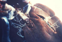
Identify the following images and give characteristics
Stilesia adult, Only proglottids are passed in the faeces without being digested in the intestinal tract, making diagnosis difficult.
Discuss the pathogenesis and pathology of Stilesia
Largely non-pathogenic
Infected livers may sometimes show slight cirrhosis with thickened walls of the bile ducts
What are clinical signs of Stilesia?
Even in the presence of high worm burdens with bile ducts almost occluded, infections are clinically inapparen
How is Stilesia treated and why?
Praziquantel
Only in areas where flocks are known to be commonly infected
Infected livers are condemned at meat inspection
Name species in Avitellina and their hosts
Avitellina centripunctata - DH: Sheep + Goat + Cattle
Avitellina goughi - DH: Sheep + Goat + Cattle
IH: Life cycle largely unknown, Psocids and oribatid mites are assumed
How is Avitellinosis diagnosed?
Only proglottids are passed in the faeces without being digested in the intestinal tract, sedimentation.
How is Avitellinosis treated and why?
Benzimidazoles: Albendazole + Mebendazole
Salicylanilides: Niclosamide
Isoquinolines: Praziquantel
Possible production losses
Name species in Thysaniezia and their hosts as well as their effect
Thysaniezia ovilla - DH: Cattle + Sheep. IH: Unkown.
Diagnosed through proglottids/strobilar pieces in the faeces, easy to distinguish from Moniezia. Non pathogenic, most common helminthosis of adult cattle
Name species in Dipylidium and their hosts
Dipylidium caninum - DH: Dog + Cat + Humans. IH: Ctenocephalides felis and canis, Pulex irritans, and Trichodectes canis
What is the predilection site for Dipylidium?
Small intestine

Identify the following images and give characteristics
Dipylidium egg packet. Uterus breaks up into egg capsules, Large oval, contains many spherical eggs with thin striated embryophores

Give characteristics of Diplydium
Dipylidium adult, Scolex: Armed rostellum, Gravid proglottids resembling cucumber seeds, Motile proglottids, Egg capsules, Reddish tinge
How is Dipylidiosis diagnosed?
Demonstration of motile gravid proglottids passed in faeces or spontaneously leaving host
Demonstration of egg capsules in faecal samples by means of flotation
Discuss the pathology and pathogenesis of Dipylidiosis, what clincial signs are seen
Largely non-pathogenic
Anal irritation (pruritus) caused by gravid proglottids migrating out of the anus, causing scooting
How is Dipylidiosis treated and controlled and why?
Isoquinolines: Praziquantel
Suspected resistance towards praziquantel has been reported
Control of lice and fleas
Man can act as an accidental definitive host, by swallowing adult fleas or as a result of dogs/cats licking the face and transferring crushed fleas
Most infections asymptomatic
Name species in Taenia and their hosts
Taenia solium - DH: Humans only. IH: Pigs, accidentally humans. (Cysticercus cellulosae)
Taenia saginata - DH: Humans only. IH: Cattle (Cysticercus bovis)
Taenia hydatigena - DH: Dogs. IH: Sheep + Goat + Pig (Cysticercus tenuicollis)
Taenia pisiformis - DH: Dog + Cat. IH: Lagomorphs (Cysticercus pisiformis)
Taenia ovis - DH: Dog. IH: Sheep + Goat (Cysticercus ovis)
Taenia multiceps - DH: Dog. IH: Sheep + Other (goat, antelopes, humans) (Coenurus cerebralis)
Taenia serialis - DH: Dog + Cat. IH: Lagomorphs + Rodents + Humans (Coenurus serialis)
Hydatigera taeniaformis - DH: Cat. IH: Rodent
Discuss the pathogenesis and pathology of Taenia solium/saginata. What clinical signs follow?
Pig/Cattle
- Musculature (muscular cysticercosis) (largely non-pathogenic in cattle)
Accidental IHs: Humans + Dog + Cat + Other
- Musculature + Subcutaneous connective tissue + CNS (neurocysticercosis)
Metacestode in intermediate hosts
▪ Largely non-pathogenic in pigs
▪ Highly pathogenic in accidental IHs such as humans, dog and cat but only if CNS is affected (neurocysticercosis)
Intermediate host:
- Infection by ingestion of eggs
- Oncospheres released in GIT. Migration into intestinal mucosa. Bloodstream. Larval predilection site
- Largely no CS
Definitive host:
- Infection by ingestion of metacestodes
- Epilepsy

Identify the following images and give characteristics
Taenia or Echynococcus eggs, cannot be differentiated, Spherical, Embryophore: Thick, brown, striated.

Identify the following images and give characteristics
Taenia solium (pork/armed tapeworm),

Identify the following images and give characteristics
Taenia saginata (beef/unarmed tapeworm)
How is Taenia solium/saginata diagnosed?
No diagnostic tools available in live pigs/cattle
Diagnosis only during meat inspection (post mortem)
Humans are the only source of eggs
How is Taenia solium/saginata treated and controlled?
Taeniasis in humans: Niclosamide + Albendazole
Neurocysticercosis in humans: Praziquantel + Albendazole
Control of taeniasis and cysticercosis must focus particularly on humans as the only definitive host
- Proper disposal of human faeces
- Identification and treatment of human carriers
- Prevention of human infection (meat inspection, cook meat properly)
- Infected carcasses are condemned and destroyed
What is the public health importance of taeniasis?
As DH:
- Relatively mild or absence of non-specific symptoms + Anal pruritus
- Aesthetically unacceptable
- After treatment the stool contains millions of infective eggs and special precautions in the safe disposal must be taken
As incidental IH:
- Autoinfection or Heteroinfection
- Of minor importance if CNS (neurocysticercosis) is NOT involved
- Epilepsy commonly seen in neurocysticercosis (neurocysticercosis can be fatal)
- Neurocysticercosis has been identified as the commonest cause of epilepsy in African communities in South Africa
Discuss the pathology and pathogenesis of Taenia hydatigena/pisiformis/ovis
Attached to any abdominal serous surface (serosal cysticercosis)
DH: Largely non-pathogenic, anal pruritus caused by motile gravid proglottids migrating out of the anus onto perianal area
Due to their large size all Taenia spp. are easy to demonstrate during necropsy
IH: Largely non-pathogenic in natural IHs, Hepatitis cysticercosa: Oncospheres and juvenile cysticerci of T. hydatigena migrate through the liver parenchyma causing haemorrhagic tracts, fibrosis, and rupture
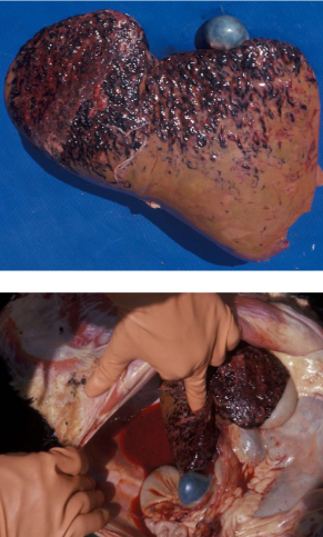

Identify the following images and give characteristics
Taeniid cysticercus

Identify the following images and give characteristics
Cysticercus pisiformis (Taenia pisiformis), IHs: Lagomorphs, Pea-sized, Attached to abdominal serous surfaces In bunches (‘grapes’)
Serosal cysticercosis

Identify the following images and give characteristics (picture is from a sheep)
Cysticercus ovis (Taenia ovis), IH: Sheep Pea-sized, Skeletal musculature, Heart + Diaphragm
Muscular cysticercosis
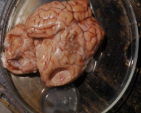
Identify the following images and give characteristics. (picture is from a sheep)
Coenurus cerebralis (Taenia multiceps), IHs: Sheep + Goat + Antelopes + Humans
Chicken egg-sized With several scoleces arranged in groups
Sheep: CNS
Cerebral coenurosis
Other IHs: Subcutaneous / intermuscular connective Tissue
Scoleces arranged in random groups
How is Taenia hydatigena/pisiformis/ovis and other carnivore Taeniosis diagnosed?
IH:
- No diagnostic tools available in live animals
- Diagnosis only during meat inspection/post mortem
DH:
- Presumptive diagnosis: ‘Scooting’
- Demonstration of motile gravid proglottids in faeces or spontaneously leaving host
- Demonstration of eggs in faecal samples by means of flotation, faecal smears
- Demonstration of eggs by means of adhesive-tape swab technique (more sensitive than faecal flotation!); eggs are mainly found under the tail and in the perianal area of dogs and cats
How is Taenia hydatigena/pisiformis/ovis treated and controlled, and why?
Larval tapeworm (metacestode) infection in IHs
- There are neither any diagnostic tools available for the diagnosis in live animals nor treatment
Taeniosis (strobilar stages) in DHs
- Isoquinolines: Praziquantel
- Diphenylmethanes: Dichlorophen
- Benzimidazoles: Fenbendazole
Prevent access to raw offal
Cook or freeze offal
Regular treatment as an individual prophylaxis to keep dogs free of patent infections
Taeniid eggs can be killed by temperatures of +80 ºC - +100 ºC within a few minutes
Common disinfectants (incl alcohol, formalin) have virtually no effect on taeniid eggs
Production losses can occur
Discuss the pathology and pathogenesis of Taenia multiceps
Sheep: Central nervous system, mostly brain (cerebral coenurosis)
Acute phase: Post-oncospheral stages migrating in CNS, Meningoencephalitis, Demonstration of immature coenuri in brain surrounded by haemorrhagic foci
Quiescent phase: Maturing growing coenurus
Chronic (terminal) phase: Space-occupying lesion, Displacement of brain tissue, Osteomalacia (pressure atrophy of bone tissue - ‘skull softening’), Demonstration of a single or several coenuri on the brain
