A&P Exam 2 TERMS AND DEFINITIONS
 Call Kai
 Learn
Learn Practice Test
 Spaced Repetition
 Match
Match Flashcards
 Knowt Play
1/251

There's no tags or description
Looks like no tags are added yet.
Name | Mastery | Learn | Test | Matching | Spaced | Call with Kai | Chat |
|---|
No analytics yet
Send a link to your students to track their progress
252 Terms
Closed fracture
Fracture that doesn’t break the skin

Open fracture
Fracture in which at least one end of the broken bone tears through the skin; carries a high risk of infection

Transverse fracture
fracture straight across the long axis of bones

Spiral fracture
fracture resulting from twisting forces applied to the bone

Comminuted fracture
Fracture in which the bone is shattered into multiple scattered into multiple fragments, difficult to repair.

Impacted fracture
fracture in which one fragment is driven into the other; usually as a result of a compression

Greenstick fracture
Fracture in which the bone breaks on one side but only bends on the other side; simillar to when a twig is bent.

Oblique fracture
fracture that occurs at an angle that is not 90 degrees

Compression fracture
fracture in which the bone is crushed under the weight it is meant to support

Avulsion fracture
Fracture in which a tendon or ligament pulls off a fragment of bone, often seen in ankle fractures.

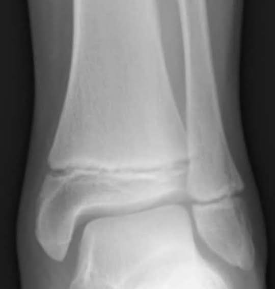
Epiphyseal Plate fracture
fracture that involves at least part of the epiphyseal plate; occurs only in children and young adults.
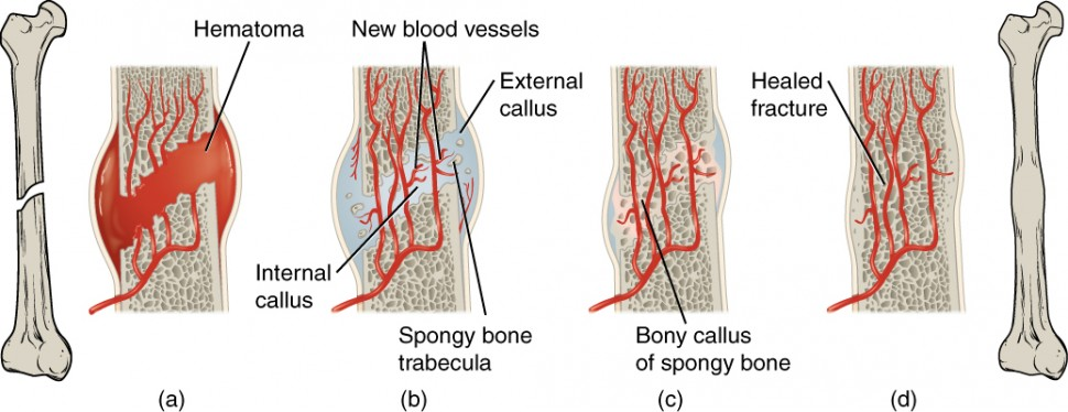
Fracture hematoma forms (blood clot)
Internal and external calli form (phagocytic cells start cleanup, fibroblasts release collagen, chondrocytes secrete cartilage = soft callous is formed)
Cartilage of calli is replaced by trabecular bone (osteoblasts turn the cartilage into spongy bone)
Remodeling occurs (spongy bone to normal bone)
Stages in fracture repair
Connective tissue features
most abundant tissue in the body
functions as strength, support, and protection
insulation
transport substance around body
degrees of vascularity
composed primarily of non-living substances
Elements of connective tissue
fibers, cells (cytes; immune cells, blood cells, fat cells, etc.)
Protein fiber types in CT
Collagen, elastic, reticular
Collagen
most common protein in body; ropes of three; high tensile strength; white fibers; stronger than steel cables
Elastic
thing fibers forming branching networks; made of elastin; coiled like a spring; yellow fibers
Reticular
short collagen fibers, has collagen; forms nets; has more give than larger collagen
blasts (cells)
create and send out proteins (create the extracellular matrix)
Cytes
“basic” cells; maintain the matrix
Immune cells
ex. macrophages, lymphocytes, mast cells
Connective tissue (overview)
cells spread out through the matrix, degrees of vascularity, functions are strength, protection, and support
All CT starts as a ____ cell
mesenchyme
Mesenchyme
source of all CT; undifferentiated; star shaped, fluid ground substance, fine protein fibrils

Areolar CT (diagram)

Areolar CT (photo)

Areolar CT characteristics
gel-like matrix, scattered fibrous proteins, most abundant CT

Areolar CT cells
fibroblasts, macrophages, mast cells, and adipocytes
Areolar CT locations
under ET, around organs and capillaries
Areolar CT functions
reservoir for salt and water, diffusion space, universal packing material
Adipose CT

Adipose Tissue characteristics
closely packed cells; chicken wire appearance; packed with lipids that push nucleus to the side; low cell division

adipose tissue functions
energy storage, insulation, supports and protects organs
adipose tissue locations
subcutaneous, surrounds kidneys, mammary glands
Reticular CT

Reticular CT
reticular fibers in web-like nets; fibroblasts
functions: supports soft tissue and small blood vessels; bed for free blood cells
locations: lymph nodes, spleen, and bone marrow

dense regular CT
matrix of parallel fibers, has a direction, more collagen than elastin, mostly fibroblasts
Functions: tensile strength and stretch resistance in ONE direction
Locations: ligaments, tendons
Dense regular CT (photo)

Dense irregular CT (photo)

Dense irregular CT
interwoven fibers, wavy/swirled, fibroblasts
functions: tensile strength and stretch resistance in multiple directions
locations: dermis, fibrous joint capsule
Elastic CT (photo)

Elastic connective tissue
similar to dense regular; slightly wave; elastin instead of collagen
functions: elastic; recoils after stretch
locations: walls of large arteries, walls of bronchial tubes
Cartilage
avascular; chondrocytes widely dispersed; matrix fairly solid; stands up to tension/compression; primarily collagen; 80% water; associated with bone
Hyaline Cartilage (photo)
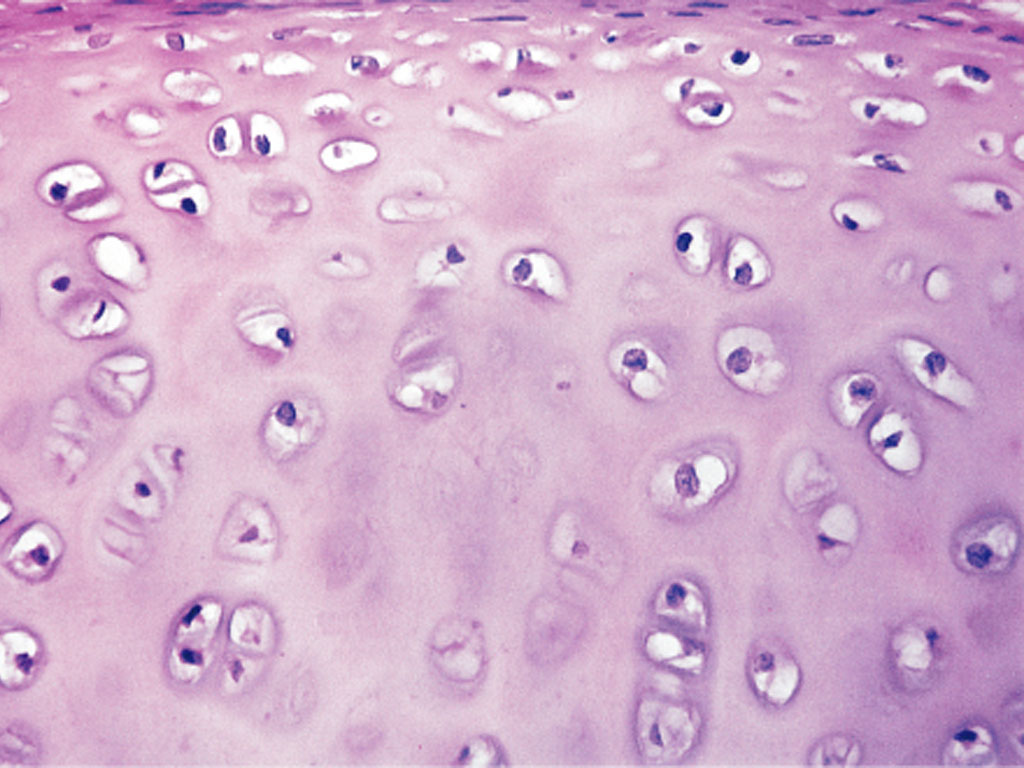
Hyaline cartilage
matrix appears transparent, chondrocytes in lacunae, chicken wire appearance
functions: rigid/smooth yet flexible, forms embryonic skeleton, allows growth of long bones
locations: articular cartilage, growth plate, costal cartilage, nose, trachea
Elastic cartilage (photo)

Elastic cartilage
similar to hyaline, but contains elastin as well; more visible fibers
functions: flexibility and strength, shape retention
locations: external ear, epiglottis
fibrocartilage (photo)

fibrocartilage
bundles of collagen fibers, chondrocytes in lacuna
functions: absorbs compression forces, tensile strength
locations: intervertebral disks, articular disks (hips, wrist, spine, etc.), pubic symphysis
bone tissue (photo)
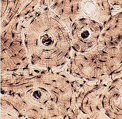
bone tissue
highly vascular; osteocytes surrounded by lacuna; matrix

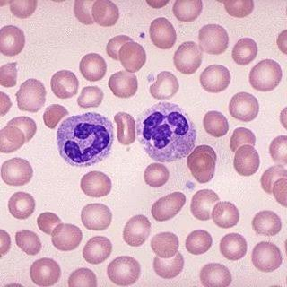
blood cells (photo)

blood
40-45% of volume is cells, fibrous proteins for clotting, mostly fluid
functions: transportation of gases, nutrients, wastes, and hormones
locations:
cardiovascular system
covering/lining membranes
thin sheet of tissue covering a structure or lining a cavity; made of epithelium and underlying CT
3 types: Cutaneous, serous, mucous
cutaneous
epithelial layer of skin
serous membranes
moist membranes in closed cavities; have 2 surfaces and serous fluid in the middle
functions of the skin
protection, temperature regulation, sensation, vitamin d synthesis, blood reservoir, excretion
vasodilate; sweat
when the body is too hot, blood vessels ___ to create ____
vasoconstrict; sweat
when the body is too cold, blood vessels ____ so that the body does not ____
Epidermis
made of ET; has keratinocytes, melanocytes, langerhans cells, and merkel cells
Dermis
made of mostly collagen fibers, has fibroblasts, gel-like ground substance, highly vascularized and innervated
Papillary layer of dermis
Areolar CT, papilla, dermal ridges
reticular layer of dermis
80% of thickness of dermis, strength/stretch/flexibility, thick collagen bundles, dense irregular CT
Hair
Functions: protect from insects, warmth (in animals), guard against physical trauma, heat loss, and sun on the head, eyelashes shield eyes from sun, nose hairs catch and filter large particles and insects
vellus hair
short, fine, unpigmented hair
terminal hair (coarse hair)
longer, pigmented; eyebrows, eyelashes, pubic, facial, chest, armpit hair
nails
heavily keratinized cells
functions: protection
Sebaceous glands
function: produce sebum (oily, lipid rich), waterproofing, antibacterial, softens skin
ducts terminate in hair follicles
Location: not in thick skin, large on face, neck, and upper chest
Acne
plugging of hair follicle by excess oil and keratin leading to infection
Eccrine sweat glands
most common type; secretes 99% water, salts, and merocrine; in dermis; NOT associated with hair follicles
Located: palms, soles, and forehead
function: temperature regulation
Apocrine sweat glands
less abundant, in axillae and anogenital regions; secretes sweat, proteins, and lipids; ASSOCIATED with a hair follicle; larger than eccrine
Functions: protection from bacteria, odor (bacteria degrading secretions/proteins)
Ceruminous glands
modified apocrine gland that produces a thick, waxy secretion; found in the ear canal
Mammary glands
modified sweat gland, produces milk, has immune function and nutritional value
Basal Cell Carcinoma
found in lowest layer of Epithelium; low malignancy; 80% of skin cancers; 99% cure rate
sun exposure, fair skin, being older than 50
risk factors of skin cancer
Squamous cell carcinoma
in stratum spinosum (keratinocytes specifically); rapid growth; 2nd most common; high cure rate if caught early
Malignant melanoma
in melanocytes; high malignancy rate; 5% of skin cancers; 30% survival if already spread far when detected
heat, chemical, electricity, light, radiation
types of burns
Superficial burn
1st degree, involves the epidermis; red, dry, painful skin; nerve endings still intact; may also be swelling
Partial thickness burn
2nd degree, damages dermis and epidermis; intense blistering forms, considerable swelling/reddening, severe pain
Full thickness burn
3rd degree, damages all layers of skin (fat/muscles/bones may also be involved); appears charred black, dry, and white; may not have any pain if nerves are severely damaged
Shock
inability to get enough blood flow to our vital organs
Burn complications
immediate loss of body fluids (blood pressure drops) and risk of infection
Abrasion
open wound where part of the epidermis/dermis is scraped off
Laceration
open wound where there is a clean cut, and likely needs a suture
Puncture Wound
Open wound where something is stabbed into the body
Avulsion
open wound where a chunk is taken out of the skin
Contusion
closed wound where blood vessels are damaged, causing blood to leak beneath the skin surface, which causes bruising.
Sesamoid bone
bone encased in a tendon
Long bone
longer than wide; almost all limb bones (except patella, carpals, and tarsals)
Short bone
Equally broad and long; cube like rounded; carpals, tarsals, sesamoids
Flat bones
thin, flat and usually curved; scapulae, ribs, and cranial bones
Irregular bones
doesn’t fit with the others; vertebrae, os coxa bones
protection, support, blood cell formation, mineral storage
Bone functions
forming a lever for our muscles to pull on
Bones create movement by….
Hematopoiesis
production of blood (happens in bone marrow)
calcium and phosphate
minerals bones store
Spongy bone tissue
location: internal
compact bone tissue
location: external
Cartilage as a bone tissue
always associated with bone;
Hyaline = ends of bones, articular
Fibrocartilage = meniscus, between vertebrae