Cellefys - uge 46: Adipose Tissue

0.0(0)
Studied by 0 people
call kaiCall Kai
learnLearn
examPractice Test
spaced repetitionSpaced Repetition
heart puzzleMatch
flashcardsFlashcards
GameKnowt Play
Card Sorting

1/63

encourage image

There's no tags or description

Looks like no tags are added yet.

64 Terms

1
New cards

How do subcutaneous and visceral white adipose tissue differ in location?

Subcutaneous white adipose tissue lies beneath the skin, for example in the abdominal wall and gluteofemoral region. Visceral white adipose tissue lies within body cavities, for example in the omentum, mesentery and around the kidneys.

<p><span><strong>Subcutaneous white adipose tissue</strong> lies beneath the skin, for example in the abdominal wall and gluteofemoral region. <strong>Visceral white adipose tissue</strong> lies within body cavities, for example in the omentum, mesentery and around the kidneys.</span></p>
2
New cards

Where are brown or beige adipocytes found in humans?

They are found in cervical (neck),

supraclavicular (above the clavicles),

paravertebral (alongside the spine) and

axillary (armpit) regions, as well as in retroperitoneal and perirenal depots.

<p><span>They are found in <strong>cervical</strong> (neck), </span></p><p><span><strong>supraclavicular</strong> (above the clavicles), </span></p><p><span><strong>paravertebral</strong> (alongside the spine) and </span></p><p><span><strong>axillary</strong> (armpit) regions, as well as in <strong>retroperitoneal</strong> and <strong>perirenal</strong> depots.</span></p>
3
New cards

What are examples of other anatomically distinct adipose tissue depots?

Retro-orbital fat behind the eyes, marrow fat within bone marrow and dermal fat within the skin.

4
New cards

How does adipose tissue store excess energy?

When energy intake exceeds energy expenditure, adipocytes store excess energy as triglycerides in lipid droplets.

5
New cards

Why are triglycerides an efficient form of energy storage?

Triglycerides are stored with very little water and yield about 9 kcal/g, compared with 4 kcal/g for carbohydrates and proteins.

6
New cards

How does the body obtain energy from triglycerides stored in adipocytes?

Triglycerides are broken down by lipolysis, releasing fatty acids. These fatty acids can be oxidized to generate ATP, with CO₂ and water as end products.

7
New cards

Why is adipose tissue considered an endocrine organ?

Adipose tissue releases signalling molecules such as leptin and adiponectin into the bloodstream. These signals act on other tissues and help regulate appetite and metabolism.

<p><span>Adipose tissue releases signalling molecules such as <strong>leptin</strong> and <strong>adiponectin</strong> into the bloodstream. These signals act on other tissues and help regulate appetite and metabolism.</span></p>
8
New cards

How does leptin help regulate energy balance?

Leptin signals the size of the body’s fat stores to the hypothalamus and helps reduce appetite.
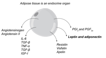
<p><span>Leptin signals the size of the body’s fat stores to the <strong>hypothalamus</strong> and helps <strong>reduce appetite</strong>.</span></p>
9
New cards

How does adiponectin affect glucose and lipid metabolism?

Adiponectin increases insulin sensitivity and stimulates fatty acid oxidation in the liver and skeletal muscle. These effects can help lower plasma glucose and triglyceride levels.
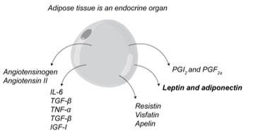
<p><span>Adiponectin <strong>increases insulin sensitivity</strong> and stimulates <strong>fatty acid oxidation</strong> in the liver and skeletal muscle. These effects can help lower plasma glucose and triglyceride levels.</span></p>
10
New cards

How do acylation-stimulating protein (ASP) and adipsin affect fat storage in adipose tissue?

ASP influences the rate of triglyceride synthesis. Adipsin promotes fatty acid storage and triglyceride synthesis.
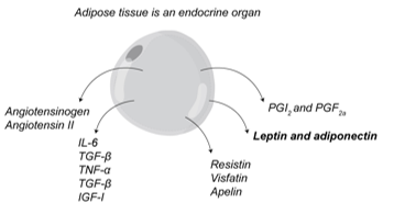
<p><span><strong>ASP</strong> influences the rate of triglyceride synthesis. <strong>Adipsin</strong> promotes fatty acid storage and triglyceride synthesis.</span></p>
11
New cards

What is adipophilin used to detect in cells?

Adipophilin (perilipin 2, PLIN2) is a protein on the surface of lipid droplets. Staining for adipophilin can reveal lipid accumulation within cells.
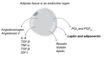
<p><span><strong>Adipophilin (perilipin 2, PLIN2)</strong> is a protein on the surface of <strong>lipid droplets</strong>. Staining for adipophilin can reveal <strong>lipid accumulation within cells</strong>.</span></p>
12
New cards

How do angiotensinogen and angiotensin II affect blood pressure and adipose tissue?

Angiotensinogen is the precursor of angiotensin II, which regulates blood pressure and electrolyte balance.

In adipose tissue, angiotensin II inhibits lipoblast differentiation and regulates lipid storage in mature adipocytes.

<p><span>Angiotensinogen is the precursor of <strong>angiotensin II</strong>, which regulates blood pressure and electrolyte balance. </span></p><p><span>In adipose tissue, angiotensin II inhibits <strong>lipoblast differentiation</strong> and regulates <strong>lipid storage</strong> in mature adipocytes.</span></p>
13
New cards

How does apelin affect the cardiovascular system?

Apelin increases cardiac muscle contractility and decreases blood pressure.
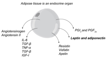
<p><span>Apelin <strong>increases cardiac muscle contractility</strong> and <strong>decreases blood pressure</strong>.</span></p>
14
New cards

What is the function of insulin-like growth factor 1 (IGF-1)?

IGF-1 stimulates cell proliferation and mediates many of the effects of growth hormone.

<p><span>IGF-1 <strong>stimulates cell proliferation</strong> and mediates many of the effects of <strong>growth hormone</strong>.</span></p>
15
New cards

What are the effects of interleukin 6 (IL-6) on immunity and metabolism?

IL-6 acts on immune cells and regulates glucose and lipid metabolism. In cancer and other wasting disorders, it can reduce adipose tissue activity.

<p><span>IL-6 acts on <strong>immune cells</strong> and regulates <strong>glucose and lipid metabolism</strong>. In cancer and other wasting disorders, it can reduce adipose tissue activity.</span></p>
16
New cards

How does plasminogen activator inhibitor 1 (PAI-1) affect blood clot breakdown?

PAI-1 inhibits fibrinolysis, the process that breaks down blood clots.

<p><span>PAI-1 <strong>inhibits fibrinolysis</strong>, the process that breaks down blood clots.</span></p>
17
New cards

What processes do prostaglandins I₂ (PGI₂) and F₂α (PGF₂α) help regulate?

These prostaglandins help regulate inflammation, blood clotting, ovulation, menstruation and acid secretion.

<p><span>These prostaglandins help regulate <strong>inflammation, blood clotting, ovulation, menstruation and acid secretion</strong>.</span></p>
18
New cards

How does resistin affect insulin sensitivity?

Resistin is associated with increased insulin resistance and has been linked to obesity and type 2 diabetes.

<p><span>Resistin is associated with <strong>increased insulin resistance</strong> and has been linked to <strong>obesity and type 2 diabetes</strong>.</span></p>
19
New cards

How does retinol-binding protein 4 (RBP4) affect glucose homeostasis?

RBP4 released by adipose tissue, particularly visceral adipose tissue, can decrease insulin sensitivity and disrupt glucose homeostasis.

<p><span>RBP4 released by adipose tissue, particularly <strong>visceral adipose tissue</strong>, can <strong>decrease insulin sensitivity</strong> and disrupt glucose homeostasis.</span></p>
20
New cards

What cellular processes does transforming growth factor β (TGF-β) regulate?

TGF-β regulates cell proliferation, differentiation and apoptosis and plays a role in development.

<p><span>TGF-β regulates <strong>cell proliferation, differentiation and apoptosis</strong> and plays a role in development.</span></p>
21
New cards

How can tumour necrosis factor (TNF) contribute to insulin resistance in obesity?

TNF can interfere with insulin receptor signalling, reducing the cell’s response to insulin.
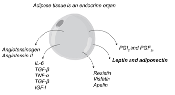
<p><span>TNF can <strong>interfere with insulin receptor signalling</strong>, reducing the cell’s response to insulin.</span></p>
22
New cards

How is visfatin associated with visceral adipose tissue?

Visfatin is produced by visceral adipose tissue, and its levels have been reported to correlate with visceral fat mass.

<p><span>Visfatin is produced by <strong>visceral adipose tissue</strong>, and its levels have been reported to correlate with <strong>visceral fat mass</strong>.</span></p>
23
New cards

How do the main functions of white and brown adipose tissue differ?

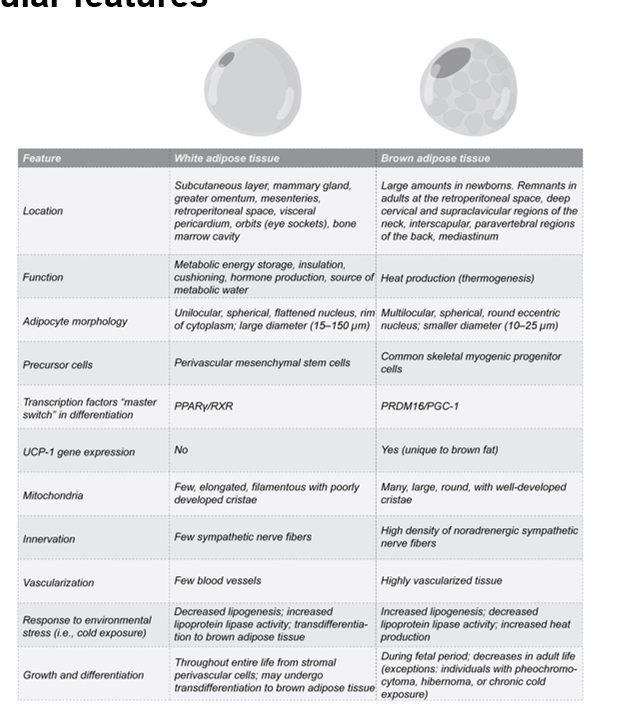
White adipose tissue stores energy, provides insulation and cushioning, and secretes hormones.

Brown adipose tissue specialises in heat production (thermogenesis).

<p><span><strong>White adipose tissue</strong> stores energy, provides insulation and cushioning, and secretes hormones. </span></p><p><span><strong>Brown adipose tissue</strong> specialises in heat production (<strong>thermogenesis</strong>).</span></p>
24
New cards

How can you distinguish white and brown adipocytes by their morphology?

White adipocytes are larger (15–150 µm) and unilocular. One large lipid droplet pushes the flattened nucleus to the edge.

Brown adipocytes are smaller (10–25 µm) and multilocular, with many lipid droplets and a round, eccentric nucleus.

<p><span><strong>White adipocytes</strong> are larger (15–150 µm) and <strong>unilocular</strong>. One large lipid droplet pushes the flattened nucleus to the edge. </span></p><p><span><strong>Brown adipocytes</strong> are smaller (10–25 µm) and <strong>multilocular</strong>, with many lipid droplets and a round, eccentric nucleus.</span></p>
25
New cards

Which precursor cells give rise to white and classical brown adipocytes?

White adipocytes can arise from perivascular mesenchymal stem cells.

Classical brown adipocytes can arise from progenitors that share a lineage with skeletal muscle cells.
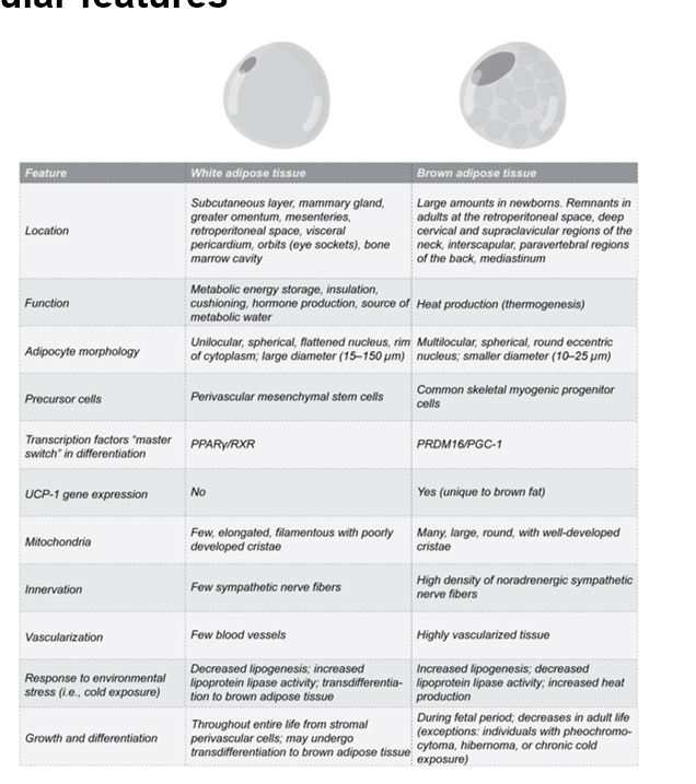
<p><span>White adipocytes can arise from <strong>perivascular mesenchymal stem cells</strong>. </span></p><p><span>Classical brown adipocytes can arise from progenitors that share a lineage with <strong>skeletal muscle cells</strong>.</span></p>
26
New cards

Which factors drive white and brown adipocyte differentiation?

PPARγ/RXR drives adipocyte differentiation.

PRDM16 and PGC-1α help establish the brown adipocyte’s thermogenic programme.
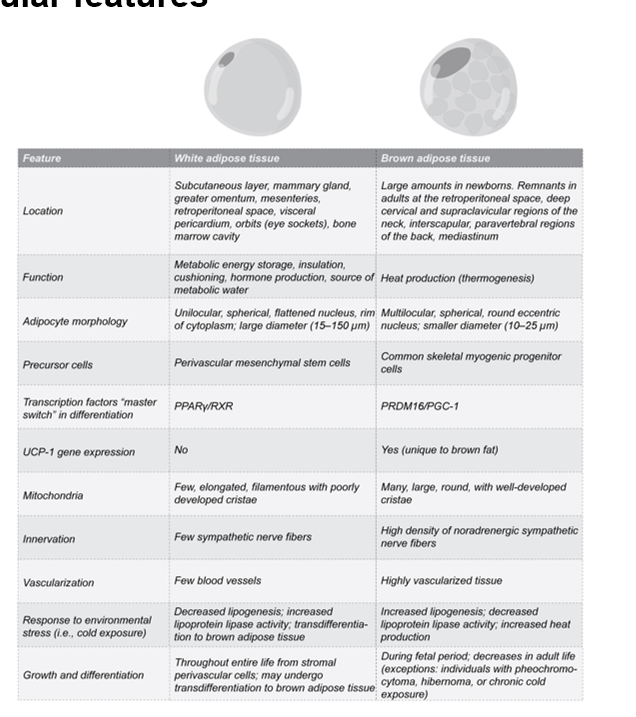
<p><span><strong>PPARγ/RXR</strong> drives adipocyte differentiation. </span></p><p><span><strong>PRDM16</strong> and <strong>PGC-1α</strong> help establish the brown adipocyte’s thermogenic programme.</span></p>
27
New cards

How does UCP1 expression differ between typical white and brown adipocytes?

Brown adipocytes express UCP1 in their mitochondria, allowing energy to be released as heat.

Typical white adipocytes lack UCP1, while beige adipocytes can express it.

<p><span>Brown adipocytes express <strong>UCP1</strong> in their mitochondria, allowing energy to be released as <strong>heat</strong>. </span></p><p><span>Typical white adipocytes lack UCP1, while <strong>beige adipocytes</strong> can express it.</span></p>
28
New cards

How do mitochondria differ between white and brown adipocytes?

White adipocytes have relatively few mitochondria.

Brown adipocytes have many large mitochondria with well-developed cristae, supporting heat production.

<p><span>White adipocytes have <strong>relatively few mitochondria</strong>. </span></p><p><span>Brown adipocytes have <strong>many large mitochondria with well-developed cristae</strong>, supporting heat production.</span></p>
29
New cards

How does sympathetic innervation differ between white and brown adipose tissue?

White adipose tissue has fewer sympathetic nerve fibres,

while brown adipose tissue has a high density of noradrenergic sympathetic fibres.

<p><span>White adipose tissue has <strong>fewer sympathetic nerve fibres</strong>, </span></p><p><span>while brown adipose tissue has a <strong>high density of noradrenergic sympathetic fibres</strong>.</span></p>
30
New cards

How does vascularisation differ between white and brown adipose tissue?

Brown adipose tissue is more densely vascularised than white adipose tissue.

Blood supplies oxygen and fuel and carries heat away from the tissue.
<p><span>Brown adipose tissue is <strong>more densely vascularised</strong> than white adipose tissue. </span></p><p><span>Blood supplies oxygen and fuel and carries heat away from the tissue.</span></p>
31
New cards

How do white and brown adipose tissue respond to cold exposure?

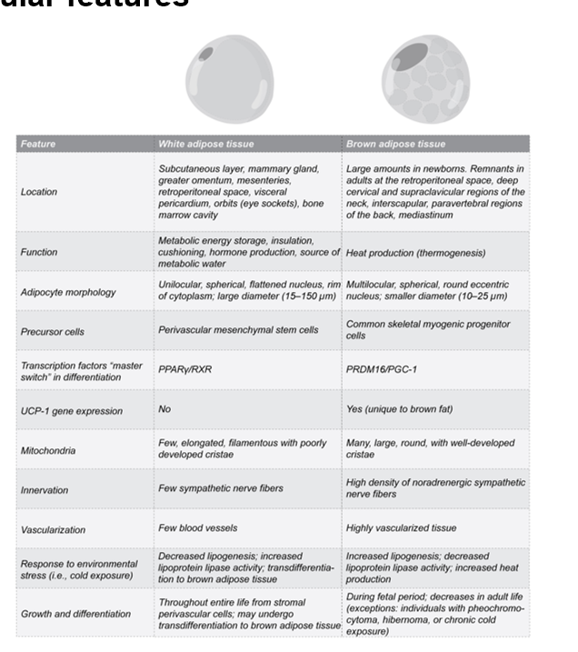
Brown adipose tissue increases heat production.

With prolonged cold exposure, some white fat depots can develop beige adipocytes, which also contribute to thermogenesis.

<p><span>Brown adipose tissue increases <strong>heat production</strong>. </span></p><p><span>With prolonged cold exposure, some white fat depots can develop <strong>beige adipocytes</strong>, which also contribute to thermogenesis.</span></p><p></p>
32
New cards

How does the development of white and brown adipose tissue differ across life?

White adipocytes can form throughout life.

Classical brown adipose tissue develops before birth, is abundant in newborns and generally becomes less abundant in adulthood

<p><span>White adipocytes can form <strong>throughout life</strong>. </span></p><p><span>Classical brown adipose tissue develops <strong>before birth</strong>, is abundant in newborns and generally becomes less abundant in adulthood</span></p>
33
New cards

How can you recognise an early lipoblast during white adipocyte differentiation?

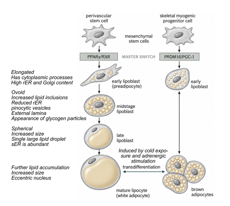
The early lipoblast (preadipocyte) is elongated, has cytoplasmic processes and contains abundant rough ER (rER) and Golgi apparatus.

<p><span>The early lipoblast (<strong>preadipocyte</strong>) is <strong>elongated</strong>, has cytoplasmic processes and contains abundant <strong>rough ER (rER)</strong> and <strong>Golgi apparatus</strong>.</span></p>
34
New cards

What changes occur at the midstage of white adipocyte differentiation?

The lipoblast becomes ovoid and accumulates more lipid inclusions.

Rough ER decreases, while pinocytic vesicles, an external lamina and glycogen particles appear.
<p><span>The lipoblast becomes <strong>ovoid</strong> and accumulates more <strong>lipid inclusions</strong>. </span></p><p><span>Rough ER decreases, while <strong>pinocytic vesicles</strong>, an <strong>external lamina</strong> and <strong>glycogen particles</strong> appear.</span></p>
35
New cards

How does a late lipoblast become a mature white adipocyte?

The late lipoblast becomes larger and spherical, with one large lipid droplet and abundant smooth ER (sER).

As more lipid accumulates, the droplet enlarges and pushes the nucleus towards the cell periphery.

<p><span>The late lipoblast becomes <strong>larger and spherical</strong>, with one large lipid droplet and abundant <strong>smooth ER (sER)</strong>. </span></p><p><span>As more lipid accumulates, the droplet enlarges and pushes the nucleus towards the cell periphery.</span></p>
36
New cards

What is the difference between adipocyte differentiation and transdifferentiation in response to cold?

In differentiation, a precursor cell develops into a mature adipocyte.

In transdifferentiation, an already differentiated adipocyte changes its phenotype.

he figure shows how cold exposure and adrenergic stimulation can promote a brown-like (beige), multilocular phenotype in white adipose tissue.

<p><span>In <strong>differentiation</strong>, a precursor cell develops into a mature adipocyte. </span></p><p><span>In <strong>transdifferentiation</strong>, an already differentiated adipocyte changes its phenotype. </span></p><p><span>he figure shows how <strong>cold exposure and adrenergic stimulation</strong> can promote a <strong>brown-like (beige), multilocular phenotype</strong> in white adipose tissue.</span></p>
37
New cards

How do subcutaneous and visceral fat depots change in obesity?

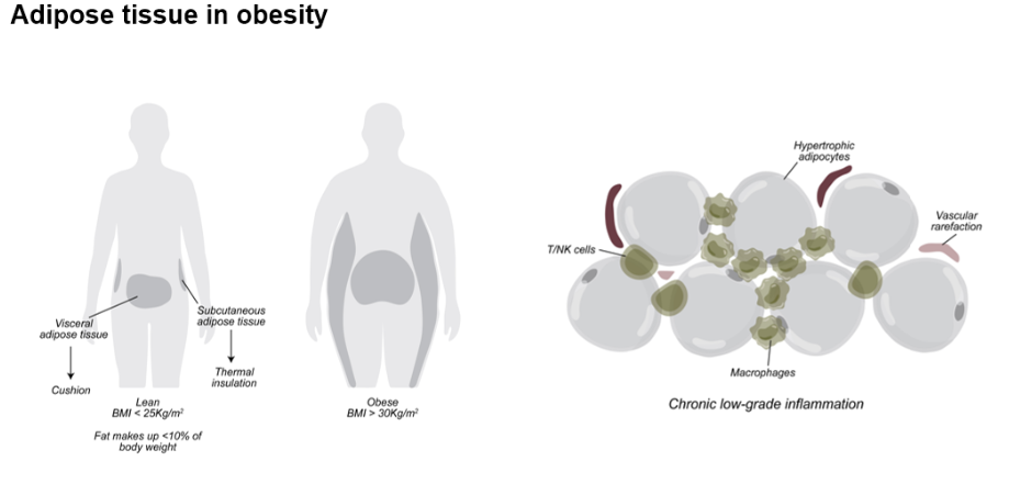
Both subcutaneous adipose tissue beneath the skin and visceral adipose tissue around internal organs can expand.

Adipocyte hypertrophy means that individual adipocytes become larger as they store more lipid.
<p><span>Both <strong>subcutaneous adipose tissue</strong> beneath the skin and <strong>visceral adipose tissue</strong> around internal organs can expand. </span></p><p><span><strong>Adipocyte hypertrophy</strong> means that individual adipocytes become larger as they store more lipid.</span></p>
38
New cards

What is vascular rarefaction in expanding adipose tissue?

Vascular rarefaction is a reduction in blood vessel density. When the tissue expands faster than its blood supply, some regions may receive too little oxygen and become hypoxic.

<p><span><strong>Vascular rarefaction</strong> is a reduction in blood vessel density. When the tissue expands faster than its blood supply, some regions may receive too little oxygen and become <strong>hypoxic</strong>.</span></p>
39
New cards

How can adipose tissue expansion contribute to chronic low-grade inflammation in obesity?

Enlarged adipocytes and inadequate oxygen supply can stress the tissue. Immune cells, including macrophages, T cells and natural killer (NK) cells, can contribute to persistent inflammatory signalling.

<p><span>Enlarged adipocytes and inadequate oxygen supply can stress the tissue. Immune cells, including <strong>macrophages, T cells and natural killer (NK) cells</strong>, can contribute to persistent inflammatory signalling.</span></p>
40
New cards

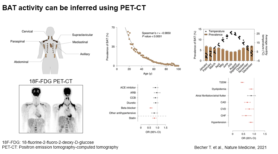
How can ¹⁸F-FDG PET-CT be used to infer brown adipose tissue (BAT) activity?

¹⁸F-FDG is a radioactive glucose analogue.

PET detects where the tracer accumulates, and CT shows whether the signal is located in adipose tissue.

Uptake in typical BAT depots, such as the supraclavicular region, indicates glucose uptake and is an indirect measure of BAT activity.

<p><span><strong>¹⁸F-FDG</strong> is a radioactive glucose analogue. </span></p><p><span><strong>PET</strong> detects where the tracer accumulates, and <strong>CT</strong> shows whether the signal is located in adipose tissue. </span></p><p><span>Uptake in typical BAT depots, such as the <strong>supraclavicular region</strong>, indicates glucose uptake and is an indirect measure of BAT activity.</span></p>
41
New cards

How does the prevalence of PET-detectable brown adipose tissue change with age?

The proportion of people with detectable BAT decreases markedly with age.

The graph shows a strong negative correlation between age and BAT detection.

<p><span>The proportion of people with <strong>detectable BAT</strong> decreases markedly with age. </span></p><p><span>The graph shows a strong negative correlation between age and BAT detection.</span></p>
42
New cards

How does outdoor temperature relate to the detection of brown adipose tissue on PET-CT?

BAT is detected more often during colder months and less often during warmer months.

This pattern is consistent with cold-induced activation of BAT.
<p><span>BAT is detected more often during <strong>colder months</strong> and less often during warmer months. </span></p><p><span>This pattern is consistent with <strong>cold-induced activation</strong> of BAT.</span></p>
43
New cards

How is beta-blocker use associated with brown adipose tissue detection on PET-CT?

People taking beta blockers had lower odds of detectable BAT.

This is consistent with BAT activation by adrenergic signalling.
<p><span>People taking <strong>beta blockers</strong> had lower odds of detectable BAT. </span></p><p><span>This is consistent with BAT activation by <strong>adrenergic signalling</strong>.</span></p>
44
New cards

How is PET-detectable brown adipose tissue associated with cardiometabolic disease?

People with detectable BAT had lower odds of several conditions, including type 2 diabetes, dyslipidaemia and hypertension.

The study shows an association. Whether BAT itself causes the lower odds remains uncertain.

<p><span>People with detectable BAT had <strong>lower odds</strong> of several conditions, including <strong>type 2 diabetes, dyslipidaemia and hypertension</strong>. </span></p><p><span>The study shows an association. Whether BAT itself causes the lower odds remains uncertain.</span></p>
45
New cards

How does cold exposure activate brown adipose tissue (BAT)?

Cold exposure increases sympathetic norepinephrine release.

Norepinephrine binds β-adrenergic receptors on brown adipocytes, increasing cAMP and activating protein kinase A (PKA).

<p><span>Cold exposure increases <strong>sympathetic norepinephrine release</strong>. </span></p><p><span>Norepinephrine binds <strong>β-adrenergic receptors</strong> on brown adipocytes, increasing <strong>cAMP</strong> and activating <strong>protein kinase A (PKA)</strong>.</span></p>
46
New cards

How does UCP1 generate heat in brown adipocytes?

The electron transport chain pumps H⁺ into the mitochondrial intermembrane space.

UCP1 lets H⁺ flow back into the matrix, bypassing ATP synthase.

This dissipates the proton gradient as heat and reduces the amount of ATP produced per unit of fuel oxidised.

<p><span>The electron transport chain pumps <strong>H⁺</strong> into the mitochondrial intermembrane space. </span></p><p><span><strong>UCP1</strong> lets H⁺ flow back into the matrix, bypassing <strong>ATP synthase</strong>. </span></p><p><span>This dissipates the proton gradient as <strong>heat</strong> and reduces the amount of ATP produced per unit of fuel oxidised.</span></p>
47
New cards

How do long-chain fatty acids and purine nucleotides regulate UCP1?

Long-chain fatty acids activate UCP1, while purine nucleotides inhibit it.

Fatty acids can also serve as fuel for mitochondrial oxidation.

<p><span><strong>Long-chain fatty acids activate UCP1</strong>, while <strong>purine nucleotides inhibit it</strong>.</span></p><p><span>Fatty acids can also serve as fuel for mitochondrial oxidation.</span></p>
48
New cards

How does β-adrenergic signalling increase thermogenic gene expression in brown adipocytes?

The cAMP–PKA pathway activates p38 MAPK, which promotes transcription of thermogenic genes such as Ucp1, Cidea and Dio2.

<p><span>The <strong>cAMP–PKA pathway</strong> activates <strong>p38 MAPK</strong>, which promotes transcription of thermogenic genes such as <strong>Ucp1, Cidea and Dio2</strong>.</span></p>
49
New cards

How do glucose and circulating fatty acids enter brown adipocytes?

Glucose enters through GLUT1 and GLUT4, while CD36 facilitates fatty acid uptake. This glucose uptake is what ¹⁸F-FDG PET-CT uses as an indirect indicator of BAT activity.

<p><span>Glucose enters through <strong>GLUT1 and GLUT4</strong>, while <strong>CD36</strong> facilitates fatty acid uptake. This glucose uptake is what <strong>¹⁸F-FDG PET-CT</strong> uses as an indirect indicator of BAT activity.</span></p>
50
New cards

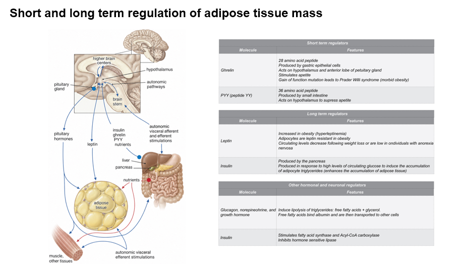
How do ghrelin and peptide YY (PYY) regulate appetite in the short term?

Ghrelin is released mainly by the stomach and stimulates appetite.

PYY is released by the intestine after a meal and suppresses appetite. Both act on the hypothalamus.

<p><span><strong>Ghrelin</strong> is released mainly by the stomach and <strong>stimulates appetite</strong>. </span></p><p><span><strong>PYY</strong> is released by the intestine after a meal and <strong>suppresses appetite</strong>. Both act on the hypothalamus.</span></p>
51
New cards

How do circulating leptin levels change with obesity and weight loss?

Leptin levels are generally higher in obesity and fall after weight loss.

In obesity, the brain can become less responsive to leptin’s appetite-suppressing signal, a phenomenon called leptin resistance.
<p><span>Leptin levels are generally <strong>higher in obesity</strong> and <strong>fall after weight loss</strong>. </span></p><p><span>In obesity, the brain can become less responsive to leptin’s appetite-suppressing signal, a phenomenon called <strong>leptin resistance</strong>.</span></p>
52
New cards

How does insulin promote triglyceride storage in adipocytes after a meal?

A rise in blood glucose stimulates the pancreas to release insulin.

Insulin stimulates acetyl-CoA carboxylase and fatty acid synthase, promoting fatty acid synthesis.

It also inhibits hormone-sensitive lipase, reducing triglyceride breakdown.

<p><span>A rise in blood glucose stimulates the <strong>pancreas</strong> to release insulin. </span></p><p><span>Insulin stimulates <strong>acetyl-CoA carboxylase</strong> and <strong>fatty acid synthase</strong>, promoting fatty acid synthesis. </span></p><p><span>It also inhibits <strong>hormone-sensitive lipase</strong>, reducing triglyceride breakdown.</span></p>
53
New cards

How does norepinephrine mobilise stored fat from adipocytes?

Norepinephrine stimulates lipolysis, releasing free fatty acids and glycerol from stored triglycerides. The fatty acids bind to albumin in the blood and can be transported to other tissues for use as fuel.

<p><span>Norepinephrine stimulates <strong>lipolysis</strong>, releasing <strong>free fatty acids and glycerol</strong> from stored triglycerides. The fatty acids bind to <strong>albumin</strong> in the blood and can be transported to other tissues for use as fuel.</span></p>
54
New cards

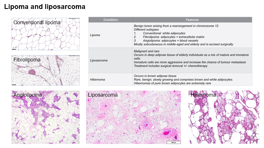
What is a conventional lipoma, and where is it usually found?

A conventional lipoma is a benign tumour composed of mature white adipocytes.

It is usually subcutaneous and can be associated with a chromosome 12 rearrangement.
<p><span>A conventional lipoma is a <strong>benign tumour composed of mature white adipocytes</strong>. </span></p><p><span>It is usually <strong>subcutaneous</strong> and can be associated with a <strong>chromosome 12 rearrangement</strong>.</span></p>
55
New cards

How can you distinguish fibrolipoma from angiolipoma histologically?

A fibrolipoma contains mature adipocytes mixed with abundant fibrous connective tissue.

An angiolipoma contains mature adipocytes mixed with numerous small blood vessels.

<p><span>A <strong>fibrolipoma</strong> contains mature adipocytes mixed with <strong>abundant fibrous connective tissue</strong>. </span></p><p><span>An <strong>angiolipoma</strong> contains mature adipocytes mixed with <strong>numerous small blood vessels</strong>.</span></p>
56
New cards

What is a liposarcoma, and how does its location typically differ from that of a lipoma?

A liposarcoma is a malignant tumour with adipocytic differentiation that often occurs in deep soft tissues. A conventional lipoma is benign and typically lies in the subcutaneous tissue.

<p><span>A <strong>liposarcoma</strong> is a <strong>malignant tumour with adipocytic differentiation</strong> that often occurs in <strong>deep soft tissues</strong>. A conventional lipoma is benign and typically lies in the <strong>subcutaneous tissue</strong>.</span></p>
57
New cards

What is a hibernoma, and what do its cells look like?

A hibernoma is a rare, benign, slow-growing tumour composed of brown fat cells, often mixed with white adipocytes. The brown adipocytes have multiple small lipid droplets, while the white adipocytes have one large droplet.

<p><span>A hibernoma is a <strong>rare, benign, slow-growing tumour</strong> composed of brown fat cells, often mixed with white adipocytes. The <strong>brown adipocytes</strong> have <strong>multiple small lipid droplets</strong>, while the white adipocytes have one large droplet.</span></p>
58
New cards

Where does circulating irisin come from during exercise?

Skeletal muscle can release irisin in response to exercise. Irisin enters the bloodstream and can act on other tissues, including adipose tissue.
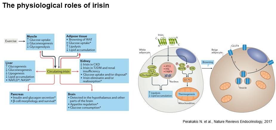
<p><span>Skeletal muscle can release <strong>irisin</strong> in response to exercise. Irisin enters the bloodstream and can act on other tissues, including <strong>adipose tissue</strong>.</span></p>
59
New cards

What does irisin-induced browning of white adipose tissue mean?

White adipose tissue develops beige adipocytes with thermogenic features, including increased UCP1 expression.

UCP1 allows mitochondria to release more energy as heat.

<p><span>White adipose tissue develops <strong>beige adipocytes</strong> with thermogenic features, including increased <strong>UCP1</strong> expression. </span></p><p><span>UCP1 allows mitochondria to release more energy as <strong>heat</strong>.</span></p>
60
New cards

How can irisin affect lipid storage in white adipocytes?

Irisin can stimulate lipolysis, the breakdown of stored triglycerides, and reduce lipid accumulation.

The figure shows a proposed pathway involving cAMP, PKA and hormone-sensitive lipase (HSL).

<p><span>Irisin can stimulate <strong>lipolysis</strong>, the breakdown of stored triglycerides, and reduce <strong>lipid accumulation</strong>. </span></p><p><span>The figure shows a proposed pathway involving <strong>cAMP, PKA and hormone-sensitive lipase (HSL)</strong>.</span></p>
61
New cards

How might irisin increase glucose uptake in beige adipocytes?

Irisin may promote the movement of GLUT4 from intracellular vesicles to the cell membrane, where it facilitates glucose uptake. The question marks in the figure indicate that this mechanism is proposed.

<p><span>Irisin may promote the movement of <strong>GLUT4</strong> from intracellular vesicles to the <strong>cell membrane</strong>, where it facilitates glucose uptake. The question marks in the figure indicate that this mechanism is proposed.</span></p>
62
New cards

How did the researchers test the effect of increasing circulating irisin in mice?

They injected AAV8–irisin into the tail vein. The vector mainly delivered the irisin gene to hepatocytes, which then released irisin into the blood.

The researchers compared these mice with mice given AAV8–GFP to control for the injection and vector delivery.

<p><span>They injected <strong>AAV8–irisin</strong> into the tail vein. The vector mainly delivered the irisin gene to <strong>hepatocytes</strong>, which then released irisin into the blood. </span></p><p><span>The researchers compared these mice with mice given <strong>AAV8–GFP</strong> to control for the injection and vector delivery.</span></p>
63
New cards

What question did the global Fndc5 knockout address?

By comparing Fndc5 knockout mice with controls, the researchers could test what happens when mice lack endogenous FNDC5-derived irisin.

Because the knockout was global, the comparison could reveal a whole-animal effect but could not identify which tissue normally supplied the relevant irisin.

<p><span>By comparing <strong>Fndc5 knockout</strong> mice with controls, the researchers could test what happens when mice lack <strong>endogenous FNDC5-derived irisin</strong>. </span></p><p><span>Because the knockout was global, the comparison could reveal a whole-animal effect but could not identify which tissue normally supplied the relevant irisin.</span></p>
64
New cards

How did the researchers use Cre–LoxP to remove ST2 specifically from regulatory T cells?

They crossed Foxp3-Cre mice with mice carrying floxed Il1rl1, which encodes ST2. The loxP sites defined the DNA segment to be deleted, and Foxp3-driven Cre restricted deletion to regulatory T cells (Tregs).

<p><span>They crossed <strong>Foxp3-Cre mice</strong> with mice carrying <strong>floxed Il1rl1</strong>, which encodes ST2. The <strong>loxP sites</strong> defined the DNA segment to be deleted, and <strong>Foxp3-driven Cre</strong> restricted deletion to regulatory T cells (<strong>Tregs</strong>).</span></p>



