The Head
 Call Kai
 Learn
Learn Practice Test
 Spaced Repetition
 Match
Match Flashcards
 Knowt Play
1/214

There's no tags or description
Looks like no tags are added yet.
Name | Mastery | Learn | Test | Matching | Spaced | Call with Kai | Chat |
|---|
No analytics yet
Send a link to your students to track their progress
215 Terms
Outline the functions of the nasal cavity
Conduction and preparation of inspired air
Moistens air
Warms air
FIlters particles
Mucus traps particles, which is then swallowed
Heat exchange for cooling the brain
Airflow cools venous blood
Subsequent countercurrent flow with arteries will cool arterial blood going to the brain
Olfaction
Caudal regions of the turbinates covered by olfactory epithelium
Outline the landmarks of the nasal cavity
Dorsally: nasal bones
Ventrally: hard palate
Laterally: incisive bone and maxilla
Rostrally: nares (nostrils)
Caudally: nasopharynx
Axially: nasal septum

Label the bones on this diagram
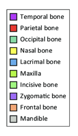
Describe the structure of the nares
Surrounded by hairless skin
Sometimes highly modified, depending on the species
Supported by nasal cartilages which are attached to the nasal septum
Describe the species differences in bovine nares
Nostrils are surrounded by a smooth, hairless nasolabial plate
Stratified cornified epithelium
Serous glands create moisture (nasolabial glands)
Describe the species differences in equine nares
Horses cannot breathe through their mouth
No ventral nasal cartilage
Incomplete cartilaginous ring: distensible nostrils to improve airflow
Alar cartilages form comma-shaped nostrils
There is a ventral ‘true’ nostril and a dorsal ‘false’ nostril
False nostril is a skin-lined diverticulum within the nasoincisive notch
Describe the species differences in canine and feline nares
Divided by medium groove (philtrum)
Secretions are from the lateral nasal gland
Describe the species differences in porcine nares
Small nostrils on a flat, mobile snout
Highly sensitive
Rostral bone provides the nostril with protection
Describe the species differences in birds
Slit-like openings (not in diving birds)
Operculum: overhanging bony flap
Describe the species differences in canine/feline BOAS
Stenotic nares
Elongated and thickened soft palate
Enlarged tongue
Narrow trachea
Outline the muscles of the nares
Nostril dilation
Levator nasolabialis
Caninus
Transversus nasi
All are innervated by the facial nerve, and supplied by the facial artery
What is the role of the nasal vestibule?
This is the opening of the nasal cavity
Contains the opening of the nasolacrimal duct
Receives nasal gland secretions in dogs
Describe the borders of the nasal cavity
Dorsal: nasal bones
Lateral: incisive bones and maxilla
Ventral: palatine bones
Caudal: ethmoidal bone
Axial: nasal septum
Describe the structure of the nasal cavity
Extends from the nostrils to the cribiform plate of the ethmoid bone
Divided into left and right sides by the nasal septum
Each side is then further divided by nasal conchae
What is the difference between choanae and conchae?
Choanae: paired caudal openings of the nasal cavity
Conchae: scrolls of turbinate bone covered in mucosa
Describe the structure and function of conchae
Structure
Scrolls of turbinate bone covered in mucosa
Highly vascular
Function
Further increase nasal surface area

What is shown by these arrows?
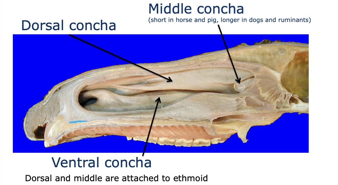
Which meatus should you intubate?
A tube should always be placed in the ventral meatus to intubate
Describe the species differences in the equine nasal meatus
4 nasal meatus
Dorsal meatus: to olfactory mucosa
Middle meatus: to paranasal sinuses
Common meatus
Ventral meatus: to the pharynx
Describe the species differences in the canine nasal meatus
Complex turbinates
More resistance to airflow
Describe the species differences in the bovine nasal meatus
Very small middle meatus
Describe the species differences in the avian nasal meatus
Rostral concha
Middle concha
Caudal concha

What is shown by these numbers?
Rostral concha
Middle concha
Caudal concha
Which epithelium is found in:
The vestibule
The nasal cavity
Vestibule: stratified squamous epithelium
Nasal cavity: respiratory epithelium, olfactory epithelium
Describe the structure of the respiratory epithelium in the nasal cavity
Pseudostratified
Ciliated
Columnar
Goblet cells
Mucous and serous glands in the lamina propria
Respiratory mucosa covers most parts of the nasal cavity
Mucosa: epithelium and lamina propria
Describe the function of the respiratory epithelium in the nasal cavity
Regulation of air flow by erectile tissue
Cleaning by cilia
Humidification
Warming (variable blood perfusion)
Protecting reflexes (e.g sneeze reflex)
Describe how olfaction occurs in the domestic animal, including the role of:
Olfactory cells
Vomeronasal organ
Ethmoturbinates extend rostrally from the ethmoid bone
These are covered with respiratory epithelium, and also contain olfactory sensory neurons
Sniffing: alters the normal airflow to bring the air into contact with the ethmoturbinates
Olfactory cells
Non-motile, cilia-like structures
Olfactory receptors
Detects chemicals
Innervated by cranial nerve I
Vomeronasal organ
Accessory olfactory sense organ
Contained within the hard palate
Has unique chemoreceptors distinct from other olfactory organs
May play a role in pheromone detection
Describe how airflow occurs in the domestic animal, and how resistance affects this
More convoluted turbinates: more resistance
The nasal cavity, pharynx and larynx cause over 60% of flow resistance
Because horses are obligate nasal breathers, they have less complex turbinates to reduce resistance
Flow is aided by straighter head-neck-thorax alignment
Resistance = length / radius⁴
Outline the structure of the paranasal sinuses
These are air-filled diverticula of the nasal cavity
Invaginate skull bones
Lined by the respiratory epithelium
Innervated from the ophthalmic and maxillary branches of the trigeminal nerve
All species have frontal and maxillary systems, but there are differences between them
These sinuses separate on the left and right side of the head
What is the function of the paranasal sinuses?
Resonating cavities (voice)
Insulation/cooling
Reduces weight of the skull
Increased insertion surfaces
Space for teeth
How do the paranasal sinuses of birds differ to other species?
Birds have an infraorbital sinus: a triangular cavity under the skin, rostroventral to the eye
How do the paranasal sinuses of cattle differ to other species?
Frontal sinus extends into the horn
How do the paranasal sinuses of dogs differ to other species?
Maxillary recess (sinus in other spp.)
Frontal sinus
How do the paranasal sinuses of horses differ to other species?
2 frontal sinuses: frontal, dorsal conchal
4 maxillary sinuses: rostral maxillary, caudal maxillary, ventral conchal, sphenopalatine

Which sinus is represented by each colour? This diagram is taken from a horse
Orange: frontal
Light green: rostral maxillary
Dark green: ventral conchal
Yellow: sphenopalatine
Dark red: dorsal conchal
Red: caudal maxillary
Draw a flow chart to show sinus drainage in the horse

What are the tooth routes of:
106 and 107
108 and 109
110 and 111
106 and 107: embedded in the maxilla
108 and 109: in rostral max. sinus
110 and 111: in caudal max. sinus
Outline the structure and function of the nasolacrimal duct
Allows drainage from the medial canthus of the eye to the nasal cavity
This helps to drain excess tears
Starts at the nasal puncta
Runs within the infraorbital canal, through the maxillary sinus
Continues within the maxilla and exits at
Nostril (horses and cattle)
Nasal cavity (dogs)

What does this diagram show?
The nasolacrimal system

What is shown by these arrows on a CT scan of the horse?

How does sinus disease usually present?
This will usually present as chronic unilateral purulent nasal discharge, and possibly facial swelling
Primary sinusitis: bacterial
Secondary sinusitis: dental, cyst, neoplasia
Describe how you can investigate sinus disease
Sinus trephination
Sinus endoscopy
Radiography can be used to detect fluid lining the sinuses (this may be blood or pus)
You can also tilt the head and take a second radiograph to see if the fluid has shifted with gravity
What are horns? How do they relate to the sinus system?
Horns are bony structures covered by keratin
In cattle, the frontal sinus goes to the back of the skull (unlike in other spp.), and extends into the base of the horn
Describe how dehorning should be performed across cattle, goats and kids. Include the nerve blocks that should be used
Disbudding (cauterisation) is preferred in young stock, using a cornual nerve block
For cattle and goats, cornual and cornual branch of infratrochlear nerve blocks should be used, possibly also the region caudal to the horn
In goats and kids, the frontal sinus is very small, and excess heat can damage the brain
During dehorning the frontal sinus will be opened, allowing a route for infection
Describe the innervation of the horn, including a labelled diagram
Cornual (‘horn’) nerve (in 100% of animals with horns)
Cornual branch of infratrochlear nerve (in 90% of animals with horns)
Frontal nerve (in 15% of animals with horns)
Branches from C1 and C2 (in 5% of animals with horns)

Describe the blood supply of the horn
Ruminants: branch off maxillary artery (superficial temporal artery and cornual artery)
The horn is very vascular, therefore dehorning can produce a lot of bleeding if the fetotomy wire is not moved quickly
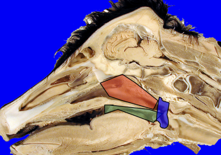
Describe the general function and structure of the pharynx, including a diagram
Common cavity for food and ingesta
Oral cavity ventrally (with oesophagus dorsally)
Nasal cavity dorsally (with larynx and trachea ventrally)
There are three parts of the pharynx:
Nasopharynx (red)
Oropharynx (green)
Laryngopharynx (blue)
Outline the margins of the nasopharynx
Ventral margin: soft palate (soft tissue continuous with palatine bone)
Rostral margin: choanae (opening of nasal cavity into nasopharynx)
Caudal margin: laryngopharynx
Lateral and dorsal margins: pharyngeal wall
Which type of epithelium lines the nasopharynx?
The nasopharynx is lined with respiratory epithelium
What is the nasopharyngeal recess?
Caudodorsal extension of the nasopharynx
Found in most ungulates
Blind ending
Outline the borders of the oropharynx
Ventral margin: tongue
Rostral margin: oral cavity
Caudal margin: laryngopharynx
Dorsal margin: soft palate
Which type of epithelium lines the oropharynx?
The oropharynx is lined with stratified squamous epithelium
Outline the borders of the laryngopharynx
Rostral margin: free tip of the soft palate
Caudal margin: larynx and opening of oesophagus
Lateral and dorsal margins: pharyngeal wall
Which type of epithelium lines the laryngopharynx?
The laryngopharynx is lined with stratified squamous epithelium
What is critical about the laryngopharynx?
This is where the ‘food’ and ‘wind’ pipes cross over
Describe the structure of the soft palate
Flexible muscular structure
Palatinus muscle: within the main body
Tensor veli palatini: within the rostral side wall
Levator veli palatini: within the caudal side wall
Innervated by the mandibular branch of the trigeminal nerve
Describe the function of the soft palate, and the muscles within it
Divides the pharynx
Palatinus muscle: shortens the palate
Tensor veli palatini: causes lateral traction and thus tension
Levator veli palatini: raises the palate during swallowing and mouth breathing

Label this diagram

Which muscles make up the pharyngeal wall?
Made up of striated muscle
This allows for constriction and shortening
Rostral: palatopharyngeal
Middle: hypopharyngeal
Caudal: thyropharyngeal
All insert onto the roof of the pharynx (dorsal and lateral arches)
Dilation is regulated by one single muscle: stylopharyngeus caudalis
What innervates the pharyngeal wall?
Innervation is by the vagus and hypoglossal nerves

Describe the process of swallowing, using this diagram as an aide
Soft palate is elevated to obstruct the nasopharynx and open the oropharynx
Palatopharyngeal arch constricts
Hyoid apparatus pulls the larynx forward
The epiglottis flips back, to cover the tracheal opening
The tongue pushes food into the oesophagus
Describe the structure of the equine guttural pouch
The auditory tube runs from the nasopharynx to the middle ear, in all species
The guttural pouch is a large diverticulum of the auditory tube in horses
The nasopharynx can be entered through slits into the nasopharynx
Stylohyoid bone: divides into lateral and medial compartments
Cranial nerves: glossopharyngeal, vagus and hypoglossal
Blood vessels: internal carotid artery and external carotid artery
Longus capitis muscle
Pharyngeal lymph nodes
Outline the margins of the equine guttural pouch
Dorsal: base of skull
Ventral: laryngopharynx and oesophagus
Lateral: skin
Medial: septum
What is the function of the equine guttural pouch?
Function: unknown, but thought to cool the blood moving to the brain
What kind of disease may occur in the equine guttural pouch?
Bacterial: strangles, strep.equi, sbps.equi
Fungal: mycosis (signs depend on location of plaques- over the artery may cause epistaxis, and over the nerve may cause neurological signs)
How can you surgically access the equine guttural pouch? Draw a labelled diagram
Via Vilborg’s triangle, made up of:
Sternomandibularis m. (beige)
Ramus of mandible
Linguofacial vein (purple)

What is BOAS?
BOAS: brachycephalic obstructive airway syndrome
This is a group of congenital anatomical defects present in brachycephalic breeds, that cause upper airway obstruction- leading to breathing difficulties
Give examples of brachycephalic dog breeds
Pekingese
Pug
Shih Tzu
French/British Bulldog
Boston Terrier
Give examples of brachycephalic cat breeds
Persian
Scottish Fold
Himalayan
British Shorthair/Longhair
Give examples of brachycephalic rabbit breeds
Netherland Dwarf
Lionhead
Miniature Lop
French/German Lop
How can you diagnose BOAS?
Examining airways under a GA using an endoscope, usually if surgery is planned
CT scanning can also be carried out to further assess
Draw a table to show the anatomical abnormalities associated with BOAS, and their mechanism and relevant clinical sign
Anatomical Abnormality | Mechanism | Clinical Signs |
Stenotic nares | Narrowed, pinched or collapsed nostrils | Labored, open-mouth breathing |
Elongated, hyperplastic soft palate | The roof of the mouth extends too far back, dangling directly over the larynx and obstructing the rima glottidis | Stertor: heavy snoring/snorting |
Aberrant nasal turbinates | The delicate bones inside the nose are crowded together, and extend too far back into the throat | Severe heat intolerance and hyperthermia |
Hypoplastic trachea | An abnormally narrow, underdeveloped trachea | Exercise intolerance and syncope |
Macroglossia | A disproportionately large, thick tongue relative to the shortened mouth | Exacerbates sleep apnea |
Describe the surgical interventions that can help animals with BOAS
Stenotic nares correction: removing a small wedge of tissue from nostrils to widen the openings and improve airflow
Soft palate resection: palate is surgically shortened so it no longer blocks the entrance to the larynx
Laryngeal saccule removal: excess tissue may be trimmed if it contributes further to airway obstruction
Tonsillectomy: tonsils are removed if they are enlarged and blocking the throat
Describe the conservative management that can help animals with BOAS
Weight control: reduces respiratory effort and internal airway pressure
Avoiding strenuous exercise, high humidity and high temperatures
Attach leashes to a body harness instead of a neck collar, to eliminate pressure on the trachea
Anti-inflammatories, corticosteroids or gastroprotectants help to manage airway swelling and secondary acid reflux
What are the possible radiographic changes associated with feline asthma?
Bronchial thickening: walls of the bronchial tubes appear more pronounced than usual
Overinflation: the lungs expand beyond their normal resting size
Air trapping: abnormal retention of air in the lungs during exhalation
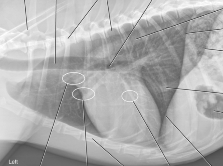
Label this diagram

What is feline asthma?
Feline asthma is a chronic respiratory condition, where airborne allergens cause inflammation, swelling and mucus buildup
Give examples of breeds prone to feline asthma
Siamese
Himalayan
Balinese
Oriental
Korat
Seychellois
Describe how feline asthma can be managed pharmacologically
Glucocorticoids (fluticasone, prednisolone)
Anti-inflammatory action
They bind to intracellular glucocorticoid receptors to alter gene transmission
This decreases the production of pro-inflammatory cytokines, reduces airway eosinophil infiltration and minimises mucosal oedema
They also help to prevent or reverse the down-regulation of beta-2 receptors
Bronchodilators (albuterol, salbutamol, terbutaline, theophylline)
Airway smooth muscle relaxation
Beta-2 agonists stimulate beta-2 adrenergic receptors
This increases intracellular cAMP to relax smooth muscle
Methylxanthines inhibit phosphodiesterase (PDE) enzymes
This prevents the breakdown of cAMP to relax smooth muscle
Describe how feline asthma can be managed non-pharmacologically
Reducing environmental triggers
Remove smoking, vaping, incense, scented candles, household cleaners etc
Transition to dust-free litter alternatives
Install air purifiers where the cat spends the majority of their time
Weight management
Keep an optimal body condition score to lower oxygen demand and reduce the effort required to breathe
Diet
Omega-3 fatty acid supplementation has strong clinical backing for natural anti-inflammatory effects
Feeding a moisture-rich diet supports hydration, and keeps the mucosal lining of the respiratory tract moist, helping the airways to better clear mucus
Probiotics can be used to stabilise immune responses
Why might flare-ups of feline asthma occur?
Missing doses or discontinuing treatment
Obesity
Environmental triggers
Dusty or clay-based cat litters
Tobacco smoke, vaping byproducts, fireplace smoke
Aerosol sprays, household cleaning chemicals, perfumes and candles
Pollen, mold spores, dust mites
Give the clinical signs of lower airway disease
Coughing
Expiratory dyspnea
Tachypnea and respiratory distress
Exercise intolerance
Abnormal lung sounds (wheezes and crackles)
How can you differentiate between lower and upper airway disease?
Breathing pattern
Upper airway: inspiratory dyspnea (difficulty breathing in)
Lower airway: expiratory dyspnea (difficulty breathing out)
Respiratory noises
Upper airway: loud, audible noises without a stethoscope
Lower airway: quiet breathing at a distance, audible noises require auscultation (wheezes and crackles)
Coughing
Upper airway: harsh, loud, honking and dry coughing, often easily induced by gentle tracheal palpation
Lower airway: soft, deep, productive or wheezy coughing
How can you evaluate lower and upper airway disease
Radiography
Upper airway: cervical and thoracic radiographs needed to evaluate the nasal passages, pharynx, larynx and extrathoracic trachea
Lower airway: full thoracic radiographs can show bronchointerstitial patterns, lung lobe consolidation or hyperinflation (air trapping)
Endoscopy
Upper airway: rhinoscopy and laryngoscopy assesses structural collapse, laryngeal paralysis or elongated soft palates
Lower airway: bronchoscopy evaluates the lower trachea and principal bronchi for collapse, inflammation and mucus plugs
Airway sampling
Upper airway: nasal flushes or brushings
Lower airway: tracheal wash or bronchoalveolar lavage
Describe the 3-part loop of the cough reflex
Regardless of the disease location, all coughs utilise a three-part reflex loop
Afferent pathway: sensory receptors in the respiratory epithelium transmit electrical signals via sensory branches of the vagus nerve up to the brainstem
Central pathway: these signals synapse in the nucleus tractus solitarius (nTS), located in the medulla oblongata and pons
Efferent pathway: once the threshold is met, the medulla coordinates motor output via the vagus, phrenic and spinal motor nerves to the effector muscles (diaphragm, intercostals, abdominal wall and larynx)
How is a cough initiated during upper airway disease?
Mechanical stimulus deforms the membranes of rapidly adapting receptors (RARs)
Myelinated A𝛿 fibres fire rapidly in response to physical changes
When the upper trachea/larynx is directly stimulated, it triggers a sudden forced expiration against a closed glottis to move the obstruction
How is a cough initiated during lower airway disease?
Inflammation/chemical stimulus causes the local release of inflammatory mediators along with tachykinins
These mediators bind to unmyelinated C-fibre receptors, which act locally to release more tachykinins, which further activate neighbouring mechanical RARs
This mandates an inspiratory phase to expand the lungs fully before an expulsive phase
How should you treat a patient in respiratory distress? Include initial triage, assessment and advanced intervention
Initial triage
Provide oxygen as soon as possible
Administer sedation if stress is high
Assessment
Observe breathing pattern and effort from a distance
Check mucous membrane colour and listen for upper airway noise
Utilise fast, non-stressful tests such as ultrasound rather than radiography
Advanced intervention
Intubate, or perform a tracheostomy immediately if the patient is fatiguing, cyanotic or suffering from a complete upper airway obstruction
Perform thoracentesis if pleural space disease is suspected and contributing to restrictive breathing
Actively cool hyperthermic patients
Describe the effect of restrictive bronchial disease
Increased airway resistance
Reduced lung compliance → reduced functional residual capacity
Hypoxemia (low blood oxygen)
Hypercapnia (high carbon dioxide)
Increased risk of bronchospasm
Anatomical dead space decreases due to narrower airways
Alveolar dead space increases due to poorly ventilated alveoli
How can you manage the effects of restrictive bronchial disease in a patient under anaesthesia
Administer 100% oxygen via mask or flow-by for several minutes before induction to build an oxygen reserve
Be ready to provide mechanical or positive-pressure ventilation, since anaesthetic agents depress the respiratory centre
Premedicate with targeted bronchodilators if reactive airway spasms are anticipated
Closely monitor oxygenation, breathing patterns and mucous membrane colour
Describe the effects of anaesthetic agents on respiratory function
Alpha-2 agonists
Can significantly alter pulmonary mechanics and potentially contribute to hypoxemia or pulmonary issues
Opioids
Cause centrally-mediated respiratory depression
Propofol and thiopental
Rapid IV administration frequently causes transient apnea or profound hypoventilation
Isoflurane, sevoflurane, desflurane
Reduce the sensitivity of the respiratory centre to carbon dioxide
Decreases respiratory rate and/or tidal volume
Ketamine
Dissociative anaesthetics cause minimal direct respiratory depression, though combinations with sedatives like alpha-2 agonists can still compromise overall ventilation
What are the common causes of coughing in cats?
Feline asthma
Chronic bronchitis
Respiratory infections (feline herpesvirus, calicivirus)
Parasites (heartworms or lungworms)
Foreign objects
Chest tumors, lung cancer or severe trauma
What are the common causes of coughing in dogs?
Kennel cough
Pneumonia
Canine influenza
Chronic bronchitis
Parasites (heartworms or lungworms)
Foreign objects
What are the common causes of coughing in horses?
Equine asthma
Equine influenza
Strangles
Pneumonia
Environmental irritants
What are the common causes of coughing in cows?
Infectious pneumonia
Lungworm
Environmental irritants
Outline the structure of the larynx
Stratified squamous epithelium at the epiglottis of the larynx, and on the folds
Other areas are pseudostratified ciliated epithelium
Cartilaginous tube
Made up of elastic and hyaline cartilage
Hyaline: thyroid (U-shaped), cricoid (signet ring-shaped), paired arytenoids (triangular-shaped)
Elastic: epiglottis (leaf-shaped)
Suspended by the hyoid bones, which articulate with the temporal bone
Linked by synovial or cartilaginous joints
Cavities
Entrance to the larynx → vestibule (medianus recess in the horse and pig)
Glottis
Infraglottal cavity

Label this diagram

Describe the cartilage connections within the larynx
There are connections between the cartilage themselves, and also with the trachea and hyoid bones
This is by:
Joints (synovial or cartilaginous)
Collagenous and elastic ligaments
Striated musculature
Ligaments of arytenoid cartilage
Rostral: vestibular ligament
Caudal: vocal ligament
These are moved when the arytenoids move
Describe the function of the different laryngeal muscles
Glottis: involved in inspiration, swallowing and phonation
Intrinsic muscles
Between laryngeal cartilages
Involved in respiration and phonation
Extrinsic muscles
Connect the larynx to hyoid bones, pharynx and sternum
Involved in swallowing
Which intrinsic laryngeal muscles are involved in
Widening the glottis
Narrowing the glottis
Widening the glottis
Cricoarytenoideus dorsalis
Supplied by the recurrent laryngeal nerves
Relevant for left laryngeal hemiplegia- left nerve becomes diseased → inspiratory noise, poor performance, fails the slap test
Narrowing the glottis
Cricothyroideus
Cricoarytenoideus lateralis (can close the glottis)