Chp 9 - Joints
 Call Kai
 Learn
Learn Practice Test
 Spaced Repetition
 Match
Match Flashcards
 Knowt Play
1/109

There's no tags or description
Looks like no tags are added yet.
Name | Mastery | Learn | Test | Matching | Spaced | Call with Kai | Chat |
|---|
No analytics yet
Send a link to your students to track their progress
110 Terms
abduction
movement in the coronal plane that moves a limb laterally away from the body; spreading of the fingers

acetabular labrum
lip of fibrocartilage that surrounds outer margin of the acetabulum on the hip bone

adduction
movement in the coronal plane that moves a limb medially toward or across the midline of the body; bringing fingers together

amphiarthrosis
slightly mobile joint


annular ligament
intrinsic ligament of the elbow articular capsule that surrounds and supports the head of the radius at the proximal radioulnar joint
anterior cruciate ligament
intracapsular ligament of the knee; extends from anterior, superior surface of the tibia to the inner aspect of the lateral condyle of the femur; resists hyperextension of knee

anterior talofibular ligament
intrinsic ligament located on the lateral side of the ankle joint, between talus bone and lateral malleolus of fibula; supports talus at the talocrural joint and resists excess inversion of the foot


articular capsule
connective tissue structure that encloses the joint cavity of a synovial joint

articular cartilage
thin layer of hyaline cartilage that covers the articulating surfaces of bones at a synovial joint

articular disc
meniscus; a fibrocartilage structure found between the bones of some synovial joints; provides padding or smooths movements between the bones; strongly unites the bones together

articulation
joint (any place where adjacent bones or bones cartilage come together (articulate with each other) to form a connection.
atlanto-occipital joint
articulation between the occipital condyles of the skull and the superior articular processes of the atlas (C1 vertebra)

atlantoaxial joint
series of three articulations between the atlas (C1) vertebra and the axis (C2) vertebra, consisting of the joints between the inferior articular processes of C1 and the superior articular processes of C2, and the articulation between the dens of C2 and the anterior arch of C1

ball-and-socket joint
synovial joint formed between the spherical end of one bone (the ball) that fits into the depression of a second bone (the socket); found at the hip and shoulder joints; functionally classified as a multiaxial joint
Ex: Shoulder and hip joints

biaxial joint
type of diarthrosis; a joint that allows for movements within two planes (two axes)
Ex: metacarpophalangeal joint (knuckle joint)
bursa
connective tissue sac containing lubricating fluid that prevents friction between adjacent structures, such as skin and bone, tendons and bone, or between muscles

calcaneofibular ligament
intrinsic ligament located on the lateral side of the ankle joint, between the calcaneus bone and lateral malleolus of the fibula; supports the talus bone at the ankle joint and resists excess inversion of the foot

cartilaginous joint
joint at which the bones are united by hyaline cartilage (synchondrosis) or fibrocartilage (symphysis)

circumduction
circular motion of the arm, thigh, hand, thumb, or finger that is produced by the sequential combination of flexion, abduction, extension, and adduction

condyloid joint
synovial joint in which the shallow depression at the end of one bone receives a rounded end from a second bone or a rounded structure formed by two bones; found at the metacarpophalangeal joints of the fingers or the radiocarpal joint of the wrist; functionally classified as a biaxial joint
Ex: Metacarpophalangeal (knuckle) joints of fingers; radiocarpal joint of wrist; metatarsophalangeal joints for toes

coracohumeral ligament
intrinsic ligament of the shoulder joint; runs from the coracoid process of the scapula to the anterior humerus

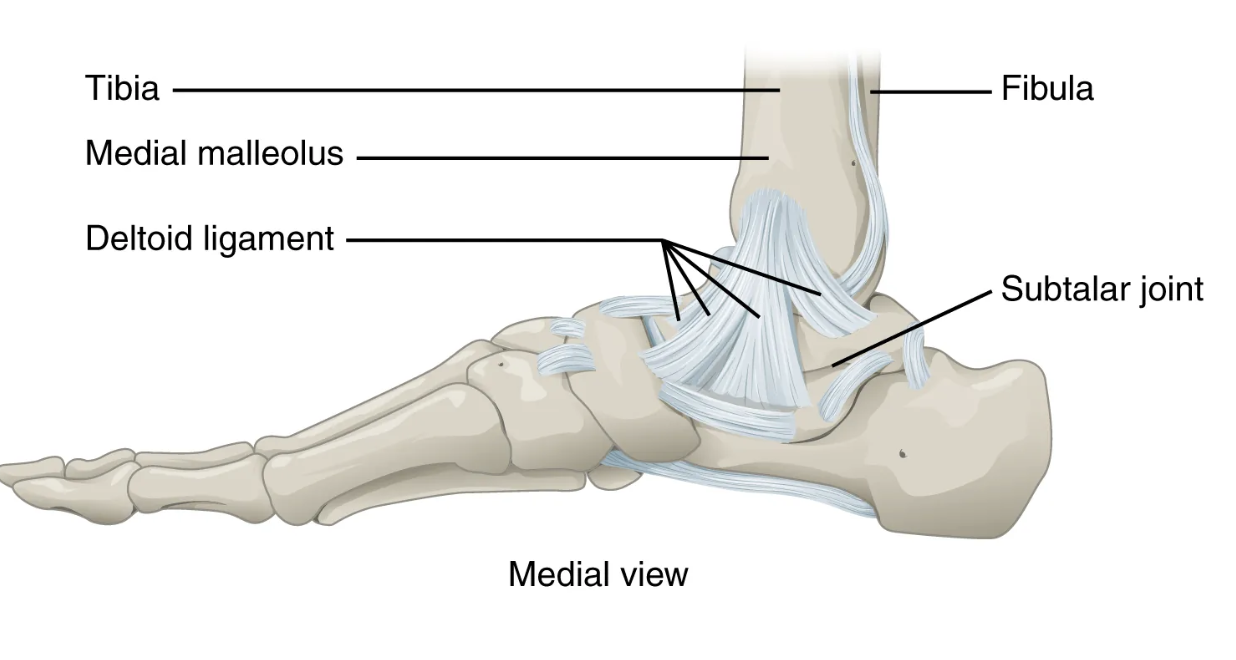
deltoid ligament
broad intrinsic ligament located on the medial side of the ankle joint; supports the talus at the talocrural joint and resists excess eversion of the foot

depression
downward (inferior) motion of the scapula or mandible

diarthrosis
freely mobile joint (all synovial joints)
dorsiflexion
movement at the ankle that brings the top of the foot toward the anterior leg

elbow joint
uniaxial joint formed by the humeroulnar joint
Humeroulnar joint, proximal radioulnar joint, humeroradial joint - all three of these joints are enclose within a single articular capsule

elevation
upward (superior) motion of the scapula or mandible

eversion
foot movement involving the intertarsal joints of the foot in which the bottom of the foot is turned laterally, away from the midline

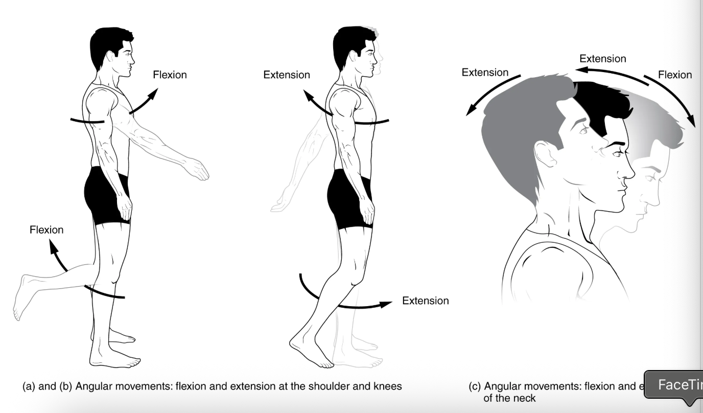
extension
movement in the sagittal plane that increases the angle of a joint (straightens the joint); motion involving posterior bending of the vertebral column or returning to the upright position from a flexed position
femoropatellar joint
portion of the knee joint consisting of the articulation between the distal femur and the patella

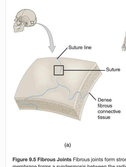
fibrous joint
joint where the articulating areas of the adjacent bones are connected by fibrous connective tissue
Examples: suture
fibular collateral ligament
extrinsic ligament of the knee joint that spans from the lateral epicondyle of the femur to the head of the fibula; resists hyperextension and rotation of the extended knee

flexion
movement in the sagittal plane that decreases the angle of a joint (bends the joint); motion involving anterior bending of the vertebral column

glenohumeral joint
shoulder joint; articulation between the glenoid cavity of the scapula and head of the humerus; multiaxial ball-and-socket joint that allows for flexion/extension, abduction/adduction, circumduction, and medial/lateral rotation of the humerus
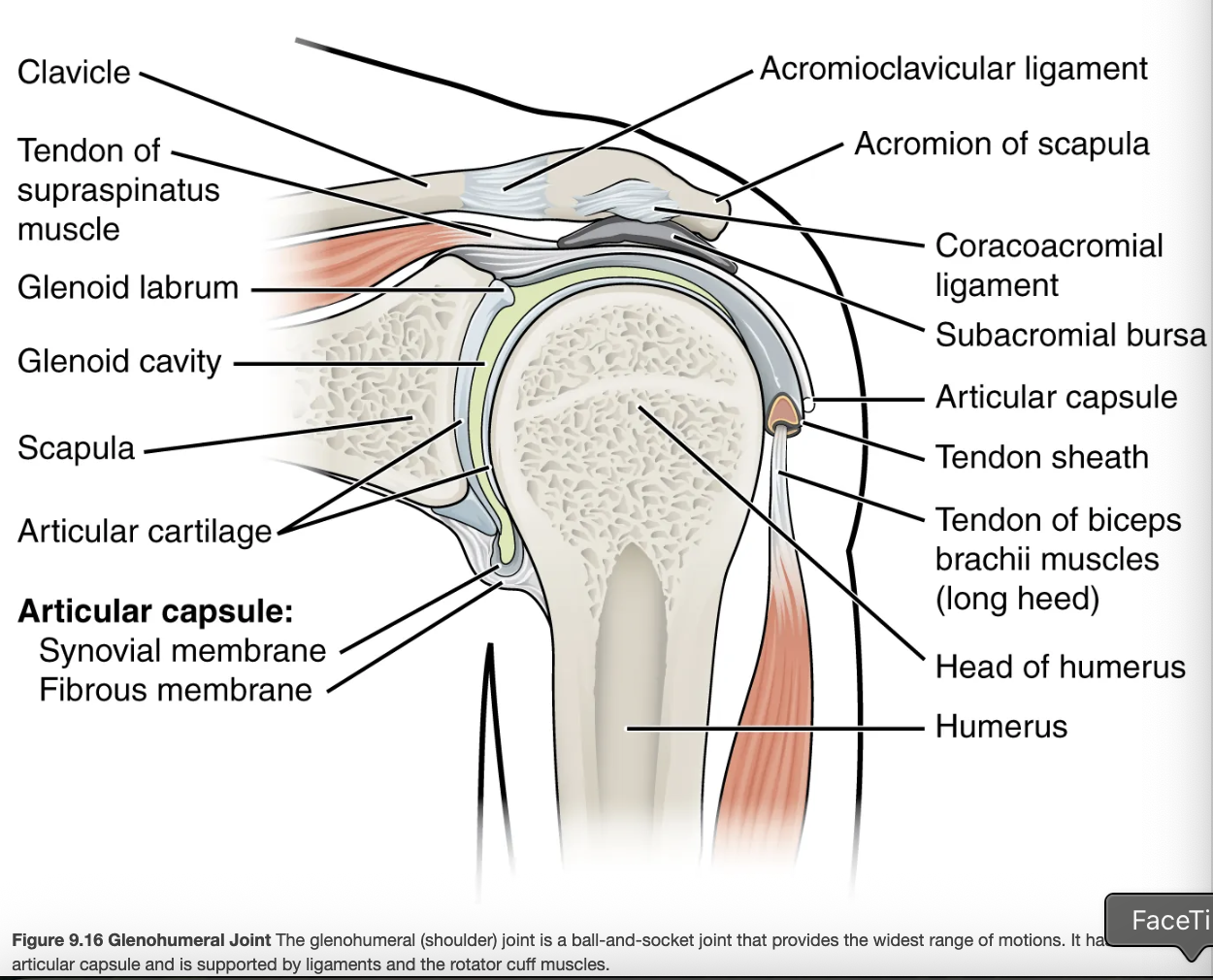
glenohumeral ligament
one of the three intrinsic ligaments of the shoulder joint that strengthen the anterior articular capsule
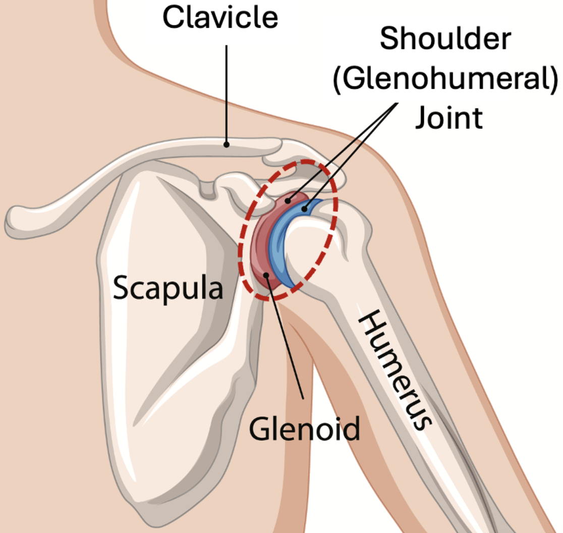
glenoid labrum
lip of fibrocartilage located around the outside margin of the glenoid cavity of the scapula

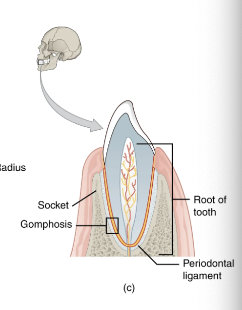
gomphosis
type of fibrous joint in which the root of a tooth is anchored into its bony jaw socket by strong periodontal ligaments
hinge joint
synovial joint at which the convex surface of one bone articulates with the concave surface of a second bone; includes the elbow, knee, ankle, and interphalangeal joints; functionally classified as a uniaxial joint
Ex: Elbow; ankle; interphalangeal joints of fingers and toes

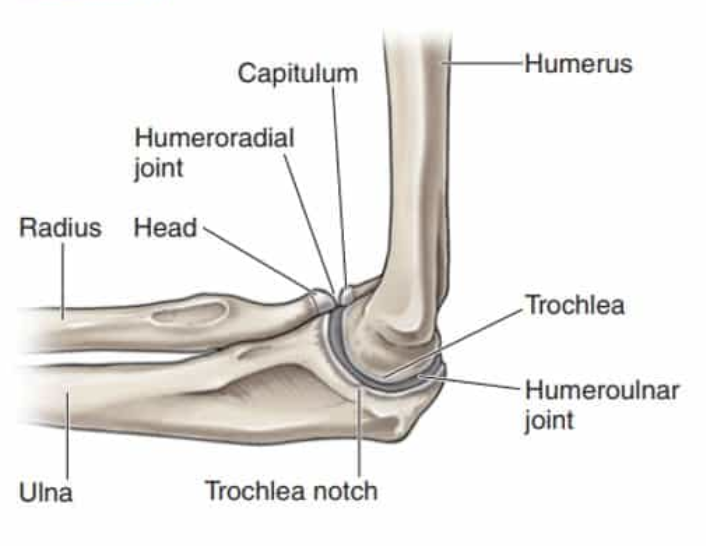
humeroradial joint
articulation between the capitulum of the humerus and head of the radius
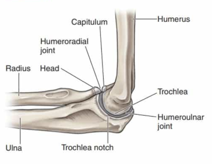
humeroulnar joint
articulation between the trochlea of humerus and the trochlear notch of the ulna; uniaxial hinge joint that allows for flexion/extension of the forearm
iliofemoral ligament
intrinsic ligament spanning from the ilium of the hip bone to the femur, on the superior-anterior aspect of the hip joint

inferior rotation
(scapular downward rotation) movement of the scapula during upper limb adduction in which the glenoid cavity of the scapula moves in a downward direction as the medial end of the scapular spine moves in an upward direction

interosseous membrane
wide sheet of fibrous connective tissue that fills the gap between two parallel bones, forming a syndesmosis; found between the radius and ulna of the forearm and between the tibia and fibula of the leg

inversion
foot movement involving the intertarsal joints of the foot in which the bottom of the foot is turned toward the midline

ischiofemoral ligament
intrinsic ligament spanning from the ischium of the hip bone to the femur, on the posterior aspect of the hip joint

joint cavity
space enclosed by the articular capsule of a synovial joint that is filled with synovial fluid and contains the articulating surfaces of the adjacent bones

joint interzone
site within a growing embryonic limb bud that will become a synovial joint
Cells in the center of the interzone undergo programmed cell death (cavitation) to create the open joint cavity.
Cells at the margins/periphery differentiate to form the articular cartilage, articular capsule, and supporting ligaments
lateral (external) rotation
movement of the arm at the shoulder joint or the thigh at the hip joint that moves the anterior surface of the limb away from the midline of the body

lateral excursion
side-to-side movement of the mandible away from the midline, toward either the right or left side

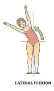
lateral flexion
bending of the neck or body toward the right or left side
lateral meniscus
C-shaped fibrocartilage articular disc located at the knee, between the lateral condyle of the femur and the lateral condyle of the tibia

lateral tibiofemoral joint
portion of the knee consisting of the articulation between the lateral condyle of the tibia and the lateral condyle of the femur; allows for flexion/extension at the knee

ligament of the head of the femur
intracapsular ligament that runs from the acetabulum of the hip bone to the head of the femur

medial (internal) rotation
movement of the arm at the shoulder joint or the thigh at the hip joint that brings the anterior surface of the limb toward the midline of the body

medial excursion
side-to-side movement that returns the mandible to the midline

medial meniscus
C-shaped fibrocartilage articular disc located at the knee, between the medial condyle of the femur and medial condyle of the tibia

multiaxial joint
type of diarthrosis; a joint that allows for movements within three planes (three axes)

opposition
thumb movement that brings the tip of the thumb in contact with the tip of a finger

patellar ligament
ligament spanning from the patella to the anterior tibia; serves as the final attachment for the quadriceps femoris muscle

periodontal ligament
band of dense connective tissue that anchors the root of a tooth into the bony jaw socket

pivot joint
synovial joint at which the rounded portion of a bone rotates within a ring formed by a ligament and an articulating bone; functionally classified as uniaxial joint
Ex: Atlantoaxial joint (C1–C2 vertebrae articulation); proximal radioulnar joint

plane joint
synovial joint formed between the flattened articulating surfaces of adjacent bones; functionally classified as a multiaxial joint
Ex: Intertarsal joints of foot; superior-inferior articular process articulations between vertebrae

plantar flexion
foot movement at the ankle in which the heel is lifted off of the ground

posterior cruciate ligament
intracapsular ligament of the knee; extends from the posterior, superior surface of the tibia to the inner aspect of the medial condyle of the femur; prevents anterior displacement of the femur when the knee is flexed and weight bearing

posterior talofibular ligament
intrinsic ligament located on the lateral side of the ankle joint, between the talus bone and lateral malleolus of the fibula; supports the talus at the talocrural joint and resists excess inversion of the foot

pronated position
forearm position in which the palm faces backward

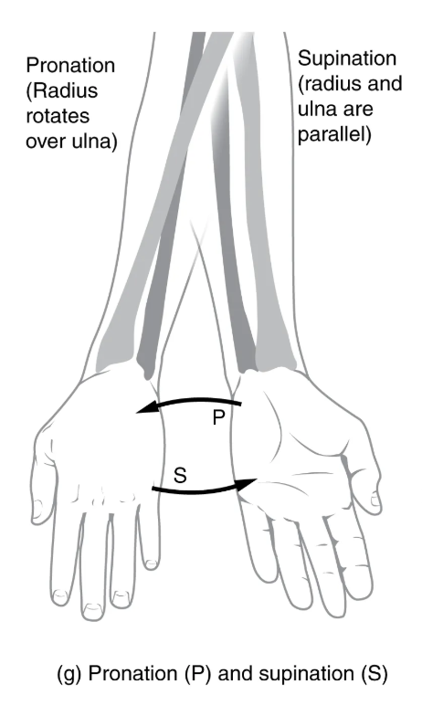
pronation
forearm motion that moves the palm of the hand from the palm forward to the palm backward position

protraction
anterior motion of the scapula or mandible

pubofemoral ligament
intrinsic ligament spanning from the pubis of the hip bone to the femur, on the anterior-inferior aspect of the hip joint

radial collateral ligament
intrinsic ligament on the lateral side of the elbow joint; runs from the lateral epicondyle of humerus to merge with the annular ligament

reposition
movement of the thumb from opposition back to the anatomical position (next to index finger)

retraction
posterior motion of the scapula or mandible

rotation
movement of a bone around a central axis (atlantoaxial joint) or around its long axis (proximal radioulnar joint; shoulder or hip joint); twisting of the vertebral column resulting from the summation of small motions between adjacent vertebrae

rotator cuff
strong connective tissue structure formed by the fusion of four rotator cuff muscle tendons to the articular capsule of the shoulder joint; surrounds and supports superior, anterior, lateral, and posterior sides of the humeral head

saddle joint
synovial joint in which the articulating ends of both bones are convex and concave in shape, such as at the first carpometacarpal joint at the base of the thumb; functionally classified as a biaxial joint
Ex: First carpometacarpal joint of the thumb; sternoclavicular joint

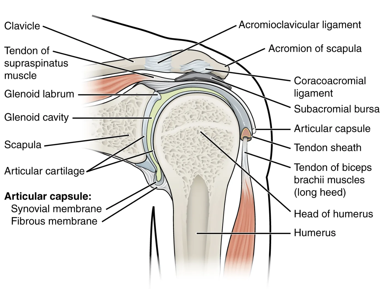
subacromial bursa
bursa that protects the supraspinatus muscle tendon and superior end of the humerus from rubbing against the acromion of the scapula
subcutaneous bursa
bursa that prevents friction between skin and an underlying bone

subtalar joint
articulation between the talus and calcaneus bones of the foot; allows motions that contribute to inversion/eversion of the foot
supination
forearm motion that moves the palm of the hand from the palm backward to the palm forward position
syndesmosis
type of fibrous joint in which two separated, parallel bones are connected by an interosseous membrane
