CHAPTER 5: CLEFT LIP AND PALATE
 Call Kai
 Learn
 Practice Test
 Spaced Repetition
 Match
 Flashcards
 Knowt Play
1/47

Earn XP
Description and Tags
books & sas
48 Terms
cleft lip
occurs when there is a split or opening in the lip
This opening can be small or large enough to connect the upper lip and nose.
cleft palate
occurs when the roof of the mouth does not close properly during a baby’s early development inside the womb
3 conditions in Pierre Robin Syndrome
Micrognathia
Glossoptosis
cleft palate
Pierre Robin sequence
named after Dr. Pierre Robin, who described this in the 1920s
occurs in a number of syndromes
refers to the triad which means that Children with cleft lip and palate may suffer from a number of other complications and conditions
It has become common for children with Pierre Robin sequence to have mandibular distraction osteogenesis at a very young age to prevent upper airway obstruction
major concern: Airway obstruction due to the malposition of the tongue
Dental Anomalies of cleft px
Rotation of the teeth near the cleft.
Hypoplasia of enamel, particularly in teeth adjacent to the cleft.
Missing teeth.
Supernumerary teeth.
Hypodontia
Occurs more commonly in patients with cleft lip and palate.
Occurs more frequently in the permanent dentition than in the primary dentition.
The tooth most likely to be missing is:
The lateral incisor.
The prevalence is higher:
On the side with the cleft.¹⁵
The prevalence of missing lateral incisors increases as the extent of clefting increases:
Cleft lip alone.
Cleft palate alone.
Cleft lip and palate.¹⁵
cleft
can occur as an isolated birth defect or as part of a larger genetic syndrome, such as van der Woude syndrome or velocardiofacial syndrome, which are both genetic malformation disorder
Common Syndromes and Abnormalities Associated With Cleft Lip and Palate
Van der Woude syndrome
Velocardiofacial syndrome
CHARGE syndrome
Beckwith-Wiedemann syndrome
Apert syndrome
DiGeorge syndrome
Pierre Robin sequence
Treacher-Collins syndrome
Kabuki syndrome
Klippel Feil syndrome
Goldenhar syndrome
Stickler syndrome
palate
begins to form during the 5th embryonic week continuing through the 12th embryonic week
2 divisions of palate at the incisive foramen
primary
secondary
Primary Palate
formed when the medial nasal prominences fuse, only at a deeper level
Extends anteriorly from the incisive foramen.
Includes:
Premaxilla
Lip
Nasal tip
Columella

Secondary Palate
posteriorly from the incisive foramen and includes the hard and soft palate
begins to form soon after the primary palate is fused, around the 7th embryonic week
Results from fusion of the lateral palatine shelves.
Fusion begins at the incisive foramen in the 8th week.
Fusion ends at the uvula in the 12th embryonic week.
Extends posteriorly from the incisive foramen.
Includes:
Hard palate
Soft palate
Uvula

causes of cleft lip and palate
Isolated mutations
Syndromes
Genetics (Syndromic, Nonsyndromic)
Environmental factors
Teratogen exposure (including alcohol and tobacco)
Classification of Cleft Palate
class I
class II
class III
class IV
class I Cleft Palate
Incomplete cleft of secondary palate

class II Cleft Palate
Complete cleft of the secondary palate

class III Cleft Palate
Unilateral complete cleft of primary and secondary palate

class IV Cleft Palate
Bilateral complete cleft of the primary and secondary palate

management of patients with cleft left and palate
involves a team working together to address the problem.
For example, if the child has problems with speech because of their cleft, they might work with a speech pathologist.
The team may also include a plastic surgeon, an oral surgeon, and/or an orthodontist.
Surgery to repair a cleft palate or lip is ideally performed in the child’s first year.
The role of a pediatric dentist
to provide a dental home for children with cleft lip and palate or other craniofacial abnormalities
play a role inpresurgical molding techniques(e.g NAM)
prevention of dental disease in high risk populations as well as restorations and interceptive orthodontics.
rule of 10s
This rule states that a child must:
10lb pounds.
>10 g/dL hemoglobin
10 weeks of age
<10,000 WBC (if more than, prolly naay infection)
This rule is not an absolute.
The risk of placing a very young baby under general anesthesia must be considered.
This is one reason that naso-labial (lip) adhesion has fallen out of favor.
Challenges and Anomalies Associated with Cleft Lip and Palate
Feeding problems
Hypodontia or missing teeth 12 and 22 → most common
Supernumerary teeth → 2nd most common
Otitis media and other middle ear problems
intraoral appliances for cleft lip and palate px
elastomeric tape
latham appliance
nasoalveolar molding appliance (NAM)
elastomeric tape
to apply force can cause movement and decrease the width of the alveolar cleft. It is inexpensive and less hassle
In its simplest form, the use of elastomeric tape to apply force can:
Cause movement.
Decrease the width of the alveolar cleft
Advantages:
Relatively inexpensive.
Less onerous for families compared to other molding devices.
Disadvantages:
Applies forces indiscriminately.
Palatal constriction may result.
Taping can be:
Done alone.
Used in conjunction with other molding techniques such as intraoral appliances.

Latham Appliance
Is surgically placed into a young child's mouth.
Is retained to the palate with a screw.²⁴
The appliance is adjusted regularly to:
Reduce the cleft size.
Advantages:
Compliance is not required.
Disadvantages:
Increased surgical interventions in a very young child.
Surgical intervention is required:
At the time of insertion.
At the time of removal.

nasoalveolar molding (NAM)
uses a nasal stent to shape the cartilage of the nose.
uses an acrylic plate to mold the bony alveolus, & is removable
is worn throughout the day & held in place by tape to the child's cheeks.
one less invasive method, pioneered by Dr. Barry Grayson and his team at New York University
functions in a similar fashion to:
the latham appliance.
early appliances like the Hotz plate.
goals:
improve symmetry, reduce scarring
prevent hindrance of growth
decrease the need for future invasive surgical correction
over time:
bony segments are slowly moved to a more favorable position.
amount of surgical correction needed to bring the lip segments together is lessened.
surgical approach for the nose is simplified.

Intraoral appliances
Are often more cumbersome to use.
Maintain the palatal width.
They maintain palatal width because:
Force can be applied in smaller areas.
Force can be applied with greater precision.
Recently:
These intraoral appliances have gained acceptance on many craniofacial teams worldwide.
They are a hot topic of research.
early detection of cleft lip in pregnancy
detected at your regular 20 week ultrasound scan
can be detected by ultrasound toward the end of the first trimester
infant orthopedic treatment
the earliest intervention
the goal is to:
enhance the surgical repair of the lip&nose
establish a proper skeletal base
one of the most popular approache is:
nasoalveolar molding (NAM) → initiated within the first few weeks of life
Cleft Palate Repair
A cleft palate is repaired between:
9 and 18 months of age.
Most often by age 1.
Early repair:
Advantage:
Better speech development.
Disadvantages:
Scar tissue can form.
Proper maxillary growth can be inhibited.
May lead to:
Malocclusion (typically Class III malocclusions and skeletal pattern).
Poor facial esthetics.
Late repair (after 18 months):
Growth may occur normally.
Speech will likely be negatively impacted.³³
Determining the best time for palate repair requires:
Considering these competing priorities.
Working as a team to develop an optimal surgical plan.
Velopharyngeal Surgery
If speech development is suboptimal:
Velopharyngeal surgery may be performed.
Timing:
Between 2.5 and 3 years of age.³⁴
Repair of the cleft lip and palate and velopharyngeal surgeries are performed by:
Plastic surgeon.
Pediatric otolaryngologist.
Oral and maxillofacial surgeon with advanced training in pediatric cranio-maxillofacial surgery.
Alveolar Bone Grafting
Patients with clefts of the alveolus often:
Have collapsed maxillary arches.
Require alveolar bone grafting, as shown in Fig. 5.10.
Alveolar bone grafting enables the surgeon to:
Obtain maxillary arch continuity.
Provide support for the dentition to erupt.
Stabilize the maxillary arch prior to orthodontic treatment
Timing depends on:
Presence of a viable maxillary permanent lateral incisor adjacent to the alveolar cleft area.
Proximity of the roots of central incisors to the alveolar cleft.³⁵⁻⁴¹
If:
A viable permanent lateral incisor is present, or
The root of the developing maxillary central incisor is too close to the alveolar cleft,
Alveolar bone grafting is recommended:
By 5 to 7 years of age.
Some craniofacial centers perform grafting later:
Closer to eruption of the maxillary permanent canines.

donor sites of Alveolar Bone Grafting
Most frequently used donor site:
Iliac crest.
Reasons:
Ease of access.
Abundance of bone.²⁷ ⁴⁰
Alternative donor sites:
Mandibular symphysis.
Rib, Tibia. Cranial bones.
A few craniofacial centers also use:
Bone morphogenetic protein (BMP
The procedure is usually performed by:
An oral and maxillofacial surgeon
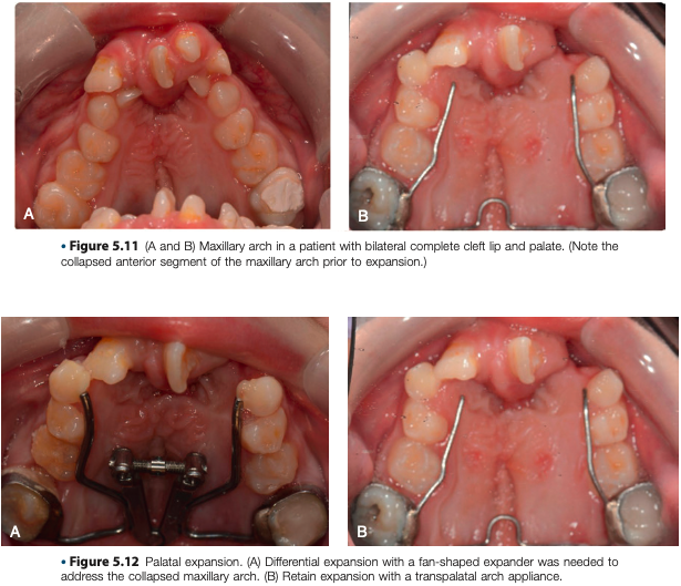
Maxillary Arch Expansion
Recommended prior to alveolar bone grafting.
Purposes:
Establish proper arch form.
Address transverse discrepancies
Maxillary arch expansion facilitates:
Better access for alveolar bone graft sites.
Maxillary expansion may be:
Differential.
Symmetric.
Performed by:
Pediatric dentist.
Orthodontist on the cleft lip and palate team.
Orthodontic Treatment During Mixed Dentition
Following alveolar bone grafting:
The patient is typically followed periodically by the cleft lip and palate team.
The eruption pattern of the permanent dentition is closely observed.
If indicated:
A limited phase of orthodontic treatment is completed during the mixed dentition phase.
Often in the maxillary arch only.
Goals:
Align and level the maxillary arch.
Correct crossbites.
Create space for facilitating eruption of permanent teeth.
Treat impacted or ectopically erupting teeth

maxillary distraction osteogenesis
indication:
If the anterior-posterior skeletal discrepancy is too large during the early teenage years:
The procedure is performed by:
An oral and maxillofacial surgeon.
Timing:
Following eruption of all permanent teeth into the arch.
The orthodontist:
Aligns and levels the maxillary arch.
Prepares the patient for the procedure
Comprehensive Orthodontic Treatment
The patient is followed during the teenage years.
The orthodontist determines the timing of a comprehensive phase of orthodontic treatment.
If there are no significant skeletal discrepancies:
Orthodontic treatment can be initiated following eruption of all permanent teeth.
If large skeletal discrepancies exist:
A comprehensive phase of orthodontic treatment in conjunction with orthognathic surgery is recommended.
Treatment is initiated following completion of growth
Types of Distractors
Previously:
Maxillary distraction was accomplished by a rigid external distractor (RED) appliance, as shown in Fig. 5.14.
Currently:
Internal distractors, as shown in Fig. 5.15, are increasingly used.
They are used in place of the more cumbersome extraoral appliances

Cleft Lip Repair
A cleft lip is repaired between:
3 and 6 months of age.
Repair is performed following infant orthopedic treatment.
Naso-labial (lip) adhesion.
This procedure is performed prior to repairing the cleft lip.
It has largely fallen out of favor in the United States.
The naso-labial (lip) adhesion procedure is purported to:
Reduce the extent of undermining.
Minimize labial tension at the time of lip repair.
normal development
4th wk → facial prominences
5th wk → nasal placodes
7th wk → fusion of the medial nasal prominences
7th wk → fusion of the maxillary prominences
facial prominences
appear at the end of the fourth embryonic week.
These prominences are derived from the neural crest.
They consist of:
Maxillary prominences laterally
A frontonasal prominence
Nasal placodes
Are located on either side of the frontonasal prominence.
Appear during the fifth embryonic week.
Eventually form the nasal pits.
borders of nasal pits
Lateral nasal prominences
Medial nasal prominences
fusion of the medial nasal prominences forms:
Nasal tip
Columella
Philtrum
A portion of the upper lip
fusion of the maxillary prominences forms:
remainder of the upper lip
Timeline of Interventions

