Chapter 10- The Muscular System
 Call Kai
 Learn
Learn Practice Test
 Spaced Repetition
 Match
Match Flashcards
 Knowt Play
1/224

Earn XP
Description and Tags
A&P 1 Lecture
Name | Mastery | Learn | Test | Matching | Spaced | Call with Kai | Chat |
|---|
No analytics yet
Send a link to your students to track their progress
225 Terms
Movement, stability, control of openings and passageways, heat production, glycemic control
functions of the muscles (5)
Movement
fucntion of muscle: place to place and throughout the body
Stability
function of muscle: stabilize the joints
Control of openings and passageways
function of muscle: sphincters- internal circular rings, controlling the movement of contents, in stomach, eyes, and mouth
Heat production
function of muscle: 85% of heat- skeletal muscles generate heat
Glycemic control
function of muscle: controlling blood-glucose levels
Endomysium
Loose CT around each muscle fiber, allows room for capillaries and nerve fibers
Perimysium
Thicker CT layer that wraps fascicles, carries nerves, blood vessels, and stretch receptors

Fascicles
Bundles of muscle fibers wrapped together
Epimysium
Fibrous sheath around entire muscle, outer surface grades into fascia, inner surface sends projection between fascicles to form perimysium
Fascia
Sheet of CT that separates neighboring muscles or muscle groups from each other and the subcutaneous tissue
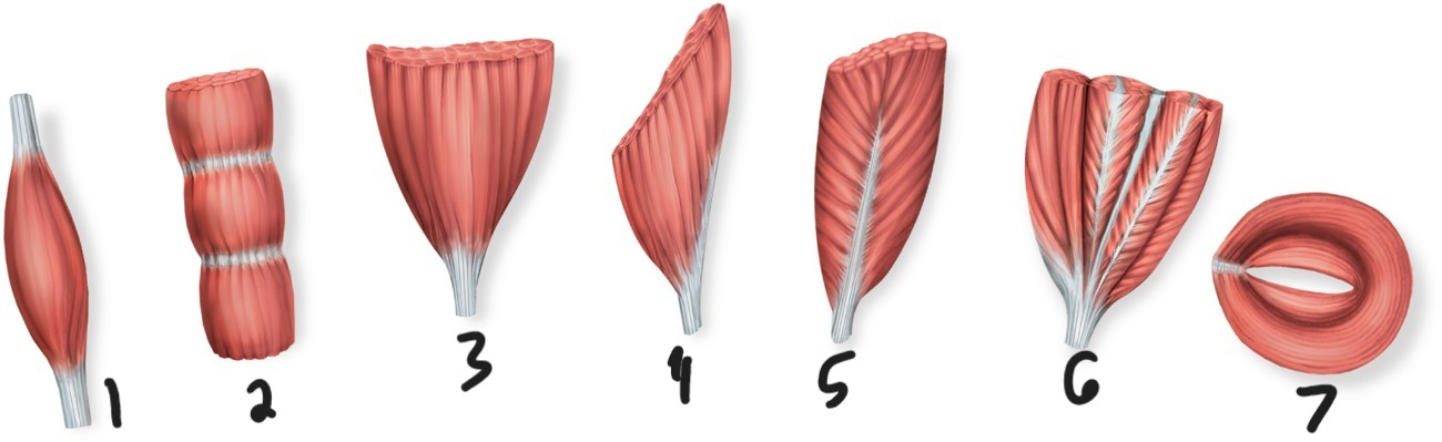
fusiform

parallel
muscle fascicles are all running parallel (image 2)

triangular
image 3

unipennate
fascicles coming from one side (image 4)

bipennate
fasicles coming from both sides (image 5)

multipennate
image 6
circular
weakest, muscle fasciles in circular motion (image 7)

Fusiform
Biceps brachii is what muscle shape
Parallel
Rectus abdominis is what muscle shape

Triangular
Pectoralis major is what muscle shape

Unipennate
Palmar interosseous is what muscle shape

Bipennate
Rectus femoris is what muscle shape

Multipennate
Deltoid is what muscle shape

Circular
Orbicularis oculi is what muscle shape

Fascicle orientation and muscle shape
Strength of a muscle and the direction of its pull are determined by what….. (2)
Muscle compartments
Fasciae package groups of functionally related muscles into compartments, contains nerves and vessels that supply the muscle group, inter muscular septa (very thick faces) separate one compartment from another
Tendons
Indirect attachment to bone
Tendons
Connect muscle to bone
Tendons
collagen fibers of the endo-, peri-, and epimysium continue into this and from there into the periosteum and the matrix of bone
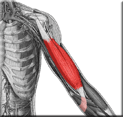
tendon
biceps brachii is a ……
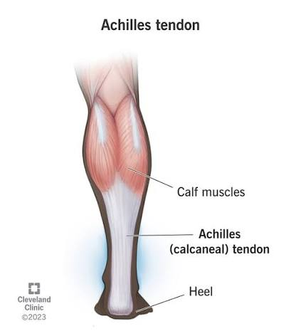
tendon
achilles tendon is a …….
Aponeurosis
Tendon is broad, flat sheet
Retinaculum
Connective tissue band that tendons from separate muscles pass under

Direct (fleshy) attachment to bone
Does not need a tendon and little separation between muscle and bone

Direct (fleshy) attachment to bone
muscle seems to emerge directly from bone
direct (fleshy) attachment to bone
margins of brachialos and lateral head of triceps brachii are exmaples of what

Attatchments of muscles
Origin (stationary end) and insertion (moving end), most skeletal muscles connect to bone, but some attach to dermis, fascia, or other soft connective tissues
origin
attachemnt that is the stationary end
insertion
attachment that is the moving end
Action
Effects of muscle → produce or prevent movement
4 action categories
Prime mover (agonist), synergist, antagonist, fixator
Prime mover (agonist)
Produces most of force during joint action
Synergist
Muscle that aids the prime mover
synergist
stabilize nearby joint; modify direction of movement; may contribute additional force
Antagonist
Opposes the prime mover
antagonist
relaxes to give prime mover control over an action, prevents excessive movement and injury
Fixator
Prevents movement of bone
Antagonistic pairs
Muscles that act on opposite sides of joint
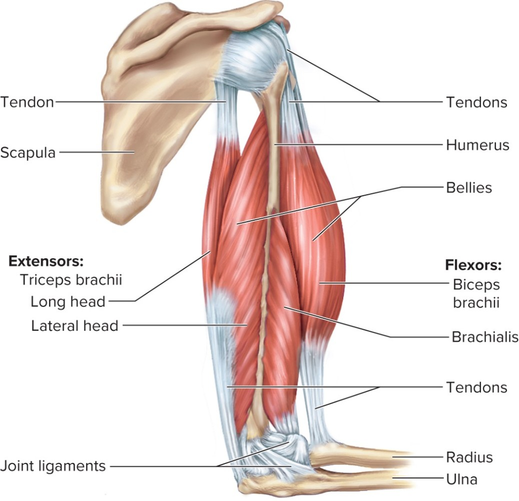
prime mover
for elbow flexion, the brachialis is the functionl group ……

synergist
for elbow flexion, the biceps brachii is the functional group ….

antagonist
for elbow flexion, the triceps brachii is the functional group …..

fixator
for elbow flexion, the muscle that holds scapula firmly in place (rhomboids) is the functional group …..
Intrinsic muscles
Entirely contained (both origin and insertion) within a region

intrinsic muscle
the hand is an exmaple of what kind of muscle

Extrinsic muscles
Act on designated region, but has its origin elsewhere

extrinsic muscles
the finger muscles are what kind of muscles

Innervation
Nerve that stimulates muscle
innervation
enables diagnosis of nerve, spinal cord, and brainstem injuries from their effects on muscle function
Spinal nerves
Arise from the spinal cord
spinal nerves
emerge through intervertebral foramina innervate muscles below the neck
spinal nerves
innervate muscles below the neck
Cranial nerves
Arise from the base of the brain (12 pairs)
cranial nerves
emerge through skull foramina
cranial nerves
innervate the muscles of the head and neck
Blood supply
Muscular system receives about 1.24 L of blood per minute at rest (one-quarter of the blood pumped by the heart), during heavy excursive, total cardiac output rises and the muscular system's share is more than three-quarters (11.6 L/min)
blood supply
capillaries branch extensively through endomysium to reach every muscle fiber
How muscles are named
Latin names describe location, shape, size, direction of muscle fibers, origin and/or insertion, action
Muscles of facial expression
Muscles that insert in the dermis and subcutaneous tissue
muscles of facial expression
tense the skin and produce facial expressions
Scalp, forehead, around the eyes, nose, and mouth, and in the neck
where are muscles of facial expression found in (6)
Muscles of the tongue
Pushes food between molars for chewing (mastication)
muscles of the tongue
forces food into the pharynx for swallowing (deglutition)
muscles of the tongue
crucial importance to speech, intrinsic and extrinsic muscles of the tongue
Depression, elevation, protraction, retraction, lateral and medial excursion
Biting and chewing movements of mandible (6)

Temporalis, masseter, medial pterygoid and lateral pterygoid
muscles of chewing and swallowing (4)
Suprahyoid group
digastric, geniohyoid, mylohyoid, stylohyoid
digastric, geniohyoid, mylohyoid, stylohyoid
suprahyoid group (4)
Suprahyoid group
function: concerned with aspects of chewing, swallowing, and speaking

Infrahyoid group
Omohyoid, sternohyoid, thyrohyoid, sternothyroid,
Omohyoid, sternohyoid, thyrohyoid, sternothyroid (4)
infrahyoid group
Infrahyoid group
Fix hyoid bone from below, allowing suprahyoid muscles to open mouth
pharyngeal constrictors
Drive food into esophagus during swallowing
Muscles acting on the head
Arise from vertebral column, thoracic cage, and pectoral girdle; end on cranial bones
Flexion, extension, lateral flexion, rotation, may cause contralateral and ipsilateral movements
Actions of muscles acting on the head (6)
Flexion
Tipping head forward
Extension
Holding the head erect (upright)
Lateral flexion
Tipping head to one side
rotation
turning the head to the left or right
Contralateral movement
Movement of head towards opposite side
Ipsilateral movement
Movement of head towards same side
Sternocleidomastoid, scalenes
neck flexors (2)
Trapezius, splenius capitols, semispinalis capitis
Neck extensors (3)
Three functional groups of the muscles of the trunk
Muscles of respiration, muscles that support abdominal wall and pelvic floor, movement of vertebral column
Diaphragm, contraction, relation, external intercostals, internal intercostals
Muscles of respiration (5)
Diaphragm
Muscular dome between thoracic and abdominal cavities
Contraction
Flattens diaphragm, enlarges thoracic cavity (inspiration)
Relaxion
Diaphragm rises, shrinks the thoracic cavity (expiration)
external intercoastals
elevate ribs and expands thoracic cavity
internal intercostals
depresses and retracts ribs, compresses thoracic cavity