Biol 200 Midterm 1
 Call Kai
 Learn
Learn Practice Test
 Spaced Repetition
 Match
Match Flashcards
 Knowt Play
1/91

There's no tags or description
Looks like no tags are added yet.
Name | Mastery | Learn | Test | Matching | Spaced | Call with Kai | Chat |
|---|
No analytics yet
Send a link to your students to track their progress
92 Terms
What defines living matter?
1. A high degree of chemical and
structural complexity
2. A capacity to extract, transform and
use energy from the environment
3. Defined functions for the organism’s
components and regulated
interactions among them
4. A capacity to sense and respond to
environment
5. A capacity to self-replicate
6. A capacity to evolve
What does life need to function? And how does it remain functional?
complex macromolecules
continuous input of energy
Lipid Barrier (cell’s plasma membrane)
serves as a selective barrier, allows greater
concentrations of nutrients and synthesized products inside the cells than outside
• Allows cells to have a distinct
intracellular environment separate
from its surroundings (to be outside of a
chemical equilibrium with its
surroundings)
Proteins
each have a specific function
very versatile in what functions it can serve
structural roles, generate movement, sense and
detect signals, catalyzing reactions (including those needed to obtain and use energy from the
environment)
DNA
contains information to self-replicate
simpler than proteins
only four building blocks vs 20 amino acids for proteins
Central Dogma
The flow of information from DNA to the production of proteins, including translation and transcription processes.
Transcription
The turning of DNA information on how to make proteins into RNA in the form of mRNA, which is later exported from the nucleus
Translation
The use of mRNA paired with tRNA by a ribosome to synthesize (make) proteins from amino acids
tRNA
a type of RNA molecule
coupled to an amino acid residue
acts an adaptor, pairing with a sequence in mRNA to convert that codon sequence into specific amino acids
Ribosomes
machines that catalyze protein synthesis
composed of proteins and RNA, called ribosomal RNA (rRNA)
How does translation occur?
Ribosomes incorporate specific amino acids into the growing protein using the genetic code
There are 4 nucleotides and 20 amino acids
By reading the sequence of RNA in groups of three consecutive nucleotide (codons), greater amount of information is conveyed in RNA (43= 64 combinations)
Each codon codes for an amino acid, but different codons can code for the same amino acid
PET
most common type of polyester
an example of a polymer
its monomer is ethylene terephthalate
cooking it requires":
mixing
two precursors
Input of energy, heat (150-290oC)
removal of byproducts (such as methanol)
Special Amino Acids
Ex:
Cysteine
contains a reactive sulfhydryl group (SH). Serves in catalysis of enzymes, reacts with other cysteines to form disulfide bonds to join two polypeptides
Glycine
smallest amino acid
Proline
has a side chain that forms a covalent bond to the N in the amino group attached to the alpha carbon kinks in the chain
Hydrophilic amino acids
• amino acids readily soluble in
water, “love water”
• either charged positively (basic), negatively (acidic), or polar (no charge)
Hydrophobic Amino Acids
• Amino acids poorly soluble in
water (hate water, fear water)
• either have hydrocarbons (linear or
branched) or aromatic rings
Nucleotides
monomers of nucleic acids (DNA and
RNA)
composed of:
pentose (five-carbon sugar), a phosphate group, and a ring-shaped molecule containing nitrogen called nitrogenous base (or just base)
• Negative charge in phosphate makes them acidic
What is the difference between DNA and RNA
The difference between the two is
the loss of an OH group at position 2’
in deoxyribose
The pentose in RNA (ribonucleic acid)
is ribose
The pentose in DNA
(deoxyribonucleic acid) is 2’-
deoxyribose

DNA & RNA bases
two types:
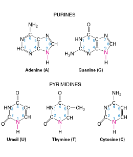
Purines, which contain a pair of fused rings
Pyrimidines, which contain a single ring
Thymine is only present in DNA
Uracil is only present in RNA (replaces Thymine)
Bases are connected to the ribose through the nitrogen atom at position 9 (in purines) or 1 (in pyrimidines)
purine Adenine (A) pairs with the pyrimidine Thymine(T)
2 hydrogen bonds (weak non-covalent bonds)
purine Guanine (G) pairs with the pyrimidine Cytosine (C)
3 hydrogen bonds
Nucleotide Ribophosphate backbone (DNA AND RNA)
joined through two phosphodiester covalent bonds where a phosphate group links two (deoxy)ribose at postions 5’ and 3’
Phosphate and (deoxy if DNA)ribose groups generate the backbone of nucleic acids
DNA and RNA have an orientation, with a 5’ end (containing a phosphate) and a 3’ end (containing a hydroxyl group)
Each monomer can contain a different base, hence, generating a unique sequence
Why is RNA more prone to breaking than DNA?
Hydroxyl group at position 2’ in the ribose of RNA can spontaneously react with the phosphate group, break the RNA molecule
DNA lacks an OH at the 2’ of ribose, making it much more stable and the choice for the genetic material in cells
DNA double Helix
The ribophosphate backbone in the outside and
the bases inside
Bases are regularly spaced (0.34nm appart)
completes a turn every 3.4-3.6 nm (i.e.
10-10.5 bases per turn)
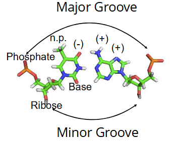
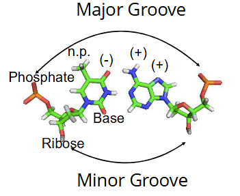
On the outside, the spaces between the strands form two helical grooves of different widths:
the major and minor groove
Atoms on the edges of each base are exposed
Major Groove
on DNA double helix
wide
contains unique chemical information
used by most protein-DNA interactions
Minor Groove
on DNA double helix
shallow
cannot distinguish all bases
What evidence do we have that all life shares a common ancestor?
Shared use of amino acids and nucleotides across life
the use of only L-amino acid stereoisomers to generate proteins (not d-amino acids even tho theyre mirrorimages of each other)
D-amino acids exist in life, but they serve other functions, such as during the cell wall synthesis in bacteria

Mirror Life/ Mirror amino acids
ex: proteins composed of D-amino acids
can have similar functions as their natural versions, but resist degradation by natural enzymes
Potentially useful in clinic, but at the same time, controversial
What is the hierarchy of protein structure?
Primary structure
Secondary Structure
Tertiary Structure
Quaternary structure
Primary Structure (sequence)
the first in the hierarchy of protein structure
covalent bonding between amino acids
Determines the size of a protein
expressed in the number of amino acid (aa) residues and their mass in daltons (Da) (1 Dalton=1 atomic mass unit)
Smallest proteins are ~40 aa in length
The ”average” protein depends on the organism. In yeast, the average is 466 aa residues. In E. coli is 329 aa. In humans is 476 aa
Largest protein known has 34,000 aa residues!
Can form:
Oligopeptides = small chains < 30 aa
Polypeptide = larger chains, >30 aa and often hundreds of aa in length
Secondary Structure
local conformations of the peptide chain backbone, generating stable arrangements of the aa residues
based on H-bonding between peptide bond carbonyl O atoms on one aa, and amide H atoms on another aa
There are three major peptide chain backbone conformations:
α-helix
β-sheet
β-turn
~60% of the length of the average polypeptide chain consists of segments of α-helix or β sheet
Several secondary structure elements per polypeptide
α-helix
secondary structure option in proteins
every NH of one amino acid at position n in the sequence forms an H- bond with the CO of a dif aa at n+4 position
Tilted axis means there are 3.6 aa residues per turn
Backbone has a straight rod structure with side chains pointing outward
Side chains determine
the probability of it being formed
its interactions with other parts of the protein and other molecules
its surface properties (ex: polarity)
Proline is not usually found in it as it creates a kink

β-strand sheets
secondary structure option for proteins
strands short (5-8 aa) nearly fully extended segments.
H-bonds are formed between two adjacent β-strands perpendicularly to the backbone
2 or more strands generate nearly 2-dimensional sheets
Side chains protrude above and below the sheet, determines:
interactions within the protein and with other molecules
likelihood of the sheet forming
two different proteins can also interact to form sheets
can be parallel or antiparallel

β-turn
secondary structure option for proteins
composed of 4 amino acids
forms a sharp U-shaped bend in the backbone
usually contains glycine and proline
The 1st and the 4th α carbon are held close by an H-Bond
helps make protein structures more compact
Motif
A combination of two or more secondary structures that form a distinct 3D structure
found in multiple proteins
associated with a specific function
ex: coiled-coil


Coiled Coil
4 different α-helices coil about one another.
aliphatic residues (hydrophobic but not aromatic) at positions 1 and 4 in a 7-aa repeat
these residues (aa) bury between the two helices.
occurs in fibrous proteins and transcription factors
Tertiary structure
overall conformation of the polypeptide (the spatial organization of the multiple secondary structure elements)
stabilized by:
hydrophobic interactions
Water molecules surrounding hydrophobic molecules in an aqueous environment surround them, like a cage ( this has low entropy)
hydrophobic molecules group together (coalescence), reducing the amount of water molecules necessary to surround them
this net increase in entropy (less low entropy water molecules) causes separate hydrophobic and aqueous phases to form
van der Walls
favors coalescence of hydrophobic molecules (grouping)
hydrogen bonds
these forces are weak, so the structure can fluctuate (change slightly now and then)
divided in domains
Oil Drop Model
a model for protein folding
Hydrophobic and hydrophilic side chains are distributed throughout the linear sequence
when the protein folds, the hydrophilic side chains tend to be exposed to water and hydrophobic residues tend to cluster together in the inner core, like drops of oil in water, hiding from the aqueous surroundings
Within this simple general model an infinite number of particular structures are possible
but, in reality, each protein adopts only one or a small number of similar, structures (conformations).

Domains
found in tertiary structures of proteins
distinct regions
They can represent a:
function
can be separated from the rest of the protein and remain functional
structure
or refer to the spatial relationship with the rest of the protein
It is common to refer to the regions near the
ends of proteins as C-terminal or N-terminal domain
similar versions can be found in different proteins
multiple copies can be found in one protein

Green Fluorescent Protein (GFP)
consist of a barrel containing 11 beta sheets and central helix that contains the chromophore
Interior of the barrel is highly crowded by side-chains of amino acid residues, limiting movement inside
Alpha-helices at the ends of the protein stabilize it
Chromophore (what gives the color) is formed by a post-translational modification that result in the cyclization of peptide backbone
Quaternary Structure
Multimeric proteins several different tertiary structures held together
can contain any number of identical or different polypeptides
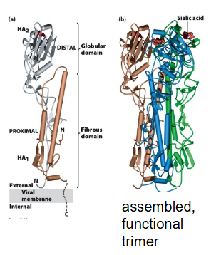
ex: Influenza virus hemagglutinin.
A trimer of identical subunits each composed of an HA1 and an HA2 domains
the three identical two-protein subunits are held together by non-covalent bonds
Supramolecular complexes
Large “molecular machines” made up of multiple distinct proteins
each protein may itself contain multiple subunits.
Ex: transcription initiation complex

How is X-ray crystallography used to determine protein structure?
passes X-rays through a protein crystal containing millions of copies of the protein of interest in a precise arrangement
X-rays are diffracted (change their trajectory) as they interact with electrons in the protein atoms
this generates a pattern on the detector that represents the distribution of electrons (and atoms) in the sample
Complex calculations are done to generate a model of the protein structure that matches the electron density.

How is Cryo-electron microscopy (Cryo-EM) used to determine protein structure?
a solution containing the protein of interest is fast frozen on a grid.
a small dose of electrons is used to prevent damage and detected using sensitive cameras.
analysis sorts particles base on orientation to make average images and further detailed structures after further refinement.

How is AI used to determine protein structure?
revolutionizing molecular biology
sequence can be converted to a structure in seconds
Capable of other predictions, like protein-protein interactions
not 100% reliable, predicted structures provide a good model that can be tested by other methods
how it works is unknown
Sequence Similarity
degree of match between two sequences
generally measured as a percentage
Homologous sequences
two or more sequences derived from a common ancestral sequence
either applicable or not, no middle ground
How to Identify Homologous Sequences
Search for maximal similarity between sequence, aligning them such that as many aa’s as possible line up
difficulty increases with length of sequence
Introduce Gaps in the shorter sequence as necessary if there is a size difference between the compared sequences
creating a dot plot representation can help
helpful to visualize changes like inversions and duplications
Protein sequences contain more information on evolutionary conservation than DNA sequences
Conservation of chemical traits in different amino acids suggest evolutionary pressures to maintain activity

How does AlphaFold identify homologous sequences? What are its pros and cons?
Protein query is compared with a database of protein sequences to look for local sequency similarity
This is then correlated to known protein structures
predictions have low confidence when no sequence similarity is found
cause this is based on evolutionary conservation, and not on calculations of the chemical properties of proteins
Protein structure prediction gets us closer to designing proteins with new functions
What is the order in which a protein begins to fold?
1st folds regions with secondary structure
Secondary structure elements group into motifs …then into domains
Finally, tertiary structure is acquired
aka: same hierarchy as protein structure
How can amino acid residue rotate?
the axis of the backbone can rotate at the bonds connecting the alpha carbon to the carbonyl and amide groups
Rotation is limited due to steric constrains imposed by the backbone and the side chains
Describe the role Proline plays in folding
Most amino acid residues result in a linear backbone
because they orient in a trans configuration relative to the peptide bond
5-7 percent of the peptide bonds with proline acquire a cis-configuration (creates a kink, no linear backbone
Isomerization between trans and cis configurations can occur spontaneously, but it is slow.
Peptidyl-proline isomerases (PPIases) are enzymes that speed up the process during folding
Isomerization (cis to trans or vice versa swap) of a single proline can dramatically change protein structure, and affect its activity
Chaperones
proteins that help guide protein folding along productive pathways
does so by permitting partially misfolded proteins to return to the proper folding pathway
recognizes exposed hydrophobic residues.
upregulated in conditions where misfolding is increased (ex. heat-shock)
can:
fold newly made proteins
refold misfolded or unfolded proteins
disassemble potentially toxic protein aggregates that form due to protein misfolding
works through ATP-dependent cycles of binding to, and release from, misfolded “client” molecules, at exposed hydrophobic patches
By blocking the exposed hydrophobic patches, it isolates the folding or refolding protein while productive folding events occur.
Two types:
chaperons (?!?!?!?, named this way because its the most common type so all of them are called chaperones 😕 , just use the e to differentiate for yourself they prolly wont)
chaperonins
Chaperonins
large complexes that isolate unfolded proteins
ex: GroEL
is composed by two stacked rings, each composed of seven subunits
Each ring interacts with a seven- subunit co-chaperone (GroES) that acts like a lid,
GroES also contains seven subunits
In the center of GroEL there are chambers where all or part of a protein enters.

Chaperons
ex: Hsp70 ( Heat-shock protein)
Binding and hydrolysis of ATP are needed for Hsp70 as they result in changes in the conformation
Binds to short segments of an unfolded proteins such as those newly synthesized as they emerge from the ribosome

If a protein is so misfolded that it cannot be fixed, then. .
it is targeted for degradation
How does the Ubiquitin/proteasome system for protein degradation work?
Poly-ubiquitin “tags” damaged or misfolded proteins for degradation
Ubiquitin-tagged proteins are fed into a multisubunit chamber
subunits of chamber form inward-facing proteases.
How it works:
1. Ubiquitin ligases recognize exposed hydrophobic residues, and add multiple ubiquitins to proteins, forming chains of 4 or more ubiquitins.
2. Polyubiquitinylated (chain of ubiquitin) proteins are recognized by Ub receptors in the proteasome
3. Deubiquitinases (Dubs) hydrolyze bonds between ubiquitins to recycle them
4. ATPase driven auxiliary proteins unfold targeted proteins and transports to the core for degradation
5. Once inside the inner chamber polypeptides are digested into short fragments of 2-24 aa in length
6.Peptide bonds of hydrophobic, acidic and basic residues are cleaved at the active site
7.The resulting peptides are further degraded into single amino acids in the cytoplasm

Ubiquitin
a small protein (76 aa residues, 8.6 KDa)
becomes covalently linked to the lysine residue of target proteins
This requires multiple steps and a specialized machinery
How it works:
1. the carboxyl terminus is activated
2. the protein is transferred to a ubiquitin conjugating subunit (E3 ubiquitin ligase)
(There are 600 E3 coded in our genome. Each with specific substrate binding.)
This allows for it to mark proteins for degradation (after forming its own chain, aka its job in the ubiquitin/proteasome system):

Protein Aggregation
Formed when misfolded proteins or incompletely degraded proteins interact with each other, hiding their hydrophobic residues
Occurs through high protein concentration and changes in environmental conditions
can be amorphous (messy) or well- organized, such as in the amyloid state.
Amyloid Fibrils
are formed by the generation of short segments (6-12residues in length) that form long arrays/ filaments, of β-sheets.
Each β-strand is oriented nearly perpendicularly to the axis of the filament
Two stacks pf beta strands twist about one another, forming protofilaments ( many of them form fibrils)
can be found in tissues
Neurodegenerative diseases such as Alzheimer’s disease and Parkison’s disease in humans, and the transmissiblespongiform encephalopathy (“mad cow” disease) in cows and sheep, all contain it
its formation formation is associated to age, but more prevalent in mutant proteins
Ligand
The molecule to which a protein binds
Diverse functions performed by proteins are based on their to engage in a common activity: binding
Proteins bind to one another, to other macromolecules, to small molecules, and to ions.
Two important properties in protein binding:
Specificity: the ability of a protein to bind only one particular ligand, even in the presence of a vast excess of irrelevant molecules
Affinity: the tightness, or strength of binding, expressed as dissociation constant (Kd). The stronger the interaction, the lower the Kd
Protein binding results from an interaction between complementary molecular surfaces
Protein binding specificity arises from interactions which are individually weak, but, if numerous, collectively strong


Antibodies
contains two (identical) heavy chains and two (identical) light chains.
Individual polypeptides (chains) in it are kept together through disulfide bonds
Molecules it recognizes are called antigens
The antigen-binding surface or CDR (complementarity determining region) involves multiple protein loops from both the heavy and the light chains
These loops are highly variable in amino acid sequence among antibody encoding genes so the total repertoire of possible CDRs in the antibody population is huge (lots of variety)

Enzymes
(proteins or RNAs)
catalyzes the making or breaking substrate covalent bonds
Ligands of enzymes include the substrates of the reactions they catalyze
accelerates the rate of a reaction by reducing the free energy of the transition state and thus ΔG‡.
Substrate specificity arises from the substrate binding site
The substrate, and the substrate binding site interact at complementary molecular surfaces.
Some amino acids in the enzyme polypeptide contribute to the substrate binding site, and others contribute to the catalytic (active) site
Vmax – the maximal rate of catalysis given saturating amounts of substrate
Depends on the amount of enzyme and how fast it can work:
the turnover number
and the enzymatic cycles per second at top speed
Km – the substrate concentration that supports a rate of catalysis equal to one-half of the Vmax
Provides a measure of the enzyme: substrate binding affinity
Enzymes free in solution: reaction intermediates diffuse from one enzyme to the next
can be slow. .
Coupling by a scaffold protein overcomes slow substrate diffusion in a metabolic pathway
Multisubunit enzyme complex formed by a scaffold protein minimizes or eliminates the substrate diffusion time
Some enzymes are fused at the genetic level, becoming domains in a single polypeptide chain

Serine protease trypsin
Proteases hydrolyze peptide bonds in polypeptides.
The serine proteases are a family of proteases whose catalytic mechanism involves a serine residue in the catalytic site.
Trypsin hydrolyzes peptide bonds adjacent to arginine and lysine (large, basic side chains).
Proper substrate binding only occurs when the substrate amino acid side chain “fits” into a negatively charged pocket within the substrate binding site
Differences in the substrate recognition pocket in the related enzymes define their differing specificities.
ex: The pocket in elastase is obstructed by bulky valine side chains, so elastase cleaves adjacent to amino acids with small side chains, e.g. alanine and glycine
How Trypsin’s (serine protease trypsin) Catalytic site works:
Protein folding brings “distant” amino acids into proximity.
Trypsin uses a 2-step catalytic mechanism involving 3 amino acid side chains: Asp-102, His-57, and Ser-195
Step 1 (a-c) cleavage of peptide bond with formation of a covalent substrate-enzyme complex (Ser195 acyl enzyme).
Step 2 (d-f) hydrolysis of acyl enzyme complex
Both subreactions depend on His-57’s ability to bind, and to release a proton.
These reactions happen best at a pH near the pK of His, i.e. ~ pH 7.

pH requirements for Acid-Base Catalysis
Catalysis requires a particular ionization state (protonated or nonprotonated) of one or more amino acid side chains in the catalytic site.
Pancreatic serine proteases (chymotrypsin) have pH 8 as activity optimum
pH <9 for required for proper protein conformation
Lysosomal hydrolases ∼4.5 pH optimum: matches the low internal pH in the lysosomes in which they function
Allosteric Regulation
A major manifestation of allosteric effects
conformational switches in regulatory proteins in response to ligand binding or post-translational modification
Allosteric Effects result from the binding of a ligand at one site on a protein, which leads to conformational changes that affect the binding of another ligand molecule at a different site of the same protein
ex: binding of ATP in chaperones,
Ca++ binding to calmodulin changes its conformation, allowing it to bind to target peptides on other proteins, thus regulating their structure and activity
G-proteins exist in “on” (GTP bound) and “off” (GDP bound) conformations that interact differently with other proteins
Switching from “on” to “off” is facilitated by GAPs and switching from “off” to “on” is facilitated by GEFs
Note: Subcellular localization also helps regulate protein function

Post-translational modification of proteins
changes in the chemistry of proteins that occur after synthesis (ex. ubiquitinylation)
Modification of protein conformation and activity can occur through phosphorylation and dephosphorylation
Protein kinases catalyze the phosphorylation of amino acid side chains
Protein phosphatases catalyze dephosphorylation of amino acid side chains
Human genome encodes >500 different protein kinases!
Kinases recognize a sequence in proteins and introduce a phosphate group at serine, threonine or tyrosine.
The reaction transfers the gamma phosphate in ATP to the protein modified

Purification of Proteins
Proteins in our cells vary widely in their properties (size, charge, hydrophobicity).
These properties are exploited for their purification.
Proteins are normally extracted from cells, by breaking them and removing big cellular debris (like membranes, RNA and DNA).
Chromatography
Separation of components based on their differential interactions with an immobile (=solid) material.
Mobile phase (liquid) moves continuously past the solid phase (paper in the experiment we did in lab).
Protein molecules are moved along in the mobile phase but at rates that depend on how much they interact with the solid phase.
generally done in columns, with a solid phase made up of ~millimeter-sized beads
3 types:
gel filtration
ion exchange
antibody affinity

Gel Filtration Chromatography
Separation based on size and shape.
The solid phase gel beads have pores (tiny, made for molecules)
The mobile phase freely enters and exits the beads (flows through)
Small proteins can enter and exit the beads through the pores (a waste of time going in and out)
Large proteins cannot enter.
They don’t “waste time” diffusing within the beads, so they migrate faster and are eluted earlier.
Shape also influences migration.
The closer to spherical the shape, the slower the migration

Ion-exchange chromatography
Separation based on electric charge
Protein molecules with a net electric charge will bind to immobile phase with the opposite charge (and thus will move slower, so whatever molecule has the same charge as the immobile phase will move faster down the column, as they will be repelled)
Electrostatically-bound proteins can be released by flowing a salt solution through the column (to collect them after the faster ones have already been collected)
The displacement of the protein by Na+ or Cl- is “ion exchange” (from the salt being flown down)

Antibody affinity chromatography
Antibodies specific for any particular protein can be covalently coupled to the solid phase
All the other proteins can be washed away and then the target protein can be released from the antibody by lowering the pH
bypasses all other properties of the protein purified, as long as it can bind to the antibody (or other molecule) in the solid phase it will work

electrophoresis
In a free solution at a given electric field, the direction of migration is determined by the net charge, and the speed of migration is determined by the net charge/mass ratio.
Protein mixture is loaded at the wells of gel (made of cross-link/bonds of the polymer polyacrylamide)
Proteins migrate through small pores in the polymer network.
Smaller proteins migrate faster
In native proteins, migration depends on charge, size and shape
Since the start point has a negative charge and end point has a positive charge, negatively charged proteins move faster (repelled by start attracted by end) than positively charged proteins

SDS-PAGE
Anionic detergent sodium docecyl sulfate (SDS).
SDS denatures proteins by the interaction of its hydrophobic tail with hydrophobic amino acid side chains, disrupting the oil drop structure (hydrophobic inner, hydrophilic outer) of proteins
The hydrophobic tail of SDS binds not only to hydrophobic residues, but also to other SDS molecules,
so it tends to coat the polypeptide chain in auniform layer of SDS molecules
Because these are all negatively charged, the various parts of the SDS polypeptide chain repel each other and this further disrupts and unfolds the protein
SDS-PAGE was developed to study of the proteins of a phage. Progress in science cannot be predicted!
Denatured proteins (proteins that lost their structure) covered by SDS are separated by size through electrophoresis (all are negative due to SDS coating)
They migrate towards the positive pole
All proteins are then coloured by using dyes that bind to them non-specifically
A pattern of bands, each band representing a different protein, can be observed
SDS-PAGE is normally used AFTER protein purification to assess degree of purity (it is not a purification technique)
Alternative specific proteins can be detected by the use of antibodies
They are then Immunoblotted
All proteins in an SDS-PAGE gel are transferred to a membrane, where proteins will be exposed in its surface.
Proteins can now be accessed by antibodies.
Normally, a primary antibody detects the protein of interest.
A secondary antibody, used for detection through fluorescence or enzymatic activity, is used to detect primary antibody.
Hence, only few ‘detection’ antibodies are needed


Immunoblot (western blot
How it works (using SDS):
All proteins in an SDS-PAGE gel are transferred to a membrane, where proteins will be exposed in its surface.
Proteins can now be accessed by antibodies.
A primary antibody detects the protein of interest.
A secondary antibody, used for detection through fluorescence or enzymatic activity, detects the primary antibody.
Hence, only few ‘detection’ antibodies are needed
Density of bands represent protein abundance
Another Example:
Experiment tests whether the presence of the G (glucocorticoid) R ligand influences the abundance of its receptor
Detection of actin, serves as reference for quantification (loading control


Co-immunoprecipitation
Use of an antibody that recognizes a specific protein to co-purify other proteins that interact with it
This technique can be used to identify proteins in a protein complex
The immunoprecipitate will contain the protein carrying the epitope (part of an antigen recognized by the antibody) and any partner proteins stably associated with that protein. (i.e. co-immunoprecipitation) (co-IP).
ex: (diagram) is testing the interaction between GR and PPARa, and the effect of GR-ligand in the interaction

Immunofluorescence
identifies cellular localization of proteins
Localization of proteins in cells and tissues studied by the use of antibodies
How it works:
Cells are killed using solvents that preserved their structure and immobilize on a microscope slide
Antibodies are then used to detect specific proteins,
Fluorescently labelled secondary antibodies are then used to mark the original antibodies (done at the fluorescence microscope)

Fluorescence
the property of certain substances of absorbing light of short wavelength and emitting light of longer wavelength
How it works:
An electron in the molecules that produce fluorescence (called fluorophores) moves to a higher energy orbital as result of the absorption of a photon (Excitation)
Excited electrons have a tendency to go back to their initial ground state
In doing so they release the excess energy in the form of light (Emmision)
Fluorophores (chemical compounds that have fluorescence) undergo thousands of excitation-emission cycles before they are chemically modified (oxidized) and stop producing fluorescence (Bleachin)
Fluorescence Microscope
4 important elements
1. Light source capable of producing intense light over a broad spectrum
2. Filter Cube
contains an emission and excitation filter
3. Objective lens
4. Sensitive camera (similar to those used in Astronomy)
also has a Dichroic mirror: Reflects one wavelength and transmits another (look at diagram lowk)
first described in jellyfish and coral, now we know it is widely distributed in animal species
How its used to label a protein of interest:
Gene fusions are generally done by cloning before being introduced to the organism with either:
a vector or by homologous recombination (to insert them in the genome).
Expression results in fluorescent protein fusions
Choice of C- or N-terminal fusion depends on the structure and activity of the protein of interest
Multiple colors, from blue to far-red are possible
Multiple fluorescent proteins can be combined in a single sample.
In contrast to immunofluorescence, where cells are dead, use of GFP allows real-time tracking of cellular events

DNA Replication (Simple basic og slide on it)
Parental strand can be used as a template for the formation of a new daughter strand (complementary to the parental strand)
Phosphate group in the incoming deoxy nucleotide triphosphate (dNTP) reacts with the 3’ hydroxyl group (OH) in the growing DNA chain
simplified: phosphate in the new nucleotide added to the growing strand binds to the 3’ hydroxyl group (OH) of the previous nucleotide to become a part of the growing DNA (phosphate becomes part of growing backbone)
The opening of the DNA duplex for replication occurs progressively in cells, forming a DNA structure called the replication fork

Replisome
the Molecular Machinery that carries out DNA Replication
Different parts:
DNA Helicase/ CGM Helicase
separates the strands in order for duplex DNA to be replicated
Hexamer of MCM
Contains accessory subunits Cdc45 and the GINS complex
Binds to the leading strand
Replication Protein A (RPA)
binds to single-stranded DNA
keeps single-stranded DNA template in optimal conformation for DNA polymerase
Prevents formation of secondary structures on single-stranded DNA
Primase/Pol alpha Complex
primase forms the RNA component of the primer
and DNA polymerase a (alpha) (Pol a) extends the primer with DNA
Pol δ replaces the Pol α/primase complex for the lagging strand and completes the synthesis of an Okazaki fragment
NOTE IMPORTANT: Pol α/primase is the primase you learned in school, it DOES NOT synthesize strands, its RNA POL, Pol ε synthesizes the leading strand and Pol δ the lagging strand
PCNA
homotrimeric protein.
Prevents the Pol ε (or δ) complex from disassociating from the template
Rfc/PCNA complex
RFC is the PCNA loader
it opens the PCNA ring and loads it at a primer on DNA
Ribonuclease H and FEN-1 displace the RNA component at the 5’ ends
of the Okazaki fragments.
➢ Pol δ replaces the RNA with DNA.
➢ DNA fragments are then ligated together by DNA ligase
Tightly regulated
Origin Recognition Complex (ORC) is a six subunit protein that
marks replication origins and loads helicase aided by other initiation factors
MCM helicase is loaded in G1 phase of the cell in an inactive form
MCM is activated in S-phase through phosphorylation and interactions with other proteins
CDK = cyclin dependent kinase
Low CDK levels in G1 → helicases is loaded but not yet activated
High CDK levels in S-phase → no loading of helicase (already loaded), helicase is activated

Supercoiling
Opening of the double helix causes an increase in the twist of DNA ahead of the replication fork
Both, under and over twisting the DNA is called supercoiling (negative or positive, respectively).
introduces torsional stress and deforms DNA.
Accumulation can inhibit DNA replication and transcription

Topoisomerase
removes supercoiling ahead of the replication fork
works by transiently cutting DNA
For example, Topoisomerase II, cuts a double stranded DNA to allow a second piece of DNA pass through the opening

DNA Polymerization
catalyzed by DNA polymerase
requires deoxynucleoside 5’-triphosphates (dNTPs, where N = A, T, C or G)
requires a primer (can be DNA or RNA)
proceeds from a 5‘ to 3’ direction
Free dNTPs are added through the formation of a new phosphodiester bond involving the 3’ hydroxyl of the terminal sugar
Primase (a specialized RNA polymerase) forms a short RNA molecule complementary to a single-stranded region of the unwound duplex DNA
DNA polymerase extends the primer to eventually form a new daughter duplex DNA.
Since the primer is RNA then the addition of dNTPs by DNA polymerase will result in a molecule that is RNA at the 5’ end and DNA at the 3’ end
DNA replication proceeds in a 5’ to 3’ direction and the two strands of duplex DNA are antiparallel.
PROBLEM: Although one new strand (called the leading-strand) can be replicated 5’ to 3’ by DNA polymerase following the movement of the replication fork, the other new strand (called the lagging-strand) must be formed in the opposite direction of the movement of the replication fork
SOLUTION: Okazaki fragments are formed
Okazaki fragments are short discontinuous fragments consisting of RNA and DNA
They are formed on the lagging strand as many RNA primers are needed to allow for it to be synthesized with DNA Pol δ, and so it creates a mix of DNA and RNA when Pol δ places DNA between them (Pol δ replaces the Pol α/primase complex for the lagging strand and completes the synthesis of an Okazaki fragment)
The RNA component of the Okazaki fragments are then replaced by DNA and the two adjoining DNA molecules are ligated together by DNA ligase
(Ribonuclease H and FEN-1 displace the RNA component at the 5’ ends
of the Okazaki fragments.
➢ Pol δ replaces the RNA with DNA.
➢ DNA fragments are then ligated together by DNA ligase)

DNA Pol Epsilon
Carries out leading strand DNA synthesis
Origins of Replication
Specific region(s) at which assembly of the replisome and initial unwinding of DNA occurs in duplex DNA.
tends to be AT-rich (Adenine and Thymine)
Replication proceeds bidirectionally from the origin
unwinding by the CMG helicase is driven by ATP hydrolysis
RPA binds to single-stranded regions
Some of the replication origins in our genome are activated first (called early origins) resulting in the initial replication of some regions of the genome
As S phase progresses, other origins activate, following a ‘program’
The replication program is established early during development and is cell type- dependent
The replication program is related to transcription and is important for genome integrity
Our genome is composed of 3.3 billion base pairs
At a rate of 2,000 bp / minute, a single replisome would take ~ 3 years to complete DNA replication
Hence thousands of replisomes originating from tens of thousands of replication origins are used at each cell cycle
This reduces the replication time in somatic cells to few hour
Mutations
If the changes these cause are permanent, they are mutations :
2,000 – 10,000 hydrolytic depurination every day / cell
~1 cytosine deamination every 5 days/cell
~1 event of Guanidine oxidation (8-oxo-guanine) every 5 days / cell
~600 events of methylation of adenine (3-meA) per day/ cell
Some of these modifications alter the original base pairing and may also block the activity of DNA polymerases use during DNA replication
DNA polymerases alone introduce 1 error every 10,000 incorporated nucleotides.
Proofreading exonuclease activityincreases fidelity by ~100 fold (1 in every million)
permanent, transmissible changes to the genetic material of a cell (or organism)
can occur spontaneously, by transposable elements, and by errors during replication
Mutagens are chemical compounds that increase the frequency of mutations
ex:ultraviolet (UV) radiation or ionizing radiation (e.g. x-rays and atomic particles)
Carcinogen is an agent that causes cancer. Many carcinogens are mutagens
The number of mutations that fathers give to their offspring increases with age due to continuous production of germ cells (at about 2 additional per year)
A curious case
T. dohrnii (a.k.a. immortal jellyfish) can revert to a sexually immature polyp stage in response to stress or ageing
Its genome contains multiple copies of MSH2 and of DNA Pol alpha and delta, which was suggested to lower its mutation rate in somatic cells
Proofreading & Repairing DNA
In eukaryotes, DNA polymerases ε and δ but not α have a 3’ to 5’ exonuclease or “proofreading” activity
Mismatch repair further increases fidelity by about 1,000 fold, reaching error rates of about 1 error in every 1,000,000,000 nt incorporated.
Adding it all, there is a probability of incorporating a single mistake of 10^9-10^10 per nucleotide in human cells
Multiple mechanisms act to repair specific types of DNA damage
Mismatches and chemical modifications in DNA are repaired by a strategy that requires the detection and removal of the affected region
The second strand is then used as a template to fill the ssDNA gap by a DNA polymerase
Mismatch Repair (MMR) (In Humans)
Error incorporated into newly synthesized strand (daughter strand), error is wrong base pairing (ex: A and C)
MSH2 and MSH6 bind to errored daughter strand
This triggers binding and activity of MLH1 endonuclease (dimerized w/ PMS2), which make cuts near the mismatch
DNA helicase unwinds and DNA exonuclease digests the cut segment of daughter strand
The gap left behind is repaired by Pol δ and DNA ligase
often acts in proximity to the relisome, correcting mismatches shortly after they have been generated
Some of the proteins involved in MMR interact with PCNA (MSH2 and MLH1)
Base Excision Repair
How it works:
DNA glycosylase hydrolyzes the bond between the mispaired base and the sugar-phosphate backbone
In the case of uracil removal, the DNA glycoysylase is called UNG
Apurinic/apyrimidinic endonuclease 1 (APE1) cuts the DNA backbone
AP lyase associated with DNA polymerase β removes the deoxyribose phosphate (backbone that was cut)
DNA polymerase β fills the gap and DNA ligase seals together the sugar-phosphate backbone
many types of base modifications possible, 11 different DNA glycosylases specifically recognize some of these types of base modifications to start repair
ex: deamination, oxidation, alkylation
Repair of bulky lesions (Nucleotide Excision Repair)
ex: Formation of thymine-thymine dimers in DNA (caused by UV lighting on outer thymines that just happened to be next to each other)
BIG problems in DNA, bulky issues
Can be caused by:
Food contaminated with fungi (for ex Aspergillus)
Smoke from organic material, such as cigarette smoke and charbroiled food
How it works:
A complex formed by proteins 23B and XP-C do the initial recognition of the problematic DNA lesion
The complex of 23B and XP-C recruit the transcription factor TFIIH, which catalyzes the unwinding of the DNA region around the lesion
This in turn recruits RPA and XP-G, which also has unwinding activity (also unwinds DNA)
Finally, XP-F is recruited.
Both XP-F and XP-G have endonuclease activities
They generate cuts 24-32 bases apart, releasing the produced fragment carrying the lesion (problem) to be degraded
The ssDNA gap generated is then filled by a DNA polymerase and joined to generate a continuous strand by DNA ligase
Translesion Synthesis
Note:
DNA repair mechanisms require that DNA is in a double-stranded form
During DNA replication, the replisome may encounter lesions on DNA that were not previously repaired
The DNA polymerase in the replisome will stop when it encounters this, preventing completion of DNA replication
Cells use a special type of DNA polymerases called translesion (TLS) polymerases
TLS polymerases are able to bypass the lesion (ignore the problem and keep synthesizing), using the damaged DNA as template
However, they will often insert the incorrect base at the position opposite to the lesion.
TLS do not have proofreading exonuclease activity. They are error prone.
In the end, the DNA lesion is left in the template DNA
Breaking in DNA strand
Single strand breaks are more common than double strand breaks
and are efficiently repaired by the ligase
DNA unwinding during DNA replication can convert a single strand break into a double strand break
Repair: Error-prone repair by non-homologous end- joining (NHEJ) or repair by homologous recombination (HR)
Singe StrandHomologous Recombination
YOU HAVE TO LOOK AT DIAGRAM, I TRY TO WRITE DOWN BUT ITS HARD
How it works:
1. After the break, the replication fork collapses, the separated bits stay with the newly made strands, the ones that were not open yet (not a part of the fork yet) stay together, creating a mix and match of strands
2. 5’ exonuclease acts on the broken end (of the new strand without continuation since the replication fork broke), the other daughter strand (one with continuation) is ligated to repaired parental strand in the unbroken chromosome (to the portion that was outside of the fork)
3. recombinase RecA or Rad51 mediated strand invasion takes place ( the new strand that got ligated together with the unopened dna strand lifts closer to the other new strand and away from the unaffected original strand it was partnered with, invading that space, the discontinued original strand/the other strand is now under it and closer to the original strand)
4. Branch Migration ( The branches criss cross to create Holliday Structure, the broken/affected original strand moves down and crosses with the lifted strand)
5. Cut strands at cross-over (the holliday structure is cut from the middle of the x)
6. Ligate ends (put closeby broken bits together to form mixed strands)
7. Rebuild Replication fork and continue DNA replication

Double Strand
High fidelity mechanism to repair DNA breaks
Requires an undamaged copy of the chromosome to be repaired
The process requires:
processing of breaks to expose ssDNA sequence,
a search for identical sequences,
using said identical sequences as template of DNA synthesis ( to repair the break)
How it works (PLEASE LOOK AT DIAGRAM THIS IS SO HARD TO JUST WRITE):
1. Ends digested by 5’-exonucleases leaving behind 3’ single stranded ends (on each opposing side of break one of the 2 strands (different on each side) has part of it eaten)
2. recombinase RecA or Rad51 mediated strand invasion of homologous chromosome (one of the uneaten longer ends moves toward the undamaged copy double strands and lifts one above itself (doesn’t have to be lift above it’d be based on pov but thats how the diagram shows it, basically swaps positions without breaking) to create an X, a Holliday structure technically but not mentioned yet)
3. 3’ end of the invading strand is extended by DNA polymerase until the displaced single-strand base pairs with the other 3’ single strand generated initially (this one is pretty nice explanation, the one that lifted keeps lifting toward the 3’ end until the strand its lifting base pairs with the other broken strand )
4. 3’ end is extended by DNA polymerase (of the other broken strand that was mentioned to have base paired)
5. ends are ligated to form holliday structures (X’s are formed from the lifted and the lifter (2 X’s including the one from earlier)
6.Holliday structures are resolved (the X is broken and the crossed strands mix together to form 2 mixed strands each)
Two Holliday junctions have to be resolved during Homologous Recombination
Alternative Resolutions are possible (need to show diagram so its on a different card)
Each Holliday junction can be resolved at any of the two orientations
described previously (on the other card)
Their combination determines if they final result is a recombinant chromosome (crossover)
During meiosis DNA recombination occurs between chromosomes from mom and dad to mingle their sequences.
Sequences of father and mother derived chromosomes are mingled, producing translocations between homologous chromosomes

Alternative Resolution of a Holliday Structure
PLS LOOK AT DIAGRAM
2 WAYS TO RESOLVE
1. the original holliday strands break & reconnect (Original)
2. outer strands break and reconnect to mixed holliday strands allowing for recombination (Recombinant)

Non-Homologous End Joining (NHEJ)
Error-prone and can introduce mutations
requires blunt ends for ligation
Hence, exonuclease and DNA polymerase activity is used so that all nucleotides are paired with the complementary strand.
This results in a loss of information, either by the removal or the addition of nucleotides.
How it works:
A complex formed by DNA-dependent protein kinase (DNA-PK) and Ku80/Ku70 heterodimer binds to the ends of the break
Ends are digested by nucleases or filled in by DNA polymerases
Ends are ligated together
A chromosomal translocation is an abnormal chromosome region that contains rearranged genetic material.
In NHEJ these comes from two non-homologous chromosomes.
Since NHEJ joining of DNA fragments is not driven by sequence similarity, these can result in mistakenly bringing together pieces of two very different chromosomes

In what part of the cycle does each type of DNA repair occur?
During G1, where only one copy of the genome is present, DNA breaks are repaired by NHEJ
During S and G2, and M phases, double strand breaks are repaired by Homologous Recombination
During S phase, Mismatch Repair and Translesion Synthesis occur
DNA repair and Cancer
Breast Cancer
Breast cancer type 1 susceptibility protein (BRCA1) binds to double strand ends and promotes their resection.
BRCA2 acts during recombination to promote the loading of Rad51 onto the resected DNA.
Mutations that inhibit the function of these proteins are associated with a predisposed development of cancer, particularly breast and ovarian.
This is likely due to an increased used of NHEJ for DNA break repair.
PARP inhibitor (PARPi)
acts by blocking the repair activity of single strand DNA discontinuities
During DNA replication, this would generate double stranded breaks
A high number of DNA breaks in cells without active DNA recombination (because of breast cancer, BRCA1&2 aren’t able to function to fix it) results in high NHEJ activity, generating too many issues in the genome and leading to cell death
There are other models proposed to explain the mode of action of PARPi
Cells in only few tissues actively divide, like skin cells, cells in the gastrointestinal tract, and blood cells in the bone marrow.
In most other parts of the body, cells do not divide often or at all, like in the brain and the muscles.
This contrasts with tumours, where cells constantly grow and divide.
This is why, treatments that damage DNA are effective against tumours
Many of the drugs and treatments used for anti-cancer therapy target DNA.
DNA damage can be repaired efficiently if DNA replication is not ongoing.