H12: Scalp and Cranial Cavity
 Call Kai
 Learn
Learn Practice Test
 Spaced Repetition
 Match
Match Flashcards
 Knowt Play
1/74

There's no tags or description
Looks like no tags are added yet.
Name | Mastery | Learn | Test | Matching | Spaced | Call with Kai | Chat |
|---|
No analytics yet
Send a link to your students to track their progress
75 Terms
Scalp
consists of 5 layers of soft tissue that cover the calvaria
Those layers include skin, close subcutaneous tissue, aponeurosis of 2 muscles, loose subaponeurotic tissue, and the pericranium

The first three layers are collectively called the ________ , why?
scalp proper
are clinically regarded as a single layer because they remain together when cut during surgery or torn off during an accident
Scalp layers
Skin
Close subcutaneous tissue
Aponeurosis
Loose subaponeurotic tissue
Pericranium

Layer 1 of scalp
LAYER 1- Skin makes up the first layer and is commonly found containing hair and sebaceous glands

Layer 2 of scalp
LAYER 2 - Close
subcutaneous tissue
(connective tissue) is richly vascularized and well supplied with nerves

Layer 3 of scalp
Aponeurosis is a flattened tendon
It is named galea aponeurotica (from Latin galea = "helmet")
The aponeurosis is found between the frontalis muscle anteriorly and the occipitalis muscle posteriorly
It is also referred to as the epicranium

Layer 3 of scalp is also referred to as
epicranium
or
aponeurosis
or
galea aponeurotica
Latin root for aponeurosis
galea aponeurotica (from Latin galea = "helmet")
the aponeurosis is found between
frontalis muscle anteriorly and the occipitalis muscle posteriorly

Layer 4 of the scalp
LAYER 4 - Loose
subaponeurotic tissue allows free movement of the scalp proper (Layers 1-3)
The loose connective tissue that allows free movement also contains many potential spaces that are capable of filling with fluid and allowing pus or blood to spread easily through here
[Emissary] veins connect this layer with the dural sinuses and can potentially lead to the spread of infection from this layer to the cranial cavity
Therefore, this is known as the DANGER LAYER
![<p>LAYER 4 - Loose</p><p>subaponeurotic tissue allows free movement of the scalp proper (Layers 1-3)</p><p>The loose connective tissue that allows free movement also contains many potential spaces that are capable of filling with fluid and allowing pus or blood to spread easily through here</p><p>[Emissary] veins connect this layer with the dural sinuses and can potentially lead to the spread of infection from this layer to the cranial cavity</p><p>Therefore, this is known as the DANGER LAYER</p>](https://assets.knowt.com/user-attachments/04297b4b-6b51-417e-a0f6-133c8398dd75.png)
Layer 4 allows free movement of _____
scalp proper (layers 1 -3)
Layer 4 is also referred to as? Why?
DANGER LAYER
The loose connective tissue that allows free movement also contains many potential spaces that are capable of filling with fluid and allowing pus or blood to spread easily through here
[Emissary] veins connect this layer with the dural sinuses and can potentially lead to the spread of infection from this layer to the cranial cavity
Layer 5 of the scalp
(deepest) - Pericranium is firmly anchored to the skull and is also known as the PERIOSTEUM of the external surface of the skull
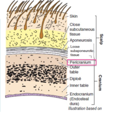
Layer 5 of the scalp is also referred to as
periosteum
or
pericranium
cranial bone
The cranial bone is made up of 3 layers that can be seen when looking at the skull
Outer table
DIPLOË
Inner table
Different cranial bones vary in thickness; the occipital and frontal regions are thick, and the temporal region is thinner
cranial bone is made up ___ layers
3
Outer table
DIPLOË
Inner table

outer table
On the external surface of the outer table is periosteum or pericranium, which is the 5th layer of the scalp
The external layer is called the OUTER TABLE and is made of compact bone
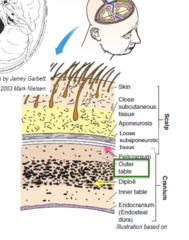
DIPLOË
The middle layer is made of spongy bone and is called the DIPLOË

inner table
The internal layer is called the INNER TABLE and is made of compact bone
Lining the inner table is ENDOCRANIUM or
ENDOSTEUM, which is the outer layer of dura

blood supply of the scalp SUMMARY
Blood supply comes from vessels below, specifically branches of the external carotid and the internal carotid arteries
Many of these vessels anastomose freely in the second layer of the scalp, close subcutaneous tissue
Branches of the internal carotid arteries supply the frontal region
Branches supplying the temporal and occipital regions come off of the external carotid artery
Veins accompany the arteries in the venae comitantes arrangement (in which 2 veins lie on either side of the artery) and have the same names as the arteries
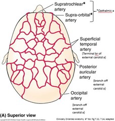
blood supply of the scalp comes from vessels below, specifically the ______ &
_________
external and internal carotid arteries
many of the vessels of the scalp anastomose freely in the _____ layer of the scalp
second layer, close subcutaneous tissue
branches of the internal carotid arteries supply what region?
frontal region
branches of the external carotid arteries supply what region?
temporal and occipital regions
venae comitantes
Blood supply of the scalp:
Veins accompany the arteries in the venae comitantes arrangement (in which 2 veins lie on either side of the artery) and have the same names as the arteries
(means vein accompanying)

Innervation of the scalp anterior to the external ear
Innervation of the scalp anterior to the external ears is through branches of the 3 divisions of the trigeminal nerve, cranial nerve V: ophthalmic (V1), maxillary (V2), and mandibular (V3)

Innervation of the scalp posterior to the ear
Posterior to the ear, nerve supply to the scalp is from the cervical spinal nerves

meninges of the brain
Similar to the spinal cord, there are 3 layers of meninges surrounding the brain: dura mater, arachnoid mater, and pia mater

Dura mater of the brain: 2 layers
1. endosteal dura
2. meningeal dura
Both of these layers adhere to each other except at certain locations where they separate, called DURAL SINUSES (C)

Dura mater outer layer
ENDOSTEAL DURA (A)
You already know about the endocranium or endosteum, the outer layer that fuses to the skull; this is also called the ENDOSTEAL DURA (A)

Dura mater inner layer
MENINGEAL DURA (B)
folders internally at four locations called dural folds

Dural sinuses and dura mater
Both of these layers of dura mater adhere to each other except at certain locations where they separate, called DURAL SINUSES (C)
Dural sinuses are spaces that contain blood and absorbed cerebrospinal fluid (CSF)

dural folds
Folded inner layer of dura mater
4 locations

4 types of dural folds
falx cerebri
tentorium cerebelli
falx cerebelli
diaphragma sellae

falx cerebri
separates the superior portion of the right and left sides of the cerebrum

Where does the falx cerebri attach?
It attaches anteriorly to the crista galli and posterioly to the tentorium cerebelli at the level of the internal occipital protuberance

Latin root for falx
falx = sickle
the fold is sickle-shaped
Tentorium cerebelli
forms a tent over the cerebellum

Latin root for tentnorium cerebelli
Latin tentorium = "tent"
falx cerebelli
nother sickle-shaped dural fold inferior to the surface of the tentorium cerebelli
It separates the right and left cerebellar hemispheres

diaphragma sellae
a small, circular, horizontal sheet of dura that forms a roof over the hypophyseal fossa in the sella turcica
a. It is pierced by they hypophyseal stalk (or infundibulum)

arterial supply of the dura
not from the same vessels supplying the scalp
Dura is supplied by the meningeal arteries, including the MIDDLE MENINGEAL ARTERY
It branches off of the maxillary artery coming off of the external carotid and courses external to the dura mater
The inner surface of the calvaria contains indentations from these vessels

middle meningeal artery
supplies dura mater
it branches off the maxillary artery coming off of the external carotid and courses external to the dura mater

Extradural or Epidural Hemorrhage (extra)
when meningeal vessels are injured
ex. blow to the side of the head
pressure from ruptured vessel separates the periosteal and dura proper
raises intracranial pressure = herniation
shape of football

dural sinuses
When the 2 layers of the dura mater separate, this is called sinus
The sinus is actually a vein lined with endothelium that is not collapsible
They drain all blood and absorbed CSF from the brain; they all flow toward the internal jugular vein

6 dural sinuses
superior sagittal sinus
inferior sagittal sinus
straight sinus
transverse sinus
sigmoid sinus
cavernous sinuses

superior sagittal sinus
lies in
the median plane on the superior portion of the falx cerebri
It connects with the diploic veins, cerebral veins, and the lateral expansions of the superior sagittal sinus called the lateral lacunae
It drains posteriorly into the confluence of sinuses (or CONFLUENCE)

superior sagittal sinus drains into
confluence of sinuses
Confulence of sinuses
Joining together of sinuses
collects blood and CSF DIRECTLY from superior sagittal sinus, straight sinus (inferior sagittal sinus drains into straight sinus) , occiptal sinus
this drains into transverse sinuses

inferior sagittal sinus
also lies in the median
plane but on the inferior portion of the falx cerebri
It drains posteriorly into the straight sinus

inferior sagittal sinus drains into
straight sinus

straight sinus
is found between the junction of the falx cerebri and the tentorium cerebelli
It receives blood from the inferior sagittal sinus and cerebral veins
It drains posteriorly into the confluence of sinuses

straight sinus receives blood from ____ and drains into _____
from inferior sagittal sinus and cerebral veins
drains into confluence of sinuses

transverse sinus
begins at the
confluence and courses laterally
It is found within the external border of the tentorium cerebelli
It passes anteriorly toward the petrous portion of the temporal bone to the sigmoid sinus

transverse sinus receives blood from ______ and drains into ______
from confluence
drains into sigmoid sinus

sigmoid sinus
extends from the transverse sinus and courses in an S-shape (S for the Greek letter sigma) along the petrous portion of the temporal bone to the jugular foramen, where it continues in the neck as the internal jugular vein

sigmoid sinus receives blood from _____ and drains into ______
from transverse sinus , superior petrossal sinus, inferior petrossal sinus
drains into internal jugular vein

cavernous sinuses
are found lateral to the sella turcica on each side
They were named cavernous because they have a cave-like appearance
pathway of blood and CSF in dural sinuses

dural sinuses image - superior

dural sinuses image 2 - side view

circle of willis also known as
cerebral arterial circle

circle of willis
(cerebral arterial circle) is a polygonal anastomosis between the right and left internal carotid arteries and the right and left vertebral arteries

the _______, after entering the cranial cavity will divide into 2 cerebral arteries: ________ and _______
The internal carotid artery, after entering the cranial cavity, will divide into 2 cerebral arteries, the ANTERIOR CEREBRAL
ARTERY, (which courses within the longitudinal fissure of the cerebrum), and the MIDDLE CEREBRAL ARTERY, (which
courses in the lateral fissure of the cerebrum)

internal carotid artery
after entering cranial cavity it will divide into 2 cerebral arteries:
1. anterior cerebral artery
2. middle cerebral artery

anterior cerebral artery
branch off internal carotid artery
courses within the longitudinal fissure of the cerebrum
(extra: least affected by strokes, leg weakness)

middle cerebral artery
largest branch off internal carotid artery
courses in the lateral fissure of the cerebrum
(extra: most common stroke, paralysis of lower face, affects upper limbs and some vision)

longitudinal fissure
separates cerebral hemispheres

lateral fissure
separates temporal lobe from frontal and parietal lobes

Veretebral artery
branches off of the subclavian artery and travels upward through the foramina in the transverse processes of the cervical vertebrae CV6-CV1
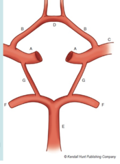
It passes through the foramen magnum and joins the vertebral artery from the opposite side to form the BASILAR ARTERY (E)
Basilar Artery
(E)
Formed by two vertebral arteries coming together after passing through the foramen magnum
This vessel courses along a groove on the pons and divides into the two POSTERIOR CEREBRAL ARTERIES (F)

posterior cerebral arteries
(F)
branches off the basilar artery
(extra: strokes in this affect vision)

circle of willis is formed by
1 Basilar artery (E)
2 Posterior cerebral arteries (F)
2 Posterior communicating arteries (G)
2 Internal carotid arteries (A)
2 Anterior cerebral arteries (B)
1 Anterior communicating artery (D)

stroke
Damage to the brain from interruption of its blood supply.
circle of willis on brain
