Developmental Biology Exam 2
 Call Kai
 Learn
 Practice Test
 Spaced Repetition
 Match
 Flashcards
 Knowt Play
1/126

There's no tags or description
Looks like no tags are added yet.
Name | Mastery | Learn | Test | Matching | Spaced | Call with Kai | Chat |
|---|
No analytics yet
Send a link to your students to track their progress
127 Terms
What type of cleavage do mouse (and many mammalian) embryos exhibit?
Rotational holoblastic cleavage: the second cleavage plane rotates 90 degrees, with one daughter cell dividing vertically and the other dividing horizontally:
Slow (12-24 hrs)
Asynchronous
Variable
Note: MZT is the same as MBT (MZT is maternal-to-zygotic transition)

What are the early and late 8-cell stages in mice? What happens, and what protein is involved?
Mouse blastomeres through the 8-cell stage form a loose arrangement; following the third cleavage, however, the blastomeres undergo compaction, one of the most crucial events of mammalian cleavage
Cell adhesion proteins such as cadherin become expressed, and the blastomeres gradually huddle together into a compact ball of cells
This tightly packed arrangement is stabilized by tight junctions that form between the outside cells, sealing off the inside of the sphere. The cells within the sphere form gap junctions that enable small molecules and ions to pass between them

What is cavitation in mouse embryos?
Initially, the morula does not have an internal cavity; however, during a process called cavitation, the trophoblast cells secrete fluid into the morula, forming a blastocoel
As the blastocoel expands, the inner cell mass becomes positioned on one side of the ring of trophoblast cells, resulting in the distinctive mammalian blastocyst
What is a key characteristic of cells of the 8-cell stage mammalian embryo? What technique can this information be used for?
They are totipotent: each cell can give rise to all cell types of the organism
Allows for pre-implantation genetic diagnosis
Take a cell from the embryo at this stage and screen it for genetic diseases
If safe, implanted back into host for development
The embryo is regulative at this stage: the early embryonic cells are flexible and can adjust their development to compensate for missing, damaged, or added cells
What is regulative development, and what is the associated type of specification?
Regulative development: when cells in an embryo can change their behavior (re-specify their fate) in response or removal or addition of other cells (humans up to the 8-cell stage)
Conditional specification: cell fate achieved through interactions with others (non-autonomous specification)
What is mosaic development, and what is the associated type of specification?
Mosaic development: when most cells in an embryo are self-differentiating (cell-cell interactions are not required); if a cell is removed, the structures it would normally give rise to will be missing from the organism
Autonomous specification: a cell achieves its fate independently of other cells
What is progressive restriction in potency? Name an example
The gradual process by which embryonic cells permanently lose their developmental potential as they divide and specialize
As development proceeds, a cell moves step-by-step down a cellular hierarchy. It transitions from a state of highest potency (where it can become absolutely any cell type) to a state of complete differentiation (where its identity is fixed to a single cell type)
Example:
The development of mammalian embryos from the early 8-cell stage to the blastocyst
Cells go from being totipotent to either pluripotent inner cell mass (ICM) or trophoblasts

What is a chimera?
An organism composed of two (or more) genetically distinct cell types
Composed of cells from two or more distinct genotypes derived from different zygotes
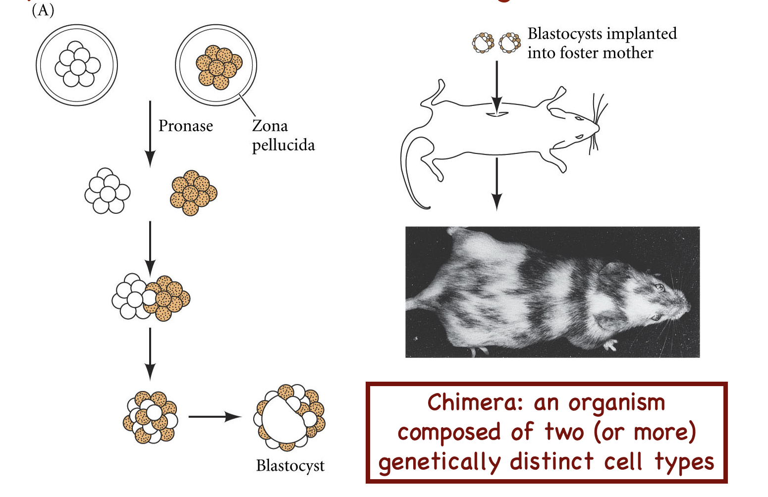
What does this experiment show?
Two 8-cell-stage embryos were mixed and implanted into the mother
A chimeric mammal resulted, where each somatic cell came from either one embryo cell type or the other
Composed of cells from two or more distinct genotypes derived from different zygotes
What is the first cell differentiation in mammalian embryos? What transcription factor plays a role? When do they differentiate?
Cells that develop into trophoectoderm (TE)
Express the transcription factor CDX2 (CDX2+)
Commit to their cell fate by the 32-cell stage
Cells that develop into inner cell mass (ICM)
Do not express CDX2 (CDX2-)
Commit to their cell fate later (32 to 64-cell stage)

What do the inner cell mass and trophoectoderm (TE) give rise to?
ICM gives rise to the embryo proper: develops into the actual tissues and organs of the new individual
TE gives rise to the extra-embryonic membranes: a protective and supportive layer of tissue that forms around a developing embryo but does not become part of the body of the baby (of placenta)
What can ICM cells be isolated and cultured for? What potency do they have, and what does that mean?
Embryonic stem cells (ES cells)
ES cells can be used to make chimeric mice
Are pluripotent, as the population that they are derived from is also pluripotent
Can differentiate into the 3 germ cell layers, but cannot make extraembryonic membranes of the placenta
What is a syncytiotrophoblast?
Makes the embryonic portion of the placenta
Induces uterine cells to make maternal part
Secretes hCG
What are the portions of the placenta?
Embryonic/fetal portion: develops from the blastocyst
Maternal portion: develops from the uterine lining

What does this show?
Lineage tracing of human embryo reveals that the first two cells contribute unequally to the epiblast
Symmetric and asymmetric cell division driven by F-actin polarity proteins at the 8-cell morula stage directs cells to remain on the outside or move inside
The cell (of these two) that next divides first makes a majority of the contributions
When the ICM differentiates, it differentiates into two things. What are they? What transcription factors do this? What is the initial organization of the two cell types?
ICM differentiates into hypoblasts and epiblasts
Hypoblasts:
Transcription factor Gata6 directs the cell to become primitive endoderm/hypoblast that eventually makes the yolk sac
Epiblasts:
Transcription factor Nanog directs the cell to become an epiblast, maintaining the cells’ pluripotency
The cell types are initially mixed/jumbled

How do the now-mixed ICM cells eventually sort out into layers? Which ends up on the inside?
Cell types sort according to adhesive properties (cadherins)
Cells with the highest surface affinities or surface tension end up at the center
If you know the surface tension for cell types (which can be measured), you can predict which cells will end up on the outside
Therefore, hypoblasts end up on the inside, epiblasts on outside


What does this show?
Cell types sorting in vitro
By day 5, the ICM forms two cell layers: epiblast and primitive endoderm (hypoblast). What does each do?
Epiblast
Makes all cells of the embryo proper: the specific part of a developing conceptus that forms the actual body of the organism, while explicitly excluding the supporting extraembryonic tissues
Makes amniotic ectoderm: the inner, single-layered cellular membrane of the amniotic sac that directly touches and regulates the amniotic fluid surrounding a developing embryo.
Later gives rise to extraembryonic mesoderm: a supportive layer of cells that forms outside the developing embryo during early pregnancy
Primitive endoderm/hypoblast
Yolk sac
Positions site of gastrulation
Extraembryonic membranes: a protective and supportive layer of tissue that forms around a developing embryo but does not become part of the body of the baby

When does the egg hatch from the zona pellucida? How? What are the consequences if it does it too early or late?
The ICM secretes an enzyme around days 4-6 to dissolve the ZP
If it hatches too soon, the egg can implant in the oviduct wall, causing lethal complications
If it does not hatch, implantation cannot occur
What is the pathway of tissue and germ layer formation?

Why is early human development hard to study?
Hard to get material
Many restrictions on studying human embryos past 14 day window
What are stem-cell-based embryo models?
Structures generated from three-dimensional cultures of pluripotent stem cells and their derivatives
Use mechanical and/or chemical cues to facilitate lineage differentiation, self-organization, and morphogenesis
Partially mimic early stages of embryogenesis that are hard to access
SCBEMs established in mice, livestock, nonhuman primates, and humans
What concepts/techniques does C. elegans allow us to study/perform?
How to make cells different from each other
Forward vs. reverse genetics (random mutagenesis)
Maternal effect genes
Laser ablation
What are the two sexes of C. elegans? How does each function?
Hermaphrodites (XX)
Produce both eggs and sperm
Temporarily produce a limited amount of sperm during larval development before switching to producing oocytes (eggs) for the rest of their lives
Lack of chemotaxis; gametes are just presented to each other
Most research uses these
Males (XO)
Important to facilitate genetics: allows crossing and generation of multiple mutant strains
>30% of cells are sexually specialized
Why are C. elegans good model organisms in developmental biology?
Small
Rapid life cycle (~2.5 days), produce many offspring
Free-living (non-parasitic)
Transparent
Genetics
>50% of worms genes have clear human homologs
~40% of described human disease genes have clear worm homologs
Cell lineage is essentially invariant
What does it mean to say that the cell lineage of C. elegans is invariant?
The developmental pathway from a single fertilized egg to the adult worm is virtually identical in every single individual
This pathway is completely mapped out (fate map shown); every somatic cell division is shown

How many somatic cells are in a C. elegans hermaphrodite? How many cells die reproducibly (programmed cell death)? What is significant about this info?
1090 somatic cells
131 die reproducibly
959 cells in each and every worm
C. elegans lead to the finding that controlled celled death is a genetic mechanism (apoptosis)
What is the difference between reverse and forward genetics?
Reverse genetics:
Use if you already have an identified gene and want to know what it does
Use CRISPR-Cas9 knockout or a dominant-negative protein and observe the phenotype change
Forward genetics:
Asks the animal which genes matter
Mutagenize the animal (random mutagenesis) and look for the phenotype you are interested in; use genetic approaches such as sequencing to identify which gene you broke
Draw out an example of how forward genetics results in homozygous mutants (ones that display the phenotype of interest)

Describe the experiment done by Leon Avery to identify phenotypes of interest in C. elegans. Is this an example of forward or reverse genetics?
Forward genetics
In C. elegans, the pharynx pumps only when food is present
Avery wanted to screen for mutants that couldn’t distinguish the presence of food and pumped all the time (even in the absence of food)
To do this, he:
Mutagenized the worms and plated them in the absence of food but in the presence of iron filings
Mutants would ingest the iron, and he was then able to use a magnet to identify the mutants
Name another example of forwards genetic techniques used on C. elegans, where screening wasn’t as easy.
Normally, the worms move in the opposite direction when touched
Mutants just curled up in a ball
Had to manually touch every worm

How do the first cell divisions of C. elegans differ from those of sea urchins?
In C. elegans, the first division is asymmetric (resulting in two cells of different sizes)
The 3rd cell divides slightly before the 4th (asynchronous divisions)
What type of cleavage does C. elegans have?
Holoblastic, rotational cleavage

What does this show?
Mitotic spindles
Shows rotational cleavage; the two cells have spindles arranged in different direction

What is the fate map for these cells?
AB makes AB.a and AB.p (a and p mean anterior and posterior)
P makes P2 and EMS
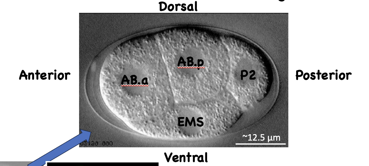
Is the anterior-posterior axis set in the unfertilized egg? What is the distribution of molecules prior to fertilization? What establishes this?
No
PKC = protein kinase C
PAR = partitioning defective

How does sperm entry break symmetry and define the egg's posterior side?
Whichever side the sperm enters becomes the posterior part; nucleus placement doesn’t matter
Aurora A (AIR-1) is already in the egg prior to fertilization
Entry of the sperm causes Aurora A to re-localize (due to the entry of the sperm centriole)
AIR-1 prevents actomyosin contractions (which would cause actomyosin to collapse)

How do the centrioles move to the nearest pole? How does AIR-1 suppress actomyosin contractions?
The sperm centriole organizes microtubules that help bring it to the nearest pole
AIR-1 phosphorylates proteins that cause actomyosin collapse

The first cell division in C. elegans yields unequal cells. What importance does this have?
The larger blastomere’s cortex is made of aPARs
The smaller blastomere’s cortex is made of pPARs, but shares an aPAR cortex where the two cells meet
The difference in cortex allows the mitotic spindle to set up in a different direction, allowing for rotational cleavage
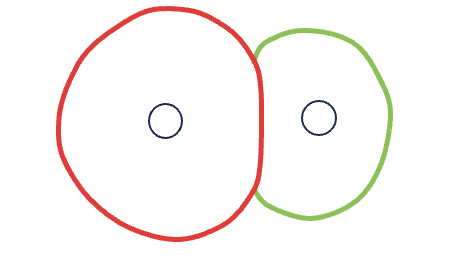
How is the study of PAR proteins relevant?
PAR proteins are conserved polarity components
These proteins are highly conserved in flies and mammals (including humans)

How would you identify genes involved in partitioning components and specifying early cell fates?
By identifying the genes active in the mother that make maternal contributions put into the egg (since these partitioning components are put into the egg by the mother and are present before fertilization)
aka maternal effect genes
What are maternal effect genes?

What are the two maternal effect lethal mutations? What screens for these?
Mutations in polarity genes (par genes and others): mediate asymmetric segregation of cell fate-determining gene products
Mutations in cell fate-determining genes: gene products that make cells different from each other
Products of the MEL genes (mRNA or proteins) are pre-packaged into the embryo by the mother required for cell fate determination and survival of the embryo
Screened for by forward genetics
What is the C.elegans fate map of the five asymmetric cell divisions?

How are MS cell fates specified in C.elegans embryos?
SKN-1 is required for EMS fate specification
What happens with SKN-1 mutants?
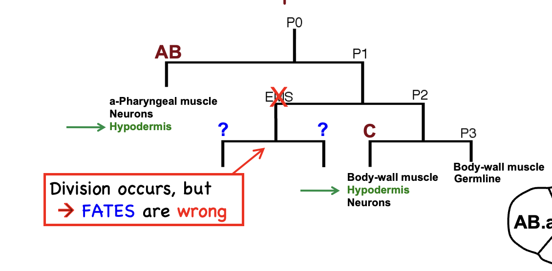
Without functional SKN-1, the EMS cell reverts to a default lineage program: Instead of forming a stomach and gut, it generates excess hypodermis (skin) and body wall muscle. This is why the gene was named skn-1 ("skin in excess")

Why do SKN-1 mutants make excess skin?
Division of EMS still occurs, but cell fates are wrong
In the absence of SKN-1, EMS cells follow cell fate C, which makes hypodermis
SKN-1 is a transcription factor EMS daughter fates require SKN-1, and MS daughter fate is instrinsic (autonomous)
Why is it important to have mechanisms that maintain germline potency?
It is important to keep the germline largely transcriptionally silent to keep cell fate-determining genes off
Mechanisms that preserve potency keep the cellular lineage uncorrupted by blocking somatic differentiation
How does SKN-1's location determine EMS cell fate?
SKN-1 is present in both EMS and P2:
In P2, PIE-1 (a CCCH zinc finger transcriptional repressor) maintains transcriptional silence in the germline; aka PIE-1 inhibits SKN-1 in P2
PIE-1 keeps cell fate genes off, blocking polymerase from elongating on those genes
What do the cell fate maps of wild-type, SKN-1 mutants, and PIE-1 mutants look like?
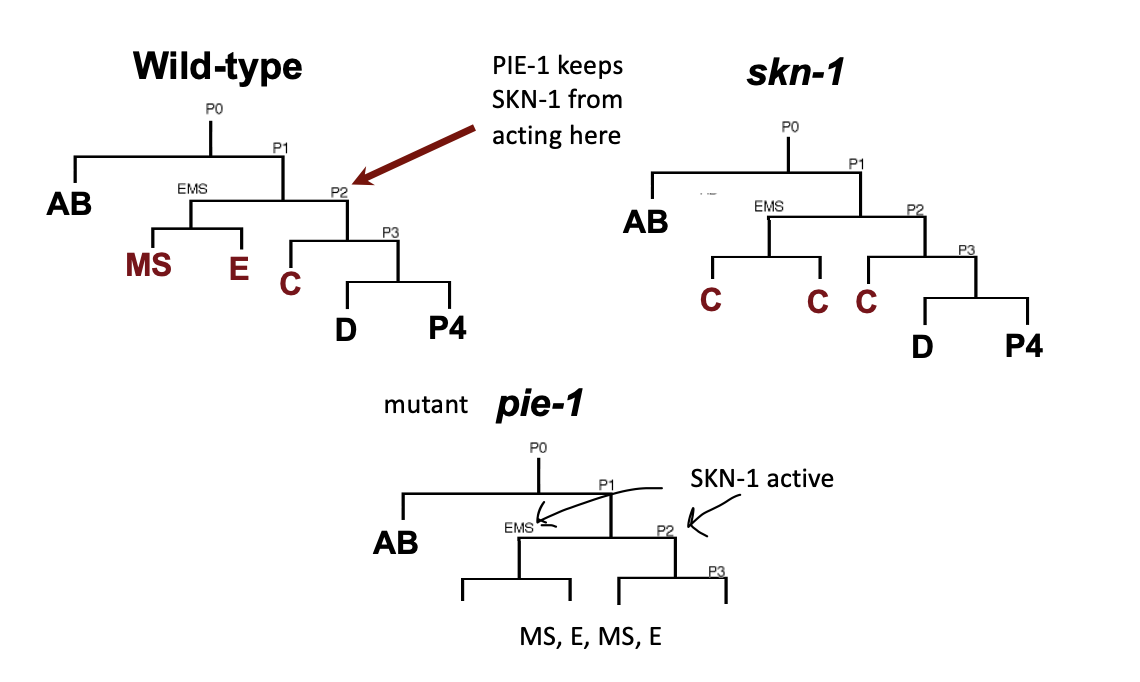
What is combinational regulation? When is this used in C. elegans?
When multiple transcription factors, signals, or genetic elements act together to determine whether a gene is turned on or off
Used to specify and properly restrict cell fate identity
PAR-1 and MEX-1 keep SKN-1 out of AB cells
PIE-1 keeps SKN-1 inactive in P2

What happens if SKN-1 is moved to AB.a and AB.p in a MEX-1 mutant?
SKN-1 is necessary for MS and E fate, but insufficient for cells to adopt gut fate (E)

How do sea urchins and C. elegans asymmetrically position components to specify fates?
In sea urchins, the mother places components into the oocyte, and they are tethered to a specific spot; cell division happens, but maternal components are already in the correct location
In C. elegans, the mother places components into the oocyte, but without a mechanism for asymmetric localization; the embryo needs to sort components/activities to the right place
Even when SKN-1 is introduced into AB cells, they do not adopt a gut fate (E). Does the missing factor act intrinsically, or is it supplied extrinsically (i.e., from another cell)? How do you know?
Extrinsic
When an EMS cell is isolated, it does not form gut, suggesting E is specified non-autonomously
Where does the extrinsic signal that specifies E cell fate come from?
Comes from P2
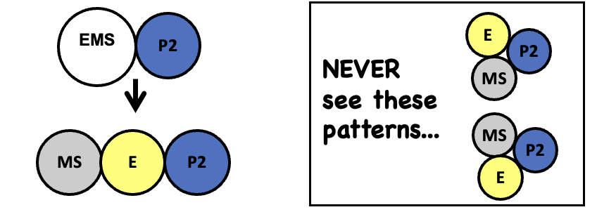
When EMS and P2 are put in contact, the pattern on the left is always the result. Why?
P2 reorients the mitotic spindle
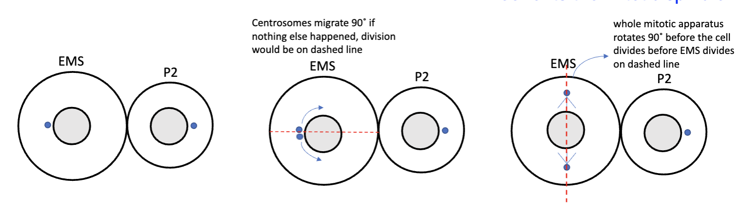
What genes specify gut fate in C. elegans?
mom-1 to -5 genes
Mutations in these lead to more mesoderm production and no gut
EMS → MS + MS fates
More mesoderm mutants
pop-1
Mutations in these lead to too much gut
EMS → E + E fates
Posterior pharynx defective mutants
What is the signaling pathway that involves gut fate genes in C. elegans?

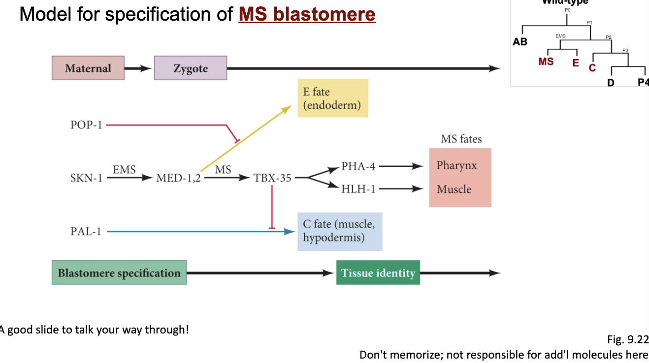
Be able to talk through this
hi
What is important about the C. elegans vulva?
The vulva forms part of the egg-laying apparatus and connects the uterus to the environment
Vulva formation occurs post-embryonically, meaning that it relies on zygotic genes rather than mRNA from the mother
Forward genetic screens easily identified many vulva defective zygotic mutants
What are some of the mutations affecting vulva formation that are easy to identify?
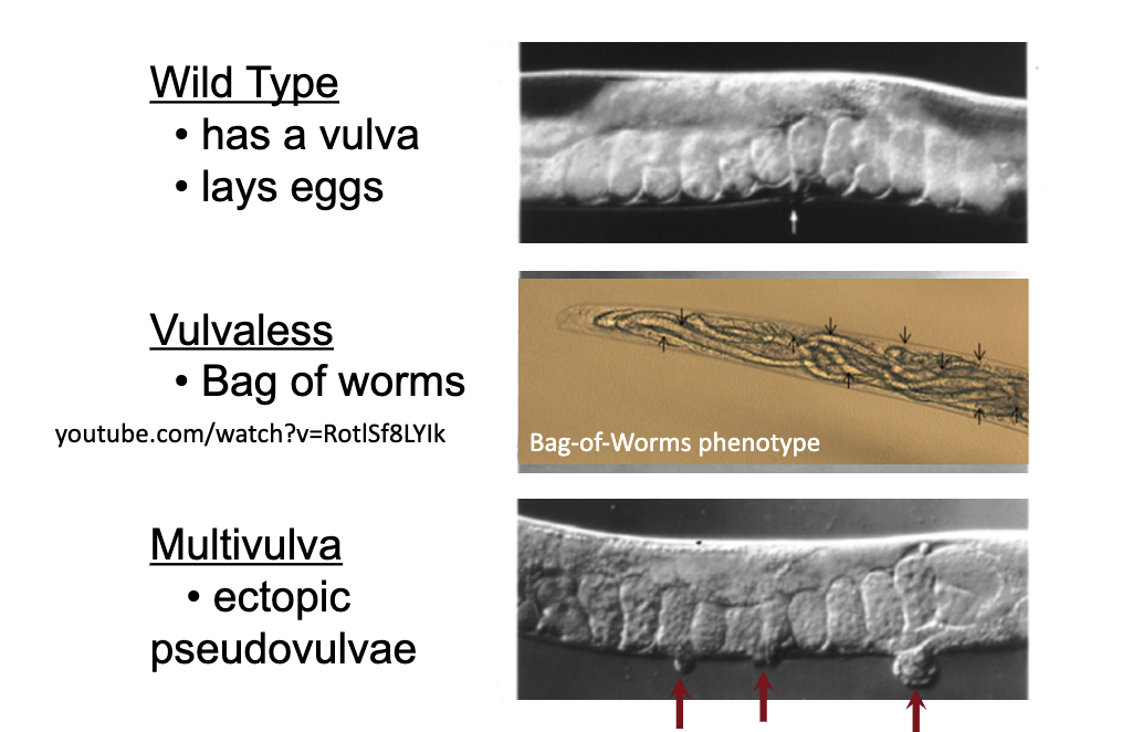
Why study vulva formation in C. elegans?
It maps how conserved molecular pathways interact, specifically the RTK/Ras/MAPK pathway
RAS genes are the most frequently mutated oncogenes in human cancers
Components originally identified in C. elegans have been found to be conserved in mammals, allowing for gene discovery, mechanism discovery, and new drug targets
What cells does wild-type vulva form from?

What do these original wild-type cells divide into in the mid-to-late L3 stage?

How is laser ablation used to determine which cells can contribute to the vulva?
Focus a laser beam through an objective lens of a microscope to kill single cells; this allows you to study what happens if a cell is removed
What happens when P6.p is killed using laser ablation?

What is the equivalence group in the vulva experiment? What quality do they have?

What happens when the anchor cell (AC) is ablated? What does this tell us?
No vulva forms
Some sort of inductive signal is coming from the AC
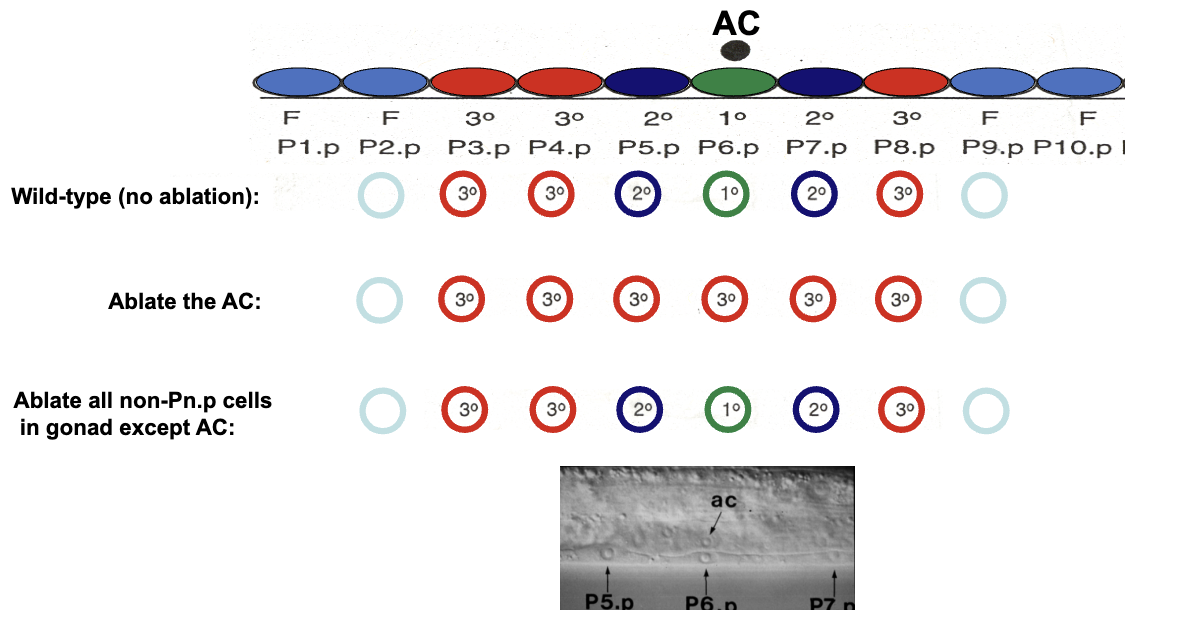
What are the 4 genes you need to know that are required for vulva formation? Are these zygotic or maternal?
Required for specification of AC fate:
lin-3 (Epidermal Growth Factor (EGF) signaling is the AC signal)
Required for specification of VPC (vulval precursor cell) fates
lin-12 ligands
lin-12
let-23 (EGF receptor, the lin-3 receptor)
All zygotic
What are two ways the AC could induce vulva formation?

What is a morphogen?
A molecule that elicits different responses depending upon its concentration
How would the AC induce vulva formation in the case of graded signaling?

What evidence is there to show that the AC uses a graded signal (and shows that lin-3 acts at a distance)?
The P3 one is further away and becomes secondary, in comparison to P3, which is closer and becomes primary
Shows that lin-3 acts at a distance
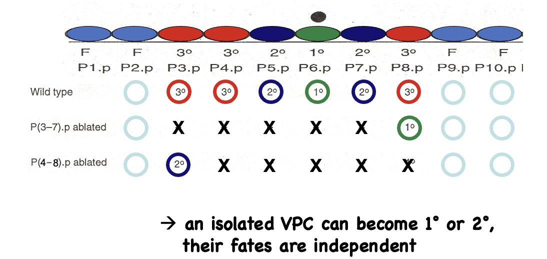
What evidence is there to show that the AC uses a graded signal (and shows that lin-3 action is concentration-dependent)?
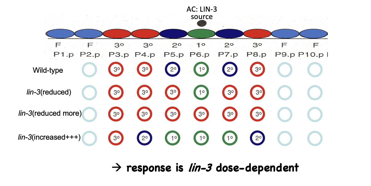
What would happen if P5.p was missing the let-23 receptor (assuming the graded signaling model is true)?

What would happen if P5.p was missing the let-23 receptor (assuming the sequential signaling model is true)?
If not receiving the lin-3 signal, it doesn’t matter if it has a receptor; only P6.p has to receive the signal from AC
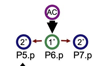
What evidence is there to show that the AC uses a sequential signal?
Shows that lin-3 acts in a relay system

What is the overall conclusion for AC signaling?
There is a combination of graded LIN-3 signal and sequential signaling
Checks and balances
What is a stem cell?
A cell that can divide indefinitely to make two types of daughter cells:
Cells of its own type/another stem cell (self-renewal)
Cells of a different, more specialized type (aka differentiation)

What does it mean to say that stem cells are immortal?
They can indefinitely produce differentiated cells while also maintaining their own identity as stem cells
Most cells can divide a limited number of times, stem cells divide indefinitely
Why do we need stem cells?
Needed for development:
Humans start from one cell and divide into 10-70 trillion cells
Needed for homeostasis:
There are 50-70 billion cell divisions in the human body per day
Stem cells replace the cells in our body that die
What are the three types of stem cell divisions?
At the population level, symmetric renewing and symmetric differentiation can maintain homeostasis (stem cell populations are doing both)

What are the levels of stem cell potency?
Totipotent: all cell types (least determined)
Pluripotent: all embryonic and adult cell types
Multipotent: multiple cell types
Unipotent: one cell type (most determined)
What cell types can’t pluripotent cells make?
Anything that comes from trophoblasts (extraembryonic cells)
Where is the only place where totipotent and pluripotent stem cells occur naturally?
In the early embryo
What do multipotent stem cells usually make? Name an example
Usually make related cell types and are tissue/organ-specific
Hematopoietic stem cells (HSCs):
Know the first division and that the two progenitor cells make different things

What is the difference between a stem cell and a progenitor cell?
Stem cells can divide indefinitely and generate specialized cells
Progenitor cells can differentiate into multiple cell types, but can only renew a limited number of times (they are a transient cell population)
Oftentimes progenitor cells have lower potency, but potency is not the fundamental difference (as in some cases they can have the same potency as stem cells)
Name an example of unipotent stem cells
Spermatogonial stem cells producing sperm
How many spermatocytes can arise from a single spermatogonial stem cell? What type of cell division is involved?
Note: this process works basically the same in the ovary to make oocytes

What is the niche hypothesis?
The niche is a special microenvironment that regulates stem cell self-renewal and survival
Leaving the niche induces differentiation
Three features:
Signaling factors keep cells alive and in multipotent states
Adherence factors keep cells in the microenvironment
Asymmetric division positions daughter cells with respect to the niche
What is a gap cell? What is a cystoblast?
Gap cell: a somatic cell that makes up the niche germ stem cell (GSC): the stem cell of the ovary (in Drosophila)
Cystoblast: the differentiating daughter of the GSC

What is the adherence factor in the fly ovarian stem cell niche?
E-cadherin keeps one daughter cell in the niche

What is the signaling factor in the fly ovarian stem cell niche?
Localized signal:
Cap cells make and secrete BMP
ECM molecules (HSPGs and collagens) keep BMPs close to the source
BMP signal is transduced to the GSC nucleus
The bam gene is repressed, which prevents differentiation

What type of signaling molecule is BMP, and how does it work?
It is a morphogen
BMP binds to a receptor, leading to phosphorylations that activate Smads that regulate gene expression

Explain the asymmetric cell division in the fly ovarian stem cell niche
One daughter cell is displaced away from the BMP signal, which physically separates it from the signal
E-cadherin anchors the centrosome to control mitotic spindle orientation (make it divide left/right)
Oocyte differentiation program is on, since the BMP signal cannot reach this far (bam is activated)

What would happen if all the cells in the ovariole contained a null mutation in the bam gene?
No oocytes would be made

What would happen if HSPGs are lost from the ovary?
There is not a high enough concentration of BMP to make GCSs (the signal diffuses out)

In AC signaling, there is a combination of a graded LIN-3 signal and sequential signaling. What is the purpose of sequential signaling, and how does it work? What experiment shows this?
When you laser ablate P cells, the closest one to the AC always becomes primary, and the one next to it becomes secondary, even when the concentration of LIN-3 presented to both cells is the same
This indicates lateral inhibition: once a cell adopts a fate, it prevents its neighbors from adopting that fate as well

Which molecule acts in the lateral inhibition of AC signaling? What happens if you remove it or add more? What is it?
lin-12
In lin-12(+++), the secondary fate happens even in the absence of the lin-12 signal
